Depending on the vessels being examined, the following types of angiography are distinguished:
- aortography;
- arteriography;
- phlebography;
- lymphadenography, or lymphography, lymphangiography;
- coronary angiography - examination of the arteries of the heart;
- angiopulmonography - study of the blood vessels of the lungs;
- cerebral angiography - examination of the blood vessels of the brain.
As a contrast agent for the angiography procedure, as a rule, water-soluble iodine compounds are used: hypaque, cardiotrast, urografin, uroselectan, triyotrast (not used for angiography of cerebral vessels!), etc. Currently, new types of contrast agents that do not contain Yoda.
This research method is most often used to detect atherosclerotic changes in blood vessels, diagnose heart diseases, assess kidney function, recognize stenoses, occlusions and aneurysms of blood vessels, tumors, blood clots, arteriovenous shunts (pathological anastomosis between arteries and veins). In addition, the angiography procedure is used as a preoperative testing method before open heart and brain surgery. Angiography of the lower extremities is used to diagnose narrowing of the arteries, blockage of the arteries. During angiography, it is possible to perform balloon dilatation and stenting of blood vessels.
What is it for?
Angiographic research is used to clarify the diagnosis and prescribe the most effective treatment. Also, such an examination is carried out in preparation for surgery, making it possible to determine the location and condition of the vessels. Angiography is combined with endovascular interventions: angioplasty and stenting. Sometimes the decision about the need to implant a stent into a vessel is made immediately during an angiographic examination - in this case, stenting is carried out simultaneously with angiography.
Subtraction angiography
Questions and answers
Possibly gongrene I injured my hand, the skin was torn off while drinking on gravel and my hand became rusty in two hours, what should I do?
Answer:
See a surgeon for an appointment.
Hypoplasia of the left vertebral artery
Hello. I have been sick for a long time. Nausea. vomiting. Impaired coordination. unsteadiness. Severe headaches. What examination should I do before someone turns to you for help?
Answer:
Do MSCT of the arteries of the neck and brain with contrast.
Thromboangiitis obliterans
Hello. My mother is 58 years old, disabled group 1. She had both legs amputated below the knees due to thromboangiitis obliterans. We underwent an ultrasound examination of the NK vessels. What additional research needs to be done...
Answer:
What about the stumps now?
Leg amputated
Hello! My brother had his leg amputated in December 2021 due to atherosclerosis, after which he developed gangrene. In January we underwent re-amputation. It is now March, but the wound is not healing. They did a CT scan of the vessels...
Answer:
What to do?
Restore blood flow. Send a link to MSCT of vessels. Gangrene
Hello, my dad had gangrene on his right leg on the big toe, his toe was amputated, the treatment prescribed by the doctor did not help, there was pain, a large crust and there was pus, they applied ointments...
Answer:
It is urgent to perform an ultrasound of the arteries of the extremities and MS CT with contraception; after receiving the examination results, we will be able to offer you the optimal treatment method.
How to treat trophic ulcers and necrosis of the fingers.
Hello. After examination at the Donetsk Institute of Emergency and Reconstructive Surgery named after. In K. Gusak (DPR), my husband was diagnosed with ischemic heart disease: atherosclerotic cardiosclerosis. CH2a. GB 2st. risk 3. Left ventricular thrombus. ...
Answer:
Good afternoon.
The left leg suffers from ischemia, i.e. lack of blood flow. To prevent it from bothering you, you need to restore blood flow. Need surgery. Perform CT angiography of the abdominal aorta and arteries of the lower extremities (to the feet)…. Red spots.
Hello, I broke my leg in September, but after 4 months, red spots in the form of bruises appeared on my leg, and they just don’t go away. WHAT COULD BE?
Answer:
Good afternoon.
Without examination it is not possible to make a diagnosis. See a traumatologist. Wet gangrene
Hello! My dad (70 years old) has wet gangrene of the leg, we live together in the same apartment with a small child (2 years old), is this situation dangerous for the baby? Thank you!
Answer:
Good afternoon.
Gangrene is dangerous if it is accompanied by an infection. Show the patient to the surgeon. Atherosclerosis of the lower extremities.
Hello, my dad is sick, he is 81 years old. atherosclerosis, calcification of blood vessels of the lower extremities. In Perm, doctors did everything they could (including angioplasty, which did not bring results). For now…
Answer:
Most likely it is possible, but you need to see the patient in person.
You cannot establish a forecast by correspondence. Occlusion of the upper limb.
My mother is 68 years old. Since August 2021, for the first time, very severe pain appeared in the elbow on the right. Gradually, the pain intensified and spread lower throughout the arm; conservative treatment had no effect. Consulted by a neurosurgeon from the Federal Center of Neurology of...
Answer:
Perform CT angiography of the arteries of the upper extremities. Send a link to the study by mail
Decoding the results
There is a certain technique for deciphering angiography results. Markers are identified for each vascular pathology. For example, if the vessels have intermittent outlines in the images, we can talk about the presence of pathologies such as thrombosis or atherosclerosis. The detection of protrusions in the walls of blood vessels in the images indicates that there are either congenital changes, an aneurysm, or the same thrombosis. We can also talk about congenital pathologies in cases where the contrast agent does not enter the capillaries, but passes directly into the vein.
If, upon administration of contrast, there is a lack of its spread, this indicates the presence of thrombophlebitis, thrombosis or thromboembolism.
Recovery after the procedure
How quickly a patient recovers after angiography depends largely on how extensive the study was. There are several recommendations that will help the patient recover faster:
- Follow bed rest and diet for several days after the procedure;
- eliminate stress and anxiety;
- exclude physical activity;
- Take antihistamines to minimize the likelihood of an allergic reaction.
As a rule, if the study was performed correctly, the patient's recovery takes approximately 2-3 days.
How is the procedure done?
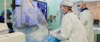
How angiography is done can be described as follows:
- administration of antihistamines (even if a preliminary test showed that there is no allergy to iodine);
- antiseptic treatment of the area of the body where the puncture will be made to introduce contrast;
- anesthesia (usually lidocaine is used);
- incision of the skin to provide access to a vein or artery;
- installation of an introducer (hollow tube) to facilitate further insertion of the catheter;
- administration of a drug that prevents vascular spasm;
- inserting a catheter into a hollow tube and moving it to the beginning of the vessel to be examined;
- administration of contrast;
- shooting.
After the shooting is completed, the catheter is removed, hemostatic measures are carried out (if there is bleeding), and then a tight bandage is applied to the puncture site.
Preparation for aortography
In preparation for angiography, the patient must donate blood for a general analysis, a biochemical analysis to exclude kidney and liver pathologies, and a coagulogram (blood test for clotting).
It is necessary to notify your doctor about allergic reactions, especially to iodine-containing drugs.
Aortography is best performed on an empty stomach to avoid nausea and vomiting. Fluid intake is not limited. If endovascular intervention is planned, you may be given antithrombotic drugs before the angiography.
Immediately before the procedure, you must sign an informed consent for the procedure.
Before the examination, the patient must go to the toilet, remove all metal objects (since they are reflected on x-rays and can distort the picture) and change into special clothes
During angiography, the patient is in a supine position and fixed to the table, since movements during the examination can distort the results.
Where can you get angiography with contrast in Moscow?
You can make an appointment with the specialists of JSC "Medicine" (clinic of Academician Roitberg) on the website - the interactive form allows you to select a doctor by specialization or search for an employee of any department by name and surname.
Each doctor’s schedule contains information about visiting days and hours available for patient visits. Clinic administrators are ready to accept requests for an appointment or call a doctor at home by calling +7 (495) 775-73-60.
Convenient location on the territory of the central administrative district of Moscow (CAO) - 2nd Tverskoy-Yamskaya lane, building 10 - allows you to quickly reach the clinic from the Mayakovskaya, Novoslobodskaya, Tverskaya, Chekhovskaya and Belorusskaya metro stations .
What happens during CTCA?
The CT-CAG test usually consists of three parts:
- Preparation,
- scanning
- recovery.
Preparation:
Before the procedure, you will be asked about your medical history (the problems or symptoms that led you to be referred by your GP or specialist for testing).
Your pulse will be checked using an electrocardiogram (or ECG) machine. About four electrode patches will be placed on your skin at the front of your chest so that the ECG leads can be attached.
The specialist doctor supervising your procedure (a radiologist or cardiologist) will look at the ECG. If your heart rate is normal, an intravenous cannula will be inserted into one of your veins, usually on the front of your elbow in a skin fold.
Depending on the type of CT scanner used, if your heart rate is above 60 beats per minute, you may be given a medicine called a beta blocker by mouth (tablets taken by mouth) or intravenously (through an intravenous cannula) to lower your heart rate. Reducing the heart rate makes images clearer and easier to understand. Your blood pressure and heart rate will be monitored, and when your heart rate has dropped to the appropriate level (normal rhythm), you will be moved from the preparation area to a room with a CT scanner.
Scanning:
The scan is performed in the supine position (see Computed tomography (CT)). The bed slides in and out while a picture of your heart is taken. It is important not to move while scanning as this will affect the quality of the images.
While you are lying in bed, you will be given a quick intravenous injection of iodine contrast material through a cannula using a pressure injector. It is often called X-ray "dye", but it is a clear and colorless liquid.
When the iodine contrast reaches the heart through the veins, a scan is initiated. You will hear the CT computer rotate around you and the bed move in and out of the scanner as images are taken.
You will be asked to hold your breath for approximately 10-12 seconds with each scan as movement will cause the images to blur. The CT scanner takes a series of “slices” of the heart from top to bottom. Your ECG is recorded at the same time these images are taken. The scanner uses an ECG recording of your heart's electrical impulses, so each time it beats, CT images are taken. The CT scan is coordinated with the ECG, and during the period or periods when the heart is moving the least, images are taken of the coronary arteries without movement, so they appear sharp rather than blurry.
The images are analyzed by a radiologist (medical imaging technologist) who performs the scan using sophisticated computer programs. Narrowing or blockage of the coronary arteries, which may be the cause of heart attacks or other symptoms, can be confirmed. Other information can also be obtained about changes in the heart muscle, the inside of the four chambers of the heart, the valves, the membranes that surround the heart (pericardium) and the rest of the chest outside the heart if it is included in the scan.
Recovery:
Once all the scans have been done (about 20 minutes), you will be taken to the recovery area for observation and the IV cannula will be removed before you are allowed to go home. If you have been taking medications that lower your heart rate, you may be asked to stay until the effects wear off.
Possible complications of carotid angiography
Complications are rare - the risk is 0.5%. The most dangerous are complications from the brain. They can develop as a result of the effect of contrast or when pieces of plaque are torn off during catheter insertion. Complications include:
- Transient ischemic attack - short-term disturbance of cerebral circulation
- Ischemic stroke is a persistent disorder of cerebral circulation
- Bleeding from the arterial access site
- Carotid artery dissection with thrombosis
- Allergic reaction to contrast
Timely diagnosis of complications allows you to minimize their consequences.
Vascular angiography: what it is and how it is done, indications, consequences
You can quite simply describe what angiography means - it is obtaining images of blood vessels. This diagnostic procedure allows you to conduct a full examination of the vessels, determine where they are blocked, identify areas of blood clots, places of narrowing and thinning of the vascular walls. For angiography
a special contrast agent is introduced, which is highlighted during scanning and helps to identify potential or existing pathologies.
Objectives of the method
Angiography is a diagnostic research method that solves the following problems:
- Determination of tumor location;
- Determination of its connection with large vessels;
- Deciding on the possibility or impossibility of tumor removal using traditional surgical methods.
Unlike other tissues, a tumor is capable of creating new vessels that feed it and enable uncontrolled growth. Moreover, the process of creating new vessels is constant.
By studying the vascular network of the tumor, the doctor can accurately determine its size and boundaries with healthy tissues. As the tumor develops, it grows into tissues and large vessels, and, narrowing their lumens, changes, i.e. turn into cancer.
Surgical removal of a tumor in the area of large vessels depends on the degree of involvement of these vessels in the process of its growth.
The angiography method is based on the introduction of a contrast agent into the blood vessels. The images taken make it possible to determine the filling of the tumor vessels and neighboring arteries with contrast.
You can determine the passage of the contrast agent:
- X-ray;
- Computed tomography;
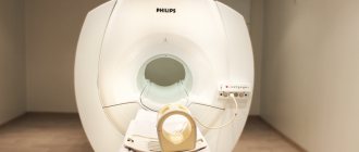
- Magnetic resonance imaging.
How can I prepare for the CTCAG?
CT images will be clearer if your heart rate is low, and you may be given medication before the test to slow your heart rate. It is recommended not to drink tea (including herbal teas), coffee, colas or other stimulants before the procedure as they contain caffeine, which can increase your heart rate.
There is no need to avoid eating or drinking before the procedure, but a full stomach is not recommended as this, along with the contrast material, may make you feel nauseous. However, each radiology facility will ask you to comply with their own requirements regarding any fasting before the examination.
When making an appointment, it is important to tell the radiology department staff if you have asthma, diabetes, kidney problems, irregular heart rhythm, or have a history of allergies to contrast agents used in a radiology procedure, or if you have a history of severe medical conditions. allergies to other things (such as food, pollen or dust).
If you have any of these conditions, you may not be able to take this test.
The procedure may require several hours of preparation after you arrive at the radiology department before you undergo CTCA.
If you are taking metformin-containing medications (Glucophage, Siofor, etc.), you must stop taking them on the eve of the procedure and in the next 24 hours to avoid unwanted reactions to the administration of contrast.
Are there any consequences of CT-CAG?
If medications have been prescribed to slow your heart rate, you will usually be monitored until any possible dizziness has subsided, which is usually about half an hour, although this may take longer.
If you have a nitroglycerin headache, it usually goes away relatively quickly in about 20 minutes, and often faster.
An allergy to the contrast agent may occur. This can range from mild effects such as sneezing, itching, rashes and hives to severe reactions. Severe reactions are rare but can lead to difficulty breathing, a drop in blood pressure, and swelling of the soft tissues of the face and throat. When this occurs in the respiratory tract, it can be life-threatening. Such reactions are very rare, but they stop immediately on the spot.
If you have previously had an allergic reaction to contrast material or have a strong history of allergies to other things (such as food, pollen, or dust), you should tell the medical staff at the radiology facility before your procedure.
The radiation dose for the procedure is approximately 2–21.5 millisieverts (a measure of radiation dose).
How long does a CT-CAG take?
The entire procedure, including preparation, scanning and recovery, can take up to 3-4 hours, especially if you were given beta blockers. The actual CT scan will take about 20 minutes.
What are the risks of CT-CAG?
Main risks of KTKA:
- Complications of the procedure and iodinated contrast agent; For example:
- Rupture of a vein from the cannula, which is rare.
- Injection of contrast material into surrounding tissue as a result of rapid injection of contrast material, which may cause the wall of a small vein to rupture.
- Air is injected into the vein, although most modern injectors have safety precautions to prevent this from happening.
- An allergic reaction to contrast material, which may include sneezing, itching, rash, and hives, which occurs in a small percentage of patients. This usually occurs within a few minutes after the injection. More serious reactions are rare and include a drop in blood pressure and soft tissue swelling, which can be life-threatening. These reactions require immediate treatment. The medical staff at the radiology department where you are having the procedure are trained to treat severe reactions if they occur.
- Patients with renal failure (kidney problems) may experience worsening kidney function after receiving iodinated contrast. This usually improves within a few days. If the impairment of kidney function is severe, the procedure is usually not performed unless the information obtained from the scan is considered so valuable that it outweighs the risk of further deterioration of kidney function.
What are the advantages of CT-CAG?
CT-CAG is a relatively new test and techniques are still evolving with the rapid development of new equipment. There is still disagreement among medical specialists (cardiologists and radiologists) regarding the benefits of the test. Published information suggests that if this test is done and no coronary artery disease is found, your doctor can use this information to treat your symptoms. When the coronary arteries show abnormalities, your doctor may change your treatment according to the details of the abnormalities shown.
The advantage of the test is that it can show the extent and location of atherosclerosis (a disease that obstructs blood flow in the arteries) in the coronary arteries, even if it does not cause obstruction to blood flow.
Along the way, during the study in our center, the calcium index is determined (according to the Agastson method) to assess general calcification and make a more correct choice of further treatment. All chambers of the heart, the mobility of their walls and the exclusion of myocardial fibrosis, and the condition of the pericardium are also assessed.
Who does CT-CAG?
A team of medical personnel is usually involved, including medical specialists (radiologists, cardiologists), radiologists and nurses. The images are usually interpreted by a radiologist and/or cardiologist, and a written report is provided to your physician.
Where is CT-CAG performed?
CTCA is performed in imaging departments of public and private hospitals, as well as in private radiology clinics.


