During the physiological formation of tissue cells, various disturbances can occur. As a result, neoplasms are formed, which can be both benign and malignant. Benign tumors do not cause discomfort to the patient and do not affect the functioning of the body. Such tumors include osteoma of the frontal bone .
Causes
Not a single theory of the origin of the disease has been reliably confirmed.
Presumably, the infectious factor plays an important role in its development. There is often a natural connection between the occurrence of arteritis and previous influenza and group B hepatitis. The genetic programming of pathology also has its supporters. Cases of this rather rare disease have been observed in close relatives and identical twins. The leading role in the formation of inflammation of the vascular wall belongs to immunological disorders, and this is recognized by adherents of all etiological concepts. With temporal arteritis, the immune system reacts inadequately to its own tissues - the process proceeds as an autoimmune one.
What is osteoma?
It is a benign tumor that appears on the bone and produces bone tissue. Osteoma grows, but very slowly and never degenerates into a malignant form. It is usually located on the bones of the skull, may be on the facial bones, and can also be located on the phalanges of the big toes, on the humerus and femur.
The tumor has different types. It can be spongy, hard, brain-like. According to another classification, there are 2 types of neoplasms: hyperplastic and heteroplastic. The first type develops from bone tissue, and the second - from connective tissue.
Osteoma is easily detected. On the skull it is located in the back of the head, on the forehead or in the maxillary sinuses. On the head, the tumor consists of bone tissue, and if it is in the tubular bones, it is made of spongy substance. Sometimes an osteoma can be located inside a bone.
Symptoms
The disease has no characteristic onset. Several options are possible: acute, subacute, but more often with a long period of precursors, which can last several weeks or more than one month.
The set of symptoms preceding the height of the disease and united under the general name polymyalgia rheumatica includes the following manifestations:
• general malaise;
• slight increase in body temperature within 37.2–37.5°C;
• excessive sweating, especially at night;
• aching joints;
• pain in the muscles;
• sleep disorders;
• weight loss.
Later, vascular disorders come to the fore; their nature and severity depend on the location and degree of damage to the artery. More than half of patients suffer from vision impairment. Patients note:
• headache of varying intensity, often sudden, in various areas (temporal, frontal, parietal, less often - occipital);
• hyperesthesia (increased sensitivity) of the scalp, making it difficult to comb and wear a hat;
• transient pain and numbness in the tongue and lower jaw, which intensifies when talking and chewing;
• painful, stringy compaction along the inflamed artery;
• visual impairment (decreased visual acuity, diplopia (double image), blindness);
• neurological, mental disorders.
When the aorta, coronary, renal, and mesenteric arteries are involved in the pathological process, which happens in severe untreated variants of the disease, the development of aneurysm, angina pectoris and heart attack, impaired blood supply and kidney and intestinal function are possible.
Could a lump on the temple be cancer?
Cancerous tumors with this localization are extremely rare. Malignant neoplasms on the temple usually arise due to a degenerated benign growth. We also cannot exclude the possibility of melanoma appearing in this area of the head.
Why can our articles be trusted?
We make health information clear, accessible and relevant.
- All articles are checked by practicing doctors.
- We take scientific literature and the latest research as a basis.
- We publish detailed articles that answer all questions.
The development of cancerous tumors is not asymptomatic. Due to this localization, growths of this type provoke neurological and mental disorders, manifested in the form of abnormal behavior of the patient. Also, the growth of a malignant lump is accompanied by constant headaches, attacks of nausea and other severe symptoms.
Diagnostics
The diagnosis of arteritis can be established by performing a histological examination of a section of the superficial temporal artery obtained by biopsy. The sample collection is carried out under local anesthesia and is not difficult. The detection of granulomatous inflammation of the vascular wall with the presence of giant cells is indisputable evidence of this pathology.
But histologists are able to identify typical changes only in half of the cases. The fragmentary nature of the lesion does not always allow for successful selection of the segment for biopsy. However, a negative result does not mean the absence of the disease, since the main criterion for diagnosing Horton’s disease is the totality of clinical manifestations.
Criteria have been formulated and generally accepted to recognize temporal arteritis. The diagnosis is reliable if three or more of the following factors are present:
• age over 50 years;
• headaches with severe intensity;
• vision problems;
• presence of complaints characteristic of polymyalgia rheumatica;
• decrease in the number of red blood cells and hemoglobin level in the blood, increase in ESR.
Sphygmography, rheovasography, and Dopplerography of the affected arteries are of auxiliary value for differential diagnosis. For the same purpose, the presence of C-reactive protein and the level of sialic acid and fibrinogen in the blood are determined.
Treatment: surgical removal of the tumor.
This disease can only be cured with surgery. The operation is prescribed for significant cosmetic defects and if the tumor causes compression of the anatomical structures that surround it. An oncologist surgeon operates.
Is surgery always necessary?
- If the tumor is small in size, does not put pressure on neighboring organs, does not hurt and is not a major cosmetic defect, then there is no rush to undergo surgery. In this case, they are limited to regular MRI, X-ray and observation by a doctor.
- If ocular symptoms, neurological disorders, high blood pressure, a feeling of fullness, or headache appear, the tumor is removed.
Patients who experience any symptoms associated with this disease should always consult a doctor. Removing a small tumor is much easier. This is done using an endoscope. During the intervention, the patient is under anesthesia, the tumor is fragmented and removed in parts. At an advanced stage, craniotomy may be necessary, removing a plate of healthy bone near the tumor and replacing it with titanium.
Rehabilitation
- The first stage is the inpatient surgical department. At this time, measures necessary to prevent the occurrence of secondary infection and to accelerate tissue regeneration are taken.
- The second stage is a special diet with a high calcium content, a rational mode of work and rest. During this period, it is very important to prevent hypothermia and the occurrence of colds for at least six months.
Treatment
At the moment, there are two directions in the treatment of temporal arteritis: therapeutic and surgical . Surgical methods are resorted to in the event of the development of complications such as aortic aneurysm and thrombosis of blood vessels, especially those supplying blood to the eyeball.
The basis of treatment for the disease, without which it is impossible to achieve positive results, is the prescription of steroid hormones (prednisolone). There is no alternative to glucocorticoids. They are prescribed as early as possible and taken for a long time. Doses and regimen are selected individually, under constant laboratory monitoring of clinical and biochemical blood parameters. A combination of hormones and drugs that suppress immune responses is possible. Symptomatic treatment is also carried out using anticoagulants, agents that improve microcirculation, and vitamins.
New bone formation - symptoms and treatment
The diagnostic algorithm includes interviewing the patient, detailing complaints and clarifying the medical history. Particular attention should be paid to fever, weight loss, loss of appetite and other general symptoms. The time that has passed since the discovery of a bone tumor, the presence and intensity of its growth are also specified.[8]
During examination, the size of the formation, its structure, localization and skin changes are assessed. The areas of regional lymph nodes are palpated to assess possible changes - enlargement of the lymph nodes, their adhesion to the surrounding tissue and to each other.
A laboratory search begins with a general and biochemical blood test: determination of alkaline phosphatase, phosphate and calcium.[1] These analyzes provide evaluative information.
There are laboratory markers of bone resorption (dissolution) that confirm the predominance of bone breakdown (possibly at the tumor site).[9]
But their diagnostic value, unfortunately, is limited, since bone resorption can also occur in other conditions not associated with a bone tumor.
Instrumental diagnostics
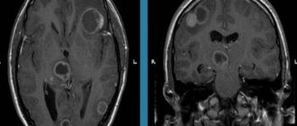
The most accessible, fastest and most revealing instrumental diagnostic method is radiography. An x-ray taken in two projections gives an idea not only of the size and location of the bone tumor, but also of its type.[3]
A more detailed and sensitive visualization of new bone formations is computed tomography.[16] It is especially relevant for joint tumors and small bone tumors that are poorly visible on ordinary X-rays.[8]
Scintigraphy is used as a screening (search evaluation method) - bone scanning after intravenous administration of a radioactive isotope. The isotope accumulates in the bone tumor, causing it to “glow” on the scanner screen. This method provides information about the presence of skeletal tumors and their number. It is also effective for the early detection of bone metastases in cancer patients.[6] However, it is impossible to distinguish a benign tumor from a malignant one, much less determine the type of tumor on a scintiogram.[16]
It is noteworthy that radiation methods for diagnosing neoplasms do not provide the right to reliably diagnose a specific type of bone tumor. Even with all the X-ray signs that speak in favor of a certain tumor, only examination of the tumor under a microscope is decisive.
For microscopy, cells are obtained using a biopsy - taking part of the tumor. For this, a puncture biopsy is used, when the bone is obtained with a wide needle, or an incisional biopsy - surgical removal of the tumor, i.e. cutting off part of it.[16]
Often a biopsy is performed immediately during surgery to obtain a preliminary result. This makes it possible to determine the volume of the operation.[2]
Forecast
Temporal arteritis is a serious disease. If not recognized in a timely manner and treated inadequately, the pathology poses a threat to the health and life of the patient. A timely diagnosis and compliance with doctors’ recommendations avoid complications and make the prognosis favorable.
In our department of vascular surgery, a full examination, qualified interpretation of the results obtained and professional implementation of all types of treatment measures are possible.
Reasons for development
Factors contributing to the development of atheroma on the head are:
- metabolic disorders leading to changes in the properties of the secretion of the sebaceous glands;
- hair dyeing, head shaving with damage to hair follicles;
- hyperhidrosis (especially in combination with seborrheic dermatitis);
- inflammatory diseases of the scalp;
- low-quality hygiene products for hair care;
- Irregular hair washing with the accumulation of viscous oil on the scalp.
Atheromas often form not only on the scalp, but also around the ears and on the chin.
Prosyanka
This is one of the most harmless bumps, the appearance of which on the eyelid does not cause any trouble other than aesthetic discomfort. Millets, also known as milia, come in different sizes. From the smallest - smaller than a poppy seed, to quite large - the size of an average grain of rice. Milia can appear on both the lower and upper eyelids with almost equal frequency. At their core, they are whiteheads localized in the eye area.
Milia can appear in anyone, even in those who have never experienced skin problems. It is better to remove millet from a cosmetologist, because only a specialist can guarantee safety.
To prevent milia, you need to monitor your diet and from time to time make masks to exfoliate dead skin particles that can close pores and clog the ducts of the sebaceous glands.
How is TMJ dysfunction treated?
While there is no single treatment for TMJ dysfunction, there are different treatments that can significantly reduce your symptoms.
Your dentist will be able to recommend one or more of the following methods:
- Try to relieve muscle spasms and pain by applying hot compresses or taking medications such as muscle relaxants, aspirin or other over-the-counter pain relievers or anti-inflammatory medications.
- Reduce the adverse effects of clenching and grinding by wearing a device sometimes called a bite block or splint. Custom-made to fit your mouth, this appliance covers your upper teeth and prevents them from rubbing against your lower teeth.
- Learn relaxation techniques to control jaw muscle tension. Your dentist may recommend that you attend training or counseling to teach you how to manage stress.
- If your jaw joints are not healthy and other treatments are not helping, jaw joint surgery may be recommended.
Our dental clinic employs a team of specialists involving neurostomatologists, osteopaths, and general orthopedists, which gives good results. Patients with this disease should be very careful, because... This is a chronic disease and, like any chronic disease, can get worse. Therefore, it is necessary to strictly follow the treatment prescribed by the dentist. It is necessary to visit your dentist regularly so that the specialist can monitor your joint and adjust treatment in time to avoid relapses and exacerbations.
Treatment tactics
If there is no arthrosis, but there is dysfunction, the tactics are as follows:
- determine the root cause and eliminate it, for example, grind teeth and fillings that impede the functioning of the joint, correct prosthetic errors, and correct the bite;
- reduce the load on the joint - the patient is recommended to switch to soft foods, massage the facial muscles and exercises to relax the jaws;
- if the defects are serious, surgical treatment is performed - bone or muscle plastic surgery of the area of dysfunction.
If the doctor determines degenerative changes in the cartilage tissue, treatment for osteoarthritis will be prescribed. With the help of painkillers, they relieve pain. If necessary, a complex of physical therapy is prescribed, for example, laser or ultrasound. Good results are achieved by intra-articular injections of the Noltrex synovial fluid prosthesis, which restore the functionality of the joint by replenishing the missing lubrication.
Arthrosis of the TMJ is successfully treated with intra-articular injections of Noltrex
Xanthelasma
It is more likely not a lump, but a flat plaque. The problem of xanthelasma is more often encountered by women with hypercholesterolemia, diabetes and a number of other diseases. Xanthelasmas are yellowish in color and rise very slightly above the surface of the skin. They can appear on the eyelids, the skin near the eyes, and on the face. A single lump of xanthelasma is rare; as a rule, they appear in groups and never go away on their own. This is due to the fact that xanthelasma is the result of a lipid metabolism disorder caused by the underlying disease. If such a problem occurs, you must notify your doctor, who will advise what to do.
Barley
This disease is widespread, almost more common than chalazion. When a stye occurs on the lower or upper eyelid, a painful lump occurs due to inflammation of the eyelash bulb (follicle). Both adults and children are susceptible to this disease.
Barley is an inflammatory disease that occurs due to blockage of the sebaceous duct, the secretion from which lubricates the eyelash, thereby protecting it from the effects of the external environment.
There are two types of stye: external, when the sebaceous gland becomes inflamed, or internal, resulting from inflammation of the meibomian gland.
At the first stage of the appearance of external barley, the patient feels the presence of a foreign body in the eye or a stabbing pain. With internal styes, the lump on the eyelid is not so noticeable, although it causes no less discomfort.
In the absence of treatment, a couple of days after the appearance of swelling and redness, a head of purulent contents appears on the lump, which spontaneously opens and goes away after a few days.
And yet barley heals better. Firstly, this will speed up recovery, and secondly, it will help avoid unpleasant complications, such as the development of chalazion. Moreover, if the stye does not go away on its own within 2 weeks, a visit to a specialist is required! An ophthalmologist will open the stye under local anesthesia and recovery will come very soon.
The complex of therapeutic measures for barley includes the use of various antibiotics in drops and ointments. For example, solutions of albucid, gentacymin, penicillin, erythromycin, as well as rithromycin and tetracycline ointments.
Self-help methods for diseases of the temporomandibular joint:
The following self-help methods may provide temporary pain relief:
- Moist Heat: Compressing with a hot pack or bottle wrapped in a warm, damp towel can improve chewing function and reduce pain. Be careful to avoid burns when using high temperature.
- Ice: Ice packs can reduce inflammation, numb pain, and promote healing. Do not place the ice pack directly on your skin. When using, wrap the bag with a clean cloth. Do not use the ice pack for more than 10-15 minutes.
- Diet - Soft Foods: Soft or mixed foods allow the jaw to rest temporarily. Remember to avoid hard, crunchy, and chewy* foods. Do not stretch your mouth when trying to bite off large pieces or the whole fruit.
- Over-the-counter analgesics: Over-the-counter (over-the-counter) analgesics are useful for temporarily reducing pain. Together with your doctor, carefully read the instructions for each medication before using it.
- Lower jaw exercises: Slow, gentle exercises of the lower jaw can help increase jaw mobility. Your doctor or physical therapist can evaluate your condition and suggest appropriate exercises based on your individual needs.
- Relaxation techniques (relaxation techniques): Relaxation can help combat the pain that accompanies TMJ dysfunction. Deep, slow breathing increases relaxation and reduces pain sensitivity.
If you find an error, please select a piece of text and hold LEFT Ctrl and press Enter.
You can send no more than 5 messages in 30 minutes!
Liked? Tell your friends!