Diagnosis of diseases affecting the brain and the vessels that supply it is difficult due to the inaccessibility of the study area. Various pathologies may have similar symptoms or occur latently, without characteristic manifestations. An informative diagnostic method is magnetic resonance imaging, which reveals changes in gray, white matter, vascular wall and allows you to differentiate diseases in the early stages.
MRI procedure of the brain and blood vessels
Do you need preparation for MRI examinations?
Magnetic resonance imaging, as a reliable and accurate diagnostic method that is not affected by harmful X-ray radiation, has virtually no health contraindications. But there are a number of factors that cannot be ignored:
- the presence of a pacemaker located under the skin;
- metal clamps on vessels;
- the presence in the body of foreign metal objects of a fragmentary nature;
- metal crowns or pins in the mouth;
- presence of an implanted cochlear prosthesis;
- internal central nervous system stimulants;
- the presence of a compression-distraction device;
- built-in metal pins;
- the presence of a stationary prosthetic limb.
The above factors prohibit you from undergoing tomography. There are also conditional rules that you just shouldn’t forget about:
- When choosing comfortable clothes for the upcoming procedure, carefully inspect the items for the absence of metal buttons, zippers, hooks, buckles and decorations.
- When going for an MRI diagnosis, remove all jewelry from your body in advance.
- Check your pockets first so as not to miss even a small thing that could interfere with the diagnosis.
In addition to external reasons that prevent MR examination, there are also psychological factors that need to be taken into account.
It is important to warn the patient that he will be immobilized for a long time. The duration of the process can be 30-60 minutes.
Preparing for an MRI with contrast
Contrast is a special dye solution administered intravenously. Before performing an MRI using a contrast agent, specialists need to make sure that the patient does not have problems with the kidneys or liver. A preliminary opinion from a urologist may be required.
It is important to exclude the presence of allergic reactions to the contrast agent. To do this, tests are carried out in advance for rejection of the composition.
Modern drugs are produced based on gadolinium salts. They do not cause allergies, but are excreted through the liver and kidneys. If you have chronic diseases of these organs, problems may arise. Therefore, preliminary consultation with a specialist is necessary.
How does the procedure work?
The patient removes all metal jewelry and takes out removable dentures containing metal.
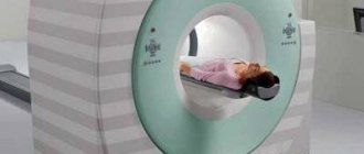
The patient is placed on a movable table and secured with special belts. This measure is necessary because you will have to lie inside the tomograph, remaining motionless, for a long time.
A device equipped with wires that transmit and receive radio signals is placed on the head. The equipment is quite noisy and tires with constant clicks and whistles. Therefore, the patient’s ears are protected with earplugs. After this, the table slides into the device, and the specialist sits down at the computer, in which the transmitted data is analyzed and processed.
The technique takes pictures, the quality of which depends on the characteristics of a particular MRI scanner. The thinner the visual slices the equipment makes, the more accurate the final images will be. The duration of diagnosis is 20-30 minutes, and when contrast is used - up to an hour.
After MRI diagnostics, you can immediately return to your normal life. No side effects subsequently or during the MRI examination occur, with the exception of an extremely rare allergy to gadolinium salts.
Finished photographs are handed out printed or recorded on a magnetic medium - disk or flash card. It can be sent to email with SMS notification.
- Standard - done without the introduction of contrasting solutions, but at the same time provides a sufficient amount of information.
- With contrast, before which drugs containing gadolinium salts are injected into the vein - gadopentetic and gadoteric acids, Omniscan, Magnevist, etc. These solutions penetrate into the bloodstream and, once in the rays of the MRI scanner, highlight the resulting “picture”. At the same time, the changed areas become better visible, which simplifies decoding. The technique is most often used to detect vascular abnormalities, multiple sclerosis and tumor formations. The dose of contrast agent is selected individually, taking into account weight.
- MR angiography – is performed to assess the condition of blood vessels in atherosclerosis, aneurysms, blood clots and pre-stroke conditions. It is done with gadolinium contrast to show blood flow problems in detail.
- MR imaging of the pituitary gland, an appendage that is an endocrine gland. The pituitary gland secretes hormones responsible for reproductive function, tissue metabolism and the regulation of human growth. An examination is prescribed if an adenoma is suspected - a benign tumor that causes migraine-like pain, hormonal imbalances, gigantism, infertility, obesity and sexual dysfunction. The same method is used to identify malignant pituitary formations that have similar symptoms and are accompanied by a pronounced deterioration in health.
Preparing for an abdominal MRI
To be prepared for diagnosing internal organs, you need to familiarize yourself with some rules:
- the procedure is carried out on an empty stomach, so it is advisable to refrain from eating 6-12 hours before the session;
- several days before the study you need to follow a diet that excludes raw fruits and vegetables, sweets, drinks containing gas, dairy products, legumes and mushrooms;
- two to three days before the tomography, take medications that reduce flatulence (Mezim or Espumisan);
- 30 minutes before the procedure you need to take an antispasmodic agent, for example No-Shpa.
Preparing for an MRI of the pelvis
This examination includes the study of organs such as the prostate gland, bladder and adjacent organs, the uterus and its appendages. To prepare for an MRI of the pelvic organs, perform the following procedures:
- cleaning the gastrointestinal tract with an enema;
- fasting for six hours;
- for the examination to be effective, it is necessary that the bladder is partially filled; for this, it is recommended to refrain from going to the toilet two to three hours before the examination and drink half a liter of clean water without gas an hour before the session;
- take the antispasmodic within 30-40 minutes.
How often can I scan with a tomograph?
During scanning of the vascular system of the brain, the body is not exposed to radiation, so there are no restrictions on the frequency of such examinations. The decision on the appropriateness of such a diagnosis is made by the attending physician. For multiple sclerosis, for example, the procedure is performed 1 to 2 times a year. It is prescribed after strokes; in such cases, preventive examinations can be done up to 4 times a year. In Alzheimer's disease, a one-time diagnosis is sufficient to confirm the presence of pathology.
Preparing for MRI in women
Additional rules exist for conducting research in women:
- If a woman is pregnant or suspects pregnancy, it is necessary to warn the doctor about this.
- If possible, avoid undergoing tomography during the menstrual cycle, when internal organs and arteries change in size and contours. Tomography results may be incorrect.
- If a session was carried out using a contrast agent, women who are breastfeeding must interrupt breastfeeding for two days until the dye is completely removed from the body.
What the results show
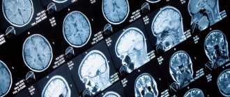
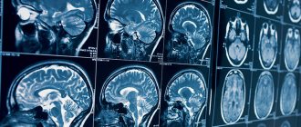
MRI studies, especially those performed with contrast, reveal numerous pathological processes. In the sections, compactions, cystic cavities, and hematomas (collections of blood) are visible in detail. Scars, parasites and their cysts, foci of degeneration, sclerosis and inflammation are identified.
Vascular changes are diagnosed, manifested by impaired patency, narrowing or dilation of blood vessels, the appearance of aneurysms (protrusion of walls) and thrombosis.
The degree of tissue damage in traumatic brain injuries, hemorrhagic and ischemic strokes is determined. The affected areas look lighter and are visible even if they are small in size and have sparse neurological symptoms.
Congenital defects are determined - underdevelopment and hypertrophy of the organ, small and incorrectly located gyri, cysts, holoprosencephaly - lack of division into hemispheres. Hydrocephalus is detected - accumulation of fluid in the ventricles, which with this anomaly are greatly enlarged.
Pathological areas and neoplasms appear as dark or lightish spots of various sizes and shapes, standing out against a grayish background. Oncological seals, especially malignant ones, have blurred, uneven edges and surrounding areas of necrosis.
MR diagnostics is recommended periodically for everyone who has been treated for oncological pathologies of any location. It detects metastases, which usually accompany cancer recurrence.
How to prepare children for MRI
Before going to the procedure with your child, relieve him of internal fears about an unfamiliar situation. The day before, you need to talk to the baby, tell him what awaits him. You need to convince him that the process is safe and painless.
MRI in children has some peculiarities. Since it is difficult for a child to remain motionless for a long time, the session is carried out with the help of mild sedatives or under general anesthesia. During the entire tomography process, the child’s condition is monitored by an experienced specialist – an anesthesiologist.
Before conducting diagnostics with a contrast agent, the presence or absence of allergic rejection to the drug is determined. A severe form of bronchial asthma in a child’s diagnosis may also be a contraindication.
Preparing for an MRI of the brain
If a doctor sends a patient for an MRI of the brain, you also need to know that:
- if there are metal crowns, bridges, dentures, or braces in the oral cavity, visit the dentist in advance and temporarily remove these objects;
- all removable electronic and metal devices must be left outside the threshold of the room with the MR installation;
- emotionally prepare yourself for the upcoming procedure in order to avoid immersion in a stressful state, remember that the diagnosis is absolutely safe;
- If you cannot get rid of your anxiety, ask your doctor to give you a sedative.
By following the above rules, the patient will help specialists conduct an effective diagnosis. The MR procedure will be comfortable and high quality.
Price
Dear patients, on October 9 and 10 there is a 20% discount on MRI!
| Description | Price | Until October 12 | From 21:00-2:00 7:00-9:00 |
| MRI of the brain | RUB 5,400 | RUB 4,590 | RUB 4,050 |
| MRI of cerebral vessels (angiography) | RUB 5,400 | RUB 4,590 | RUB 4,050 |
| Brain MRI with contrast | RUR 11,000 | RUB 10,250 | — |
| Issue of film with photograph | 500 rub.r. | 400 rub.r. | 400 rub.r. |
| Recording a photo on flash | RUR 1,000 | RUR 650 | 600 rub. rub. |
All MRI prices >>>