Ganglionitis is an inflammatory change that occurs in one of the ganglia - a collection of nerve-type nodes. If not one segment is affected, but several at once, the phenomenon is called polyganglionitis. Typically, this pathology is the result of the development of infectious processes, but it can be provoked by injuries, disruptions in metabolic processes, tumor growths, and exceeding prescribed dosages of medications. We cannot talk about the uniformity of the symptoms of the disease. Clinical manifestations may vary significantly depending on where the disorder is located. The symptom that unites all varieties of this disease is a feeling of pain, itching, as well as swelling and active sweating. If you notice similar symptoms, contact the IntegraMed clinic (formerly NDC) on Gakkelevskaya, our specialists will help you cope with the disease.
Causes of ganglioneuritis
The very first cause of this disease is an infectious process. The causative agents can be:
- acute infections: measles, dysentery, sepsis, diphtheria, tonsillitis or influenza;
- chronic infectious diseases (syphilis, tuberculosis, rheumatism).
The cause of ganglioneuritis of the pterygopalatine node can be complicated dental caries, and adnexitis or prostatitis (in men) can provoke sacral ganglionitis. In rare cases, ganglioneuritis can be provoked by a tumor (ganglioneuroma or secondary metastatic process), then the disease is toxic in nature.
Risk factors for the occurrence of ganglioneuritis will be nervous overstrain, hypothermia, constant fatigue, alcohol addiction, and operations performed in the ganglion area.
General symptoms of ganglioneuritis
Symptoms of ganglioneuritis depend on the level of damage; it has a complex clinical picture. Pain syndrome will be the main symptom in the clinical picture. The pain is characterized by a strong burning and bursting character, patients also note a feeling of pulsation.
Patients often cannot accurately indicate the source of pain because the disease is diffuse. Patients describe pain in the entire half of the body, focusing on the fact that the pain is constant and does not change with movement. Increased pain occurs when the weather changes, stressful situations, or after eating.
In addition to the pain syndrome with ganglioneuritis, there is also a loss or partial impairment of sensitivity in the form of hyperesthesia (increased sensitivity) or, conversely, hypoesthesia (decreased sensitivity). Sometimes paresthesia (a feeling of numbness, a sensation of “pins and needles”, tingling or lethargy) is also noted.
There are cases of neurotrophic and vasomotor disorders that are expressed in the area of localization of the affected ganglion and the nerve fibers associated with it. If the disease is long-term, sleep disturbances, emotional instability, and the development of neurasthenia, asthenia, and hypochondriacal syndrome may occur.
Shingles (herpes zoster)
The incubation period for herpes zoster (from the initial infection to reactivation) lasts for many years. The initial period of the disease can be manifested by prodromal signs: headache, malaise, low-grade body temperature, chills, and dyspeptic disorders. At the same time, pain, burning and itching, as well as tingling and paresthesia along the peripheral nerve trunks in the area of future rashes may occur. The intensity of these subjective local signs varies in individual patients. The duration of the initial period varies from 1 to 3-4 days; in adults it is observed more often and it is usually longer than in children.
In most cases, the disease begins acutely. Body temperature can rise to 38-39 °C; its rise is accompanied by general toxic reactions (headache, malaise, chills). At the same time, skin rashes with characteristic pain and other subjective sensations appear in the innervation zone of one or more spinal ganglia.
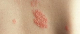
At first, the exanthema has the appearance of limited pink spots 2-5 mm in size, but on the same or the next day, small, closely grouped vesicles with transparent serous contents, located on a hyperemic and edematous base, form against their background. In most cases, exanthema is accompanied by enlargement and tenderness of regional lymph nodes; Children often show signs of upper respiratory tract catarrh.
The exanthema is localized according to the projection of one or another sensory nerve. Most often, the lesion is unilateral: along the intercostal nerves, branches of the trigeminal nerve on the face, less often along the nerves of the limbs. In some cases, skin lesions are observed in the genital area. As the disease progresses, new spots may appear at intervals of several days with the development of vesicular elements against their background. After a few days, the erythematous background on which the vesicles are located turns pale, their contents become cloudy. Subsequently, the vesicles dry out and crusts form, falling off by the end of the 3rd week of the disease, leaving light pigmentation.
The elevated body temperature lasts for several days, the symptoms of toxicosis disappear as it normalizes.
The following clinical forms of herpes zoster are distinguished: 1) gangliocutaneous; 2) ear and eye; 3) gangrenous (necrotic); 4) herpes zoster with damage to the autonomic ganglia; 5) meningoencephalitic; 6) disseminated; 7) abortifacient.
The most common gangliocutaneous form of the disease begins acutely with fever, symptoms of general intoxication and severe burning pain at the site of future rashes. After 3-4 days (sometimes only after 10-12 days) a characteristic rash appears. The localization of pain and rash corresponds to the affected nerves (usually intercostal) and has a surrounding character. The pain sometimes becomes unbearable, intensifying at the slightest touch to the skin, with cooling, or movement. At the site of the vesicular rash, infiltration and hyperemia of the skin first occurs, on which blisters then appear in groups, filled with transparent and then cloudy contents. The bubbles dry out and turn into crusts. Sometimes the disease is characterized by intoxication and neuralgic pain, but there is no rash. When skin rashes appear, the pain usually becomes less intense.
Typical clinical symptoms are the ocular and ear forms of herpes zoster. In the ocular form, the trigeminal ganglion (Gasserian ganglion) is affected and the rash is localized along the branches of the trigeminal nerve (on the mucous membranes of the eye, nose, and facial skin). In the auricular form , the geniculate ganglion is involved in the process, and rashes appear on and around the auricle, and can also be in the external auditory canal. Facial paralysis may develop. The rash is preceded by symptoms of general intoxication and fever. Trigeminal neuralgia is severe and can last for several weeks. In the ocular form, specific viral keratitis, less often iritis, and glaucoma are observed.
The gangrenous (necrotic) form of herpes zoster usually develops in weakened individuals. There is deep damage to the skin with subsequent formation of scars. One might think that the accumulation of bacterial infection plays a certain role in the genesis of these forms.
The meningoencephalitic form is relatively rare. The disease is severe, with a mortality rate above 60%. This form begins with gangliocutaneous manifestations, most often in the area of the intercostal nerves, although it can also occur in the cervical region. Subsequently, symptoms of meningoencephalitis appear (ataxia, hallucinations, hemiplegia, meningeal symptoms, coma may occur). The time from the appearance of skin rashes to the development of encephalopathy ranges from 2 days to 3 weeks.
Generalized form . Sometimes, a few days after the onset of localized exanthema, single or multiple vesicles appear on all areas of the skin and even on the mucous membranes, which is often mistakenly regarded as an addition to chickenpox herpes zoster. If the exanthema is generalized, as well as in cases where localized herpes does not go away within 2-3 weeks, immunodeficiency or the development of malignant neoplasms should be suspected.
Abortive form . Characterized by the rapid disappearance of an erythematous-papular rash and the absence of vesicles.
Any of the above forms may be accompanied by damage to the autonomic ganglia with the development of symptoms unusual for herpes zoster (vasomotor disorders, Horner's syndrome, urinary retention, constipation or diarrhea).
The severity of the disease is often directly related to the location of the exanthema. Cases with rashes located in the area of innervation of the supraorbital, frontal and nasociliary nerves are characterized by intense neuralgic pain, hyperemia and swelling of the skin, damage to the eyelids, and sometimes the cornea.
The duration of clinical manifestations of herpes zoster in the abortive form is on average several days, in the acute form - 2-3 weeks, in the protracted form - more than a month.
Pain in the area of exanthema with herpes zoster is of a pronounced vegetative nature: it is burning, paroxysmal, intensifies at night and is often accompanied by pronounced emotional reactions. Local paresthesia and skin sensitivity disorders are often observed. Possible radicular paresis of the facial and oculomotor nerves, limbs, abdominal muscles, and bladder sphincter.
The disease can occur with the development of serous meningitis; inflammatory changes in the cerebrospinal fluid are not always accompanied by severe meningeal symptoms. In rare cases, encephalitis and meningoencephalitis are observed in the acute period. Cases of polyradiculoneuropathy and acute myelopathy have been described.
The first episode of herpes zoster is usually followed by persistent remission; recurrence of the disease is observed in no more than a few percent of cases. Most patients recover without residual effects, but neuralgic pain can persist for a long time, for several months or even years.
Complications of herpes zoster: transverse myelitis, accompanied by motor paralysis.
Shingles is more severe in people with HIV and other immunodeficiencies. The duration of the appearance of the rash increases to 1 week, the crusts covering the blisters dry out no earlier than the 3rd week of the disease. Patients with lymphogranulomatosis or lymphoma are at greatest risk of developing progressive shingles, and about 40% of them may have a rash spread over the entire surface of the skin. 5-10% of people with disseminated skin manifestations develop viral pneumonia, meningoencephalitis, hepatitis and other severe complications.
Ganglioneuritis of the upper cervical ganglion
The clinical picture of ganglioneuritis of the upper cervical ganglion is characterized, first of all, by the symptoms of Bernard-Horner syndrome. The inflammatory process in this ganglion provokes the development of Pourfur du Petit syndrome (enlarged palpebral fissure, exophthalmos). The inflammatory process affects the functionality of the thyroid gland and causes the appearance of hyperthyroidism. Secretory and vasomotor disorders are observed (hyperhidrosis, redness in half of the face, decreased intraocular pressure). Sensitivity disturbances are observed in the area of the second rib. Possible voice changes or laryngeal paresis. Sometimes patients feel severe pain spreading to the jaw area. Due to the inability to pinpoint the exact source of pain, people often mistakenly resort to dental treatment, which for known reasons does not produce any results.
Treatment
The basis of the therapeutic effect is drug treatment, during which analgesics, antispasmodics, ganglion blockers and antiviral substances can be prescribed. It is often necessary to use antibiotics, sulfonamides, desensitizers, immunomodulators, antipsychotics and antidepressants. Additionally prescribed:
- nootropics;
- vitamins;
- anticholinergics;
- biogenic stimulants;
- injections of glucocorticosteroids;
- novocaine blockades.
Physiotherapy procedures demonstrate high effectiveness. Diadynamic therapy, electrophoresis, darsonvalization, UHF, amplipulse therapy, etc. may be prescribed.
Ganglioneuritis of the stellate ganglion
The clinical picture is characterized by the presence of pain in half of the chest on the side of the inflamed node. Sensitivity and motor reflex disturbances are observed in the fingers. The impairment of motor skills is especially noticeable in the fifth finger of the hand located in the affected area. The area of pain, loss or impairment of sensitivity has a so-called “half-jacket” appearance. Often the pain spreads to the chest area, therefore it resembles angina attacks and must be differentiated from coronary heart disease.
Prevention
Certain antiviral medications may help prevent or reduce the effects of shingles, thereby reducing the risk of postherpetic neuralgia:
- Chickenpox vaccine – The varicella zoster virus vaccine (Varivax) is now a routine childhood vaccine, but may also be recommended for older children and adults who have never had chickenpox. This vaccine does not guarantee that a person will not get chickenpox or shingles, but it may reduce the duration and severity of symptoms and the risk of complications such as postherpetic neuralgia.
- Shingles vaccine - (Zostavax) can be given to people over 60 years of age (who have had chickenpox but not shingles). Zostavax is not recommended for use in certain groups of people (for example, those undergoing cancer treatment or those who are immunocompromised).
- Antiviral medications – Antiviral medications such as acyclovir, valocyclovir, famciclovir, when taken within the first 72 hours after the shingles rash appears, can help shorten the duration of shingles and reduce the chance of developing postherpetic neuralgia.
Physiotherapy helps reduce pain and relieve inflammation. Various techniques are used (including transcutaneous electrical stimulation).
Exercise therapy helps restore the elasticity of ligaments and muscles. Exercises can be carried out both on simulators and in the form of gymnastics.
Acupuncture. This method is quite effective in restoring conductivity and reducing pain.
Diagnosis of ganglioneuritis
Diagnosis of ganglioneuritis is a complex process, since the symptoms of the clinical picture are similar to a number of other diseases (otitis media, coronary heart disease, oncological formations, spinal formations, circulatory disorders, various types of neurosis, etc.). The difficulty also lies in determining the variation of ganglioneuritis, since the symptoms of its various types are very similar. Untimely or incorrect diagnosis can significantly worsen the patient’s condition, slow down the treatment process and, accordingly, lead to disappointing prognoses.
If at least some of the above symptoms appear, you should consult a neurologist. The initial diagnosis of ganglioneuritis usually occurs on the basis of an analysis of the clinical picture from the patient’s words and examination of the patient, identifying signs of vasomotor and neurotrophic disorders, and sensitivity disorders.
Hardware diagnostics of ganglioneuritis
Thoracic and sacral ganglioneuritis are especially difficult to differentiate, and for the accuracy of the diagnosis, the patient undergoes a long examination for the presence of somatic diseases. For this purpose, consultations with a gynecologist, cardiologist, gastroenterologist, and phthisiatrician are prescribed. In order to exclude a number of diseases, radiography of the spine, electromyography, MRI and CT may be prescribed.
The inflammatory process often causes changes in somatic organs, so the patient undergoes MSCT or ultrasound of the abdominal organs, genital organs, and prostate in order to identify these pathologies.