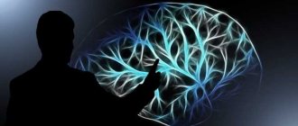
The brain is a complex and poorly understood human organ that is difficult to diagnose. However, the brain is the control center responsible for the operation of all systems in the body. Therefore, any pathological processes in it affect the physical and mental health of a person.
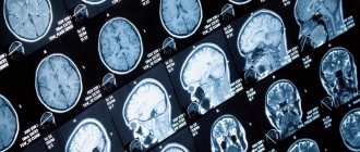
MRI is one of the methods for studying the brain to determine negative conditions in it. Diagnostics is prescribed for adults and children and is a safe examination method.
What it is
The method refers to a non-invasive examination using radio waves and a magnetic field. During the scanning process, the doctor receives an image showing parts of the brain. Diagnosis is carried out without radiography. The study reveals disturbances in the functioning of the nervous and vascular systems, aneurysms, and tumors.
Also, based on the results of the examination, the level of activity of the cerebral cortex is determined. When using a contrast agent, tissues are better visualized, which helps to more accurately identify pathologies.
Is it possible to do an MRI during menstruation?
If the planned procedure is scheduled for one of the days of the female menstrual cycle, there is no need to be afraid of the session. “Critical days” are not a direct contraindication.
There is a hypothesis among some doctors that powerful magnetic fields have the ability to influence the functioning of the pituitary gland, which is responsible for hormonal balance in the body. However, there is no direct evidence on this matter, since no experiments or studies have been conducted in this area.
Whether to go for examination during menstruation or not, the woman decides independently. In support of the safety of such a procedure, it can be said that the duration of exposure to a magnetic field during a session cannot affect the disruption of the regularity of the female cycle.
Advantages of the method
Advantages of brain tomography:
- the patient does not experience pain during diagnosis;
- During the examination, no foreign substances are introduced into the human body;
- no ionizing radiation;
- clear images help to accurately recognize pathology and prescribe treatment;
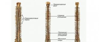
- as prescribed by a specialist, an examination of the head and cervical/upper spine is carried out to assess the functional activity of the brain - based on the results, a safe method of surgical intervention is prescribed;
- areas of the brain hidden by the bones of the skull are examined - other methods do not diagnose these areas;
- MRI helps to obtain a complete picture of the brain and vascular system even without the use of a contrast agent;
- tumors and other pathologies are detected in the early stages of development.
Make an appointment now!
Progress of the procedure: how does an MRI of the brain with contrast work?
The NMR tomography method allows you to examine and study human organs based on the saturation of tissues of various structures with hydrogen atoms. The characteristics of their magnetic properties are also important. This procedure is most often prescribed in difficult clinical situations: for cancer patients, for a detailed and clear examination of the tumor and the affected organ, for demyelinating diseases, for some types of strokes.
During the procedure, the condition of all parts of the brain is monitored, as well as the cerebellum, basal ganglia, ventricles, the ratio of gray and white matter and other structural indicators.
The method is safe, since the patient is not exposed to x-rays during diagnosis.
The contrast agent is injected into a vein using a syringe system. Within 10-30 seconds, the substance penetrates all vessels and tissues and illuminates the areas necessary for diagnosis.
The implementation consists of several stages:
- Before administering the drug, a native study is performed, without the use of contrast;
- After this, the patient is given an injection with a contrast agent in the required volume;
- Then you need to re-set the scanning programs.
Most patients do not notice any deterioration in their health or side effects.
Why is an examination prescribed?
MRI diagnostics helps to recognize pathological changes in the soft tissues of the lining of the brain, inflammatory reactions, and disorders of the central nervous system. Also, with the help of an examination, the conditions of the parts of the head are studied: the visual and occipital lobes, the pituitary gland, the cerebellum, areas of thinking and memory, and the ventricles of the brain.
Before the procedure, the patient undergoes tests, the results of which determine the type of diagnosis. For example, tests showed that the patient had an excess level of the hormone prolactin. The doctor writes a referral for examination of the cerebellum.
Examination of the brain in a magnetic field helps determine the presence of:
- Multiple sclerosis - lesions of the myelin sheath of nerve fibers. The diagnosis determines the stage of the disease, the level of damage, after which the doctor prescribes therapy.
- Benign and malignant tumors. Brain scanning is used to identify the affected area, monitor the development of the tumor, as well as the treatment process.
- Disorders in the cerebral cortex - Parkinson's disease, Alzheimer's disease. MRI determines the density of white and gray matter, the presence of pathologies in the subcortex and cortex of the head.
- Ischemic strokes, heart attacks. The images determine the area and density of ischemic lesions, the presence of cortical necrosis, the appearance of edema, and the stage of development of the disease.
- Brain injuries are damage to blood vessels and tissues due to trauma. Diagnostics reveals the first signs of VSD.
- Mental disorders of endogenous and exogenous types. The method helps to identify hereditary pathologies, changes after traumatic brain injury, viral infection, and poisoning with toxic substances. MRI diagnostics determines schizophrenia.
For children, head tomography is prescribed for the following pathologies:
- head injury, concussion after injury, intrauterine infectious processes;
- hypoxia, ischemia and other disruptions in brain development;
- epileptic seizures, cerebral hemorrhage;
- the first stages of multiple sclerosis;
- dangerous diseases such as pituitary adenoma, brain tumors, cysts and suspicions of them;
- a sharp deterioration in vision and hearing, malfunction of the inner ear;
- high intracranial pressure.
Diagnostics helps to study the state of the brain and its parts, identify the causes of headaches in children, and prevent the development of autism.
Why is an MRI of the brain done with contrast agent?
MR contrast agents are used to detect tumors, autoimmune inflammation and other pathological changes. Most often used in neurosurgery and neurology to enable more informative and high-quality research.
The substance allows you to visualize the exact boundaries, structure and localization of the tumor, the activity of the demyelinating process, the severity of the blood supply to the area under MRI observation.
The advantages of MRI of the brain are that it preserves a clear picture of all structures, even if the tissues are at different depths, and also provides the opportunity to examine all areas of the brain that are covered by the skull.
Differences in imaging with contrast-enhanced MRI
Differences between MRI and CT scan of the brain
Magnetic resonance imaging of the head differs from other diagnostic methods in the following:
- the examination is performed in 3-4 projections, which provides more information for identifying diseases and choosing treatment methods;
- pathologies are detected in the early stages of development - ischemic changes are detected 2-3 hours after a stroke;
- even minimal changes in the brain that cause multiple sclerosis are determined;
- parts of the brain that are not diagnosed by computed tomography (CT) are studied - the brain stem, the cerebellum.
How often do MRIs take place without a doctor's prescription?
As noted above, self-medication is a road to nowhere. Excessive suspiciousness or persistent laziness can cause the acquisition of additional diseases. If a person constantly projects other people’s “sores” onto himself, tries to find a problem where there is none, this is fraught with psychological dependence or mental disorder.
A dependent person risks depleting his body with constant medical manipulations and drugs. If you do not seek specialized help from a psychologist in time, there is a high probability of your health worsening.
The main rule of an exemplary patient is the correct attitude: first, a visit to the doctor, and then an examination.
When is a diagnosis prescribed?
A brain examination is prescribed for the following ailments:
- developmental anomalies and diseases of cerebral vessels;
- head injuries and bruises, cerebral hemorrhages;
- sharp deterioration of vision and hearing;
- tumor development;
- infectious reactions in the central nervous system - meningitis, HIV infections, abscesses;
- pituitary adenoma, epilepsy;
- pathologies of the base of the skull;
- multiple sclerosis, sinusitis, neurodegenerative diseases;
- aneurysms, thrombosis and other anomalies in the vessels of the head.
Diagnosis is also carried out after surgery. Prescribed for frequent migraines and fainting, tinnitus, sudden deterioration of memory and attention, impaired coordination of movements, and mental disorders.
Do's and Don'ts for MRI
Advanced diagnostic methods have significantly expanded the range of diseases for which diagnosis can be made at the earliest stages. However, along with their wide capabilities, there are situations when their use is impossible or limited.
When is it possible and when not to do magnetic resonance imaging? The executive director of Clinic Expert Orenburg LLC, Yuri Andreevich Podlevskikh, answers these and other questions.
- Yuri Andreevich, what is magnetic resonance imaging and is it always safe?
This is a modern method of obtaining medical images to assess the condition of various organs and systems, based on such a physical phenomenon as nuclear magnetic resonance.
Readers should not be confused by the concept “nuclear”: in this case we are not talking about nuclear energy, but about the nuclei of atoms of the elements that make up any organism. These nuclei, being in an external magnetic field created by an MRI machine, oscillate and emit recorded signals - on their basis, using special computer programs, digital images are compiled, from which the doctor later makes his conclusions.
Is the conclusion of an MRI examination a diagnosis? Find out here
But talking about the complete safety of magnetic resonance imaging is not always correct for the reason that there are a number of contraindications to its implementation.
- What is the indication for MRI diagnostics?
It can be used as a clarifying diagnostic method when, after other studies, some unclear, unresolved questions remain.
The next category is patients presenting some kind of complaints: the latter can be completely different and depend on which organ or organ system is involved in the pathological process.
The third category is people undergoing MRI as a screening test. For example, we recommend that everyone undergo magnetic resonance imaging of the brain for preventive purposes. In this case, a person may not complain about anything at all.
How often should an MRI of the brain be done for prevention? Find out here
- Who should not have an MRI and when?
We are talking about contraindications. It is advisable to divide them into relative and absolute. If with relative examination it is possible in some cases, then with absolute examination it is not, since MRI can be harmful to human health.
Relative contraindications to magnetic resonance imaging include:
- fear of closed spaces (claustrophobia);
— alcohol or drug intoxication;
- first trimester of pregnancy;
- psychomotor agitation, panic attack;
- conditions in which it is difficult or impossible to remain still (for example, severe pain);
- metal braces;
- decompensated heart failure;
— the need for continuous monitoring of vital physiological parameters (for example, blood pressure, respiratory rate, electrocardiogram) and/or continuous resuscitation measures;
— presence of tattoos (depending on the dye).
Absolute contraindications to MRI include:
- artificial pacemaker, pacemaker. At the same time, not only the examination itself is prohibited, but also being in the room with the MRI machine;
— neurostimulator;
— electronic and ferromagnetic metal implants;
— ferromagnetic hemostatic clips on blood vessels;
— Ilizarov apparatus;
— ferromagnetic fragments (if these fragments are located in the area of vital organs or are of significant size);
— insulin pumps;
— heart valve prostheses, with the exception of prostheses of completely biological origin or made from modern polymer materials.
-Can an MRI be done if dental crowns are installed?
Yes, you can, they are not an absolute contraindication. However, there is another point that must also be taken into account. Sometimes dental crowns on the upper jaw are quite massive. If a patient comes to us and requires examination of the maxillary sinuses, the issue is resolved individually by the doctor performing the procedure. Why? The fact is that a large number of metal objects in the oral cavity can simply make it difficult to describe the resulting images to such an extent that we will not always be able to answer the questions posed by the attending physician.
- What are the contraindications for MRI with contrast?
Magnetic resonance imaging with contrast is not performed on newborns under 4 weeks of age; in case of any previously noted reactions to the contrast agent. It should, however, be remembered that, firstly, the contrast agents used in computed tomography and magnetic resonance imaging are different. Another point: if a person has ever had an allergic reaction to some contrast agent, this does not mean that he will necessarily have a reaction to another. However, in any case, this issue is fundamentally important and requires the participation of the doctor who will conduct the study.
Read more about MRI with contrast here
Also contraindications to MRI with contrast are:
- history of drug allergies;
- liver failure, acute and chronic renal failure;
— condition after liver transplantation;
— pregnancy, breastfeeding period;
- severe bronchial asthma.
The question of the possibility of performing MRI with contrast in some cases is decided together with the patient’s attending physician, including after additional laboratory tests (for example, assessing the excretory function of the kidneys in case of pathology).
- Are the indications and contraindications for MRI in children and adults the same or different?
Indications for MRI in children are the same as in adults - of course, taking into account the characteristics of the child’s body and diseases unique to children. If we are talking about absolute contraindications, then they are the same regardless of age.
-Can MRI be done after radiation therapy?
There are no restrictions on the procedure specifically due to radiation therapy. I can assume that this question arises due to concerns about whether MRI may cause additional radiation exposure. The answer is no, because it is not based on ionizing radiation.
-Will it be harmful to health if a person undergoes magnetic resonance imaging immediately after an x-ray?
If there are no contraindications to MRI, it won’t hurt. Magnetic resonance imaging does not have any additional or independent negative effects on the body.
Are x-rays dangerous to human health? Yulia Aleksandrovna Rutskaya, head of the radiology department of the Expert Clinic Kursk, tells
- Are there weight restrictions when performing MRI diagnostics?
Technically, they vary depending on the device. On our tomographs, the permissible weight is up to 180 kg, but the person’s physique is more important, not his weight.
-Can I eat or drink before an MRI scan?
It depends on what area of the human body is being examined. If we are talking about the organs of the abdominal cavity, pelvis, and enterography (or, otherwise, hydro-MRI) is performed, then there are specific features of food and water intake.
In other cases (for example, before an MRI of the brain, spine, joints), you can eat and drink; there are no special rules or restrictions.
- How often can I have an MRI?
As often as required.
You can find out the cost of MRI examinations in your city here
For reference:
Podlevskikh Yuri Andreevich
Graduate of the Orenburg State Medical Academy in 2012 with a degree in Pediatrics.
In 2013, on the basis of the above-mentioned educational institution, he completed an internship in the specialty “Radiology”. In the same year, he underwent advanced training in magnetic resonance imaging.
Currently he holds the position of executive director of Clinic Expert Orenburg LLC.
Contraindications
Head tomography is not prescribed if the patient has absolute contraindications, but is allowed at the discretion of the doctor in case of relative contraindications. Diagnostics is prohibited if the following conditions exist:
- the patient has prosthetic heart valves, pacemakers or neurostimulators;
- presence of insulin pumps, inner and middle ear prostheses, cochlear implant;
- the patient has an Ilizarov apparatus;
- the presence of metal implants, ferromagnetic particles, fragments in the body.
Relative indications include:
- tremor, the patient’s inability to hold his breath for a long time during examination;
- presence of dental braces, dentures, stents, vena cava filters;
- heart failure, clip instead of gallbladder;
- pregnancy;
- claustrophobia;
- severe pain while standing still;
- coronary artery bypass grafting.
How does a magnetic resonance imaging scanner work?
The human body contains natural magnets - protons. Without going into technical details, the principle of operation is explained by the fact that during the study, a powerful magnet maintains a stable state of magnetism in the body, and a special program registers radiomagnetic waves. As a result, specialists receive a display of radio signals from the human body.
Additionally, for better visualization, patients undergo intravenous contrast with gadolinium-based drugs: Magnevist, Gadovist, Omniscan. This contrast is considered safer than iodine-containing contrast, since it is less likely to cause side effects.
Preparatory stage
The doctor, based on the test results, determines whether to perform a tomography with or without the introduction of a contrast agent. If the first option is chosen, then the patient does not take food or liquid 5 hours before the diagnosis. Before the procedure, you must remove all jewelry and accessories, watches and objects with metal elements.
During a consultation with a doctor, the patient reports the presence of chronic diseases, allergies to medications, claustrophobia, and pregnancy.
When preparing a child for a head tomography, it is not recommended to give him anything to drink or eat 3 hours before the diagnosis. If an MRI is performed with the administration of contrast, the examination is performed on an empty stomach. Before the procedure, the child should be shown to an allergist to check for an allergic reaction to the injected substance.
Preparing for an MRI of the brain with contrast
No special preparation is required before administering contrast, but experts advise avoiding heavy meals, regardless of which organ is being scanned. This will help avoid side effects such as nausea and vomiting.
During the procedure, you should remove all products made from ferromagnetic alloys. It is also necessary to remove any metal, even non-magnetic, from the face and head (earrings, chains, etc.) to avoid interference.
Women are advised to remove make-up from their face as some dyes contain metallic particles which may distort images.
The question of the frequency and frequency of MRI depends primarily on the disease for which this examination is prescribed. It is worth addressing this issue individually with your doctor, neurologist or neurosurgeon. The MRI procedure, regardless of the frequency of use, is completely safe and can be performed as often as necessary.
Diagnostic features
If a brain tomography is performed with the introduction of a contrast agent, the examination takes 1-1.5 hours.
Stages:
- the patient removes objects and clothing with metal elements;
- lies on the tomograph table on his back;
- the doctor administers a contrast agent intravenously;
- if there is tremors or inability to lie still, the patient takes sedatives;
- legs and arms are secured with belts, bolsters are placed under the neck;
- the table is pushed into the tomograph capsule - the doctor monitors the procedure from a special room with equipment;
- during the diagnosis, the patient hears slight noises of the tomograph operating - the procedure is safe and painless, a slight sensation is felt in the area of the injection of the substance;
- head diagnostics lasts 15 minutes - the patient is recommended to remain in a stationary position to obtain accurate scan results.
MRI of the brain with or without contrast
The difference between the procedures is the information content of the image, because the use of contrast allows a detailed examination of the pathological focus under study. Standard MRI is performed without enhancing substances. There is no preparation before the usual procedure.
MRI with the drug takes longer than tomography without enhancement, since after the end of the umbrella tomography it takes about 15 more minutes to complete the contrast protocol.
The difference in price is noticeable - the cost of the substance increases the cost of scanning. The required amount of medicine is calculated based on the patient’s weight and varies between approximately 2500-5000 thousand rubles.
The substance is completely safe for the body - scientists have proven the low toxicity of gadolinium, which is part of the drug. This drug does not cause an allergic reaction and leaves the body without difficulty. The drug plays a significant role in identifying malignant tumors.
Features of examination of children
Brain tomography in children is carried out under the drug n8a8p00ko70z0o-7m9, -6v5 Propofol is administered. Anesthesia helps keep the child still during diagnosis. Children over 5 years old are given a sedative and given psychological adjustment and support before the tomography.
During the examination, the baby is shown cartoons and toys. Some clinics use open tomographs, when only the child's head is placed in the capsule, and the parents stand nearby and hold his hand.
Before diagnosis, it is recommended to take the child to the toilet, remove electronic items and jewelry with metal inserts from clothes and body. Next, the child is dressed in special clothes, “introduced” to the tomograph and allowed to listen to how it works. This helps to calm the baby before the procedure and obtain his consent to the examination.
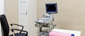
How does the procedure work?
MRI examination is completely painless. Immediately before the procedure, you must remove any metal items from yourself. The patient lies down on the tomograph couch, which is placed inside the device.
Brain MRI procedure
Before carrying out the procedure you need to know the following:
- High-resolution (high-field) magnetic resonance imaging scanners create a fairly high noise level. At our center, patients are provided with headphones to reduce discomfort from these sounds;
- the doctor conducting the study always remains in touch with the patient, the patient has an alarm button;
- The patient must remain absolutely still throughout the procedure to avoid motion artifacts.
The duration of an MRI of the brain is usually about 15 minutes. If you plan to use a contrast agent, the study will take a little longer - about 30 minutes.
Side effects
After tomography, the patient may experience nausea, dizziness, vomiting, weakness, and disorientation in space. This is a normal reaction. It occurs in people with increased sensitivity, when there are metal objects on the patient’s body, and when the rules for preparing for an MRI are not followed. Unpleasant sensations go away on their own. However, if side effects do not disappear for a long time, it is recommended to consult a doctor.
If the rules are followed, the procedure takes place without pain or side effects. The images help doctors accurately determine the cause of the disease and prescribe effective treatment.
What does a brain MRI with contrast show?
There are many symptoms of brain disorders, in which it is better not to postpone a visit to specialists and get a tomography. If you observe: atypical behavior, movement disorders, general health, often neurologists or other clinicians after examination will advise you to do an MRI as the most revealing diagnostic method. MRI of the brain with contrast is prescribed when making a diagnosis is difficult and additional examination is required.
Indications for MRI:
- tumors;
- acute vascular diseases of the nervous system;
- epilepsy;
- severe headaches;
- multiple sclerosis;
- pathology of cerebral vessels;
- bruises and injuries of the skull and brain and their consequences.
As a result of the procedure, inflammatory processes can be detected, indicating their exact location. Examinations are also prescribed after brain surgery. An MRI can help determine whether the tumor has shrunk or recurred.
For a clear and detailed image, the doctor prescribes the injection of a special substance that will help to examine the patient’s problem in more detail. It is worth noting that the contrast agent is safe for health, since it is eliminated from the body within 3-4 hours.
The main advantages of contrast in brain MRI include:
- minimum contraindications;
- obtaining slices in any plane;
- high contrast and image resolution;
- the ability to obtain images of all brain structures covered by bone tissue;
- good tolerability of the drug.
This technique uses several types of substances; they differ in composition and method of application. Most often, doctors prescribe a contrast agent, which is administered intravenously and contains the rare earth paramagnetic metal gadolinium.
The procedure has no contraindications, the exception may be a peculiarity of the body with individual intolerance. In some cases, side effects are possible: rash, nausea, slight dizziness. Complications are observed in less than one percent of clinical cases.
Preparing for an MRI with contrast