When several or all eye muscles are paralyzed, ophthalmoplegia occurs. This condition may occur due to damage to the optic nerves. In this case, one or both eyeballs may be damaged. Their motor ability is limited or lost in some types of disease. Let's look at the causes of ophthalmoplegia and methods of treatment.
In this article
- Causes of ophthalmoplegia
- Types and symptoms of ophthalmoplegia
- Diagnosis of the disease
- Treatment methods for ophthalmoplegia
- Complications of ophthalmoplegia
- Prevention
Causes of ophthalmoplegia
Various factors can serve as prerequisites for the development of this pathology. It can be either congenital or acquired as a result of certain diseases. At the same time, ophthalmoplegia occurs with equal frequency in men and women. Here are the most common causes of optic nerve palsy:
- severe infectious diseases, for example, tetanus, diphtheria, botulism;
- eye tumors and neoplasms;
- muscle dysfunction (myasthenia gravis, etc.);
- traumatic brain injuries;
- pathologies of the endocrine system - diabetes, problems with the thyroid gland;
- encephalitis;
- severe intoxication of the body;
- food or alcohol poisoning;
- chronic sinusitis;
- multiple sclerosis;
- brain diseases;
- trigeminal neuralgia;
- CNS diseases and other factors.
If any of these cases occurs, disruption of the activity of the nerves responsible for muscle contraction may occur. There are three in total:
- oculomotor, with the help of which we are able to turn the eyeball in different directions: up, down, towards the nose;
- block-shaped, providing the ability to rotate to the lower temporal angle;
- abducens - this nerve is responsible for turning the eyeball towards the temple.
Violation of the innervation of the muscles leads to a loss of their tone and the inability to move the eyes - paralysis occurs. Ophthalmoplegia has different forms.
Forecast
Prediction of successful treatment of internuclear ophthalmoplegia is based on the cause of this disease. It must be borne in mind that some diseases are incurable (multiple sclerosis), but with the help of therapy it is possible to reduce the rate of their development. After a stroke, much depends on the subsequent recovery process.
With a favorable outcome, all functions of the central nervous system are restored, including the regulation of eye movements.
A neoplasm such as a brain stem tumor can have a positive prognosis if detected early enough. The disease has a high mortality rate, and its main insidiousness is that the majority of patients are children under 15 years of age. Later stages require conservative treatment.
Thus, preventive work to prevent internuclear ophthalmoplegia comes down to general prevention of a wide range of diseases, although some of them cannot be prevented in general.
Types and symptoms of ophthalmoplegia
In modern medicine, the disease is divided into several main types:
- Internal ophthalmoplegia. It is characterized by weakening or paralysis of the muscles of the iris and ciliary body. In this case, the mobility of the eye is not impaired, but the pupil is permanently dilated, and a spasm of accommodation may also be observed.
- External ophthalmoplegia. With this type of pathology, the muscles on the outside of the eyeball are affected. At the same time, it can hardly move or is completely paralyzed. The external type of ophthalmoplegia is characterized by diplopia - double vision, but the pupil's reaction to light is preserved. Due to the uneven distribution of the tear film, the dryness of the mucous membrane of the eyes increases. Patients also complain of increased lacrimation. Internal and external ophthalmoplegia can also be partial or complete. In complete ophthalmoplegia, both the external and internal muscles of the visual organs are affected.
Total ophthalmoplegia is also characterized by immobility of the eyeballs and drooping (ptosis) of the upper eyelids - this condition is called Hutchinson's face. The patient has to tilt his head back to be able to see normally from under drooping eyelids. Partial ophthalmoplegia is a condition during which paresis of only the external or only the internal muscles of the eye develops.
- Internuclear ophthalmoplegia. This type of disease is characterized by damage to the muscles that are responsible for the simultaneous movement of the eyeballs. With unilateral internuclear ophthalmoplegia, one of them can be fixed in some position, and on the second, nystagmus occurs - frequent involuntary twitching. Most often, internuclear ophthalmoplegia is a consequence of multiple sclerosis, as well as stroke, head injuries, drug intoxication and some other risk factors. Patients complain that they cannot look in the same direction with both eyes at the same time.
- Supranuclear ophthalmoplegia. Its other name is Steele-Richardson-Olszewski syndrome. This is a rare brain disorder that causes problems with motor function and eye movements. It is also characterized by gaze paralysis - there is no simultaneous eye movement up and down or left and right. Many people complain that they are not able to direct their gaze in the right direction at will.
Treatment
Treatment of internuclear ophthalmoplegia is carried out based on the causes of the disease. And these reasons must be eliminated first. If the cause is a stroke, it is usually treated with blood thinners. In some cases, it is possible to use surgical methods to widen the arteries and remove accumulated blood clots.
Internuclear ophthalmoplegia in multiple sclerosis: in the absence of the desired effect of treatment, for example, corticosteroids, plasmapheresis is performed in multiple sclerosis. However, at this moment the use of other medications cannot be ruled out, since the resulting effect from the blood purification procedure may be lower than predicted.
When internuclear ophthalmoplegia is caused by certain neoplasms, it is necessary to approach treatment tactics very carefully. When choosing it, proceed from the level of growth and location. Most specialists choose a conservative form of treatment. This is justified by the fact that surgical removal of tumors from the brain stem is extremely difficult.
Diagnosis of the disease
Ophthalmoplegia has characteristic external signs. But in order to distinguish it, for example, from paralytic strabismus, and also to identify the exact type, a number of diagnostic hardware tests are carried out:
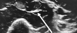
- Ultrasound of the eye allows you to detect the features of the eyeballs and study the condition of the orbital cavity;
- X-ray of the skull - the image will show internal damage, if this is the cause, as well as the condition of the sinuses;
- computed tomography of the head and neck makes it possible to detect neoplasms, which are one of the causes of ophthalmoplegia;
- proserine test for the diagnosis of myasthenia gravis;
- angiographic examination of cerebral vessels;
- perimetry to determine the boundaries of the field of view.
Depending on what is the cause of the disease, consultation and examination by highly specialized specialists is also required: in case of a neoplasm - an oncologist, in case of suspected pathology of the central nervous system - a neurologist.
Treatment methods for ophthalmoplegia
Treatment consists of eliminating the disease that caused the paralysis, as well as restoring the functionality of the muscles and nerves. Therapeutic interventions include several aspects.
Drug treatment. The patient may be prescribed various medications depending on the etiology:
- anti-inflammatory drugs;
- topical corticosteroids;
- vitamins B and C;
- nootropics to improve nervous activity;
- drugs that eliminate muscle weakness.
Hardware therapy. Along with taking medications, treatment using hardware procedures - electrophoresis, phonophoresis, acupuncture - also produces effective results. This helps strengthen the eye muscles and eliminate spasm.
Surgical intervention. It is prescribed if it is necessary to eliminate the tumor that is the cause of paralysis, as well as restore muscle activity of the eyes, and raise the upper eyelid with ptosis.
Three methods are used to influence muscles:
- myotomy (dissection);
- recession (pushing back);
- tenomyoplasty (lengthening).
Surgeries for ophthalmoplegia are aimed at restoring the correct balance between the eye muscles. The doctor decides what type of intervention is necessary, taking into account the existing form of pathology and its severity, the patient’s age and other characteristics. As a rule, several muscles are operated on at once. There are cases when a strengthening operation is performed on one eye and a weakening operation on the other.
The success of treating ophthalmoplegia depends on timely contact with specialists, as with many other diseases, and not only eye ones.
Temporary loss of vision
Eye fatigue
The most commonplace situation is called asthenopia. This is eye strain due to irrational visual load (for example, sitting for many hours in front of a monitor screen, TV, reading from a sheet in low light, driving a car at night). In this case, the muscles that regulate the functioning of the eye become overstrained. Pain in the eyes and lacrimation appear. It is difficult for a person to concentrate on small print or details of an image, and blurriness or a veil may appear before his eyes. This is often combined with a headache.
False myopia
Accommodation spasm (false myopia) often affects children and adolescents. Its clinical picture is similar to asthenopia. Transient visual impairment near or far is caused by fatigue and spasm of the ciliary muscle, which changes the curvature of the lens.
Night blindness - nyctalopia and hemeralopia
Deterioration of vision at dusk is a consequence of a deficiency of vitamins A, PP and B. This disease is popularly called night blindness, and its scientific names are nyctalopia and hemeralopia. At the same time, twilight vision suffers. In addition to hypovitaminosis, diseases of the retina and optic nerve can lead to night blindness. There are also congenital forms of pathology. At the same time, visual acuity weakens, color perception decreases, a person’s spatial orientation is disrupted, and visual fields are narrowed.
Vascular spasms
Transient visual disturbances may indicate vascular spasm in the retina or brain. Such situations are associated with hypertensive crises (sharp jumps in blood pressure), chronic cerebral circulatory disorders (against the background of atherosclerosis, vertebral artery syndrome, cerebral amyloidosis, blood diseases, vascular anomalies, vasculitis, venous hypertension). As a rule, there is blurred vision, flickering of spots before the eyes, and darkening of the eyes. Combined symptoms may also occur, for example, hearing and vision impairment or dizziness, blurred vision.
Migraine
A migraine attack may be accompanied by temporary blurred vision against the background of severe vasospasm. Often, pain in the head is accompanied by the appearance of an aura in the form of flickering scotomas (flickering or floating dark spots in front of the eyes).
Intraocular pressure
If intraocular pressure is normal from 9 to 22 mmHg, then an acute attack of glaucoma can raise it to 50-70 or higher. In this case, a sharp headache covering half the head and the eyeball accompany a one-sided process. If both eyes are affected, the whole head hurts. In addition, blurred vision, rainbow circles before the eyes, or dark spots (scotomas) may appear. Autonomic disorders (nausea, vomiting, heart pain) are often associated.
Medicines
Drug effects can also lead to transient myopia. This occurs when taking high doses of sulfonamides.
Complications of ophthalmoplegia
The consequences of advanced ophthalmoplegia can have a very negative impact on the condition of the visual organs. Paralysis of the internal muscles leads to impaired accommodation and decreased visual acuity. Internuclear ophthalmoplegia can cause the development of persistent nystagmus.
Inflammatory diseases such as conjunctivitis, keratitis, blepharitis are often present; the patient’s normal functioning of the lacrimal and meibomian glands is disrupted. Paralysis of the eye muscles is accompanied by facial asymmetry. With supranuclear ophthalmoplegia, difficulties with orientation in space and imbalance also arise. Another common complication of ophthalmoplegia is xerophthalmia, drying of the conjunctiva and cornea.
Classification
A unified classification of endocrine ophthalmopathy has not been developed. There are several main scales for assessing the activity, nature, severity and severity of the process.
NOSPECS (1977)
NOSPECS (1977) distributed abroad. Describes the severity of clinical manifestations.
Consists of 6 main classes:
- N (0) – without clinical manifestations;
- O (I) – isolated narrowing (retraction) of the upper eyelid;
- S (II) – soft tissue damage;
- P (III) – the appearance of exophthalmos of varying severity;
- E (IV) – involvement of the oculomotor muscles in the process;
- C (V) – corneal damage;
- S (VI) – damage to the optic nerve, decreased visual acuity.
CAS (1989)
CAS (1989) evaluates the activity of the process on a scale to determine the optimal tactics for patient management; it consists of 7 main and 3 additional parameters.
The main ones include:
- spontaneous pain;
- pain when rotating the eye;
- hyperemia (redness) of soft tissues;
- pinpoint hemorrhages of the conjunctiva;
- tumor (edema) of the eyelids;
- chemosis (swelling due to environmental factors) of the conjunctiva;
- redness and swelling of the caruncle (lunar fold and lacrimal month).
Additional:
- dynamics of growth of eyelid protrusion by 2 mm or more within 60 days;
- dynamics of decrease in eyeball mobility by 8° or more within 60 days;
- loss of visual acuity by more than a tenth of the previous values over 60 days.
The process is considered active if there are 3 or more basic points. Additional points evaluate the dynamics of patient treatment.
EUGOGO (2008)
- Mild degree.
Eyelid retraction less than 2 mm, exophthalmos up to 3 mm, intermittent diplopia, soft tissues are slightly involved, corneal dryness is relieved by the use of moisturizers. - Moderate/severe.
Eyelid retraction more than 2 mm, exophthalmos more than 3 mm, soft tissues are strongly involved, almost constant diplopia. - Threat of vision loss.
Severe optic neuropathy with corneal damage or ulceration.
Baranova (1983)
Baranova (1983) has become widespread in the CIS countries.
The following stages are distinguished:
- Thyrotoxic exophthalmos of mild severity.
It manifests itself as a protrusion of the eyeball by 16 mm, characterized by swelling of the eyelids, discomfort, a feeling of “sand,” and tearfulness. Oculomotor functions are performed in full. - Edematous (infiltrative) exophthalmos of moderate severity.
The eyeball protrudes by 17 millimeters or more. There are mild changes in the conjunctiva, a feeling of congestion in the eye, mild disorders of the oculomotor muscles, unstable diplopia (double vision), and lacrimation. - Severe endocrine myopathy.
Exophthalmos more than 22mm, impaired eyelid closure. Corneal ulcerations, severe diplopia, and decreased oculomotor function often occur. The oculomotor nerve is involved in the dystrophic process.
Prevention
There are no special preventive measures in this case. To reduce the risk of pathology, you need to adhere to general recommendations:
- after eye injuries, head bruises, you should monitor the condition of the visual organs; if discomfort occurs, you should visit an ophthalmologist for diagnostic purposes;
- engage in timely treatment of infections, preventing transition to the chronic stage;
- Observe precautions when frequently working with toxic substances;
- If there are factors that can lead to ophthalmoplegia, such as diseases of the nervous and endocrine systems, you should also undergo regular examinations by a specialist so as not to miss the development of pathology.
A careful and careful attitude to eye health will help to detect negative symptoms in time and begin appropriate therapy.
Breeding Spanish amphibians
As a rule, one-year-old individuals become sexually mature and ready to reproduce. At room temperature water, mating games among amphibians take place from September to May. During this period, the male transfers the spermatophore to the female.
Immediately after this, fertilization occurs, and a day or two later the female lays up to 1000 eggs over the course of two days. In order for the caviar to survive, it should be removed to another container, where the temperature is maintained at 22-24°C. Already 10 days later, larvae will appear from the eggs.
With proper feeding, juveniles of Pleurodeles waltli will reach a size of 6-9 cm in 2-3 months.