Venous encephalopathy is a common condition of the cerebral vascular system, in which there is a violation of the venous outflow of blood from the brain. Moreover, this disease can be combined with arterial problems and so-called discirculatory encephalopathy (the author uses the words “so-called” due to the fact that venous encephalopathy is also inherently a discirculatory process), or it can be an independent disease.
Information for doctors. According to ICD 10, there is no diagnosis of venous encephalopathy. Because of this, to more accurately indicate the presence of a venous problem, it is more logical to use codes M53.0 (cervicocranial syndrome) and indicate that problems with the cervical spine led to this condition, leading to the diagnosis description of chronic venous outflow disorder syndrome, or indicate this syndrome within the framework of diagnosis I67.8 - discirculatory encephalopathy - in the event that this diagnosis also occurs.
At the moment, venous dysfunction is a controversial condition, not recognized by a number of authors, however, the clinical practice of the author of the site shows the importance of the presence of disturbances in the venous outflow from the cranial cavity in the formation of various symptoms.
A little about venous encephalopathy
Venous encephalopathy develops in the presence of chronic disorders of the venous outflow of blood from the brain. Due to this condition, the production of cerebrospinal fluid changes, intracranial pressure increases, and metabolic processes are disrupted. There are many reasons for this. Among them, the most common are hereditary structural features of the vein wall, cervical compression syndromes, residual dysontogenetic background (vascular hypoplasia, etc.). Less commonly, these processes arise from such reasons as the consequences of an inflammatory process in the veins and sinuses (for example, after sinus thrombosis), compression by a brain tumor, or trauma to the skull, including birth trauma.
Venous encephalopathy itself as a disease does not pose a threat to the patient’s life. However, its presence can significantly worsen the quality of life, reduce ability to work, and worsen the prognosis in the presence of other somatic diseases.
The mechanism of pathology development
The pathogenesis of dyscirculatory encephalopathy includes factors leading to deterioration of cerebral circulation, consequently to hypoxia and disruption of trophism of cells of the central nervous system. This leads to cell death and the appearance of areas of rarefaction of brain tissue, that is, leukoaraiosis or silent infarctions - multiple small lesions.
The subcortical structures and white matter of the deep parts of the brain are most susceptible to destruction during DEP. This is due to their location on the border of the carotid and vertebrobasilar basins. Chronic ischemia provokes the phenomenon of disconnection, that is, disruption of connections between the cerebral cortex and subcortical ganglia. Experts believe that this is the main pathogenetic mechanism. It causes a characteristic symptomatic picture:
- Violation of the emotional sphere;
- Cognitive disorders;
- Pathologies of motor functions.
A distinctive feature of DEP is the reversibility of functional disorders at the first stage of the disease with appropriate treatment and the persistent nature of neurological defects leading to disability at later stages.
Symptoms and diagnosis of venous encephalopathy
The symptoms of venous encephalopathy are varied, often they resemble other diseases (dyscirculatory process, problems with the cervical spine, depression, etc.). The most typical “venous” complaints are the following symptoms:
- Morning (sometimes night) headaches of a bursting nature
- Dizziness, which is non-systemic in nature, worsens when changing body position,
- Noise in the head, especially when going to bed
- Visual disturbances (a progressive decrease in visual acuity and various photopsia may be noted)
- Feeling of discomfort in the eyes in the morning
Symptoms are aggravated by working at an incline, wearing narrow collars and ties, and sleeping on a low surface. Objectively, the following signs may also be noted:
- Pastosity (swelling) of the face and eyelids in the morning (with a pale or bluish tint)
- Mild nasal congestion
Conditions after which the complaints intensify are also characteristic: bending forward with lowering the head, horizontal position of the body, taking alcohol-containing drinks, nitrates, vasodilators, warm bath or sauna, hot drinks, being in a stuffy room, daytime sleep. Or, on the contrary, they decrease: drinking caffeine-containing drinks, rinsing in cold water, walking in the fresh air, vertical body position, sleeping strictly on a high pillow.
A neurological examination is important, which, however, also does not have cardinal diagnostic signs. The neurological status reveals the following symptoms: nystagmus is expected, there is convergence insufficiency of the eyeballs, decreased corneal reflexes (rarely tested), pain in the exit points of the branches of the trigeminal nerve, decreased sensitivity in the area of innervation of the first trigeminal nerve, dissociation (difference between) knee and Achilles reflexes, impaired coordination when walking.
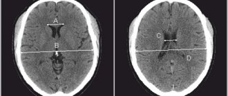
Diagnostic methods include a set of the following examinations: ophthalmological examination, which allows to identify congestion in the retina, dilated retinal veins, ultrasound examination of the veins of the neck and brain, MRI venography (if necessary, with the introduction of a contrast agent). A relative study that can help in the absence of the above methods is rheoencephalography, which allows one to evaluate microcirculation and venous outflow. The presence of diastolic index values below normal may make you think about the presence of venous disorders.
However, there are no clear diagnostic criteria. In 90% of cases, the diagnosis is made on the basis of complaints, anamnesis, and neurological examination data, focusing on the research methods carried out (if possible).
Ideally, for a reliable diagnosis, it is necessary to combine the presence of all the above-described signs of the disease along with the absence of other reliable reasons for the development of the patient’s complaints.
Classification of dyscirculatory encephalopathy
DEP is distinguished by etiology:
- Atherosclerotic;
- Hypertensive;
- Venous;
- Mixed.
According to the speed of development, it can be rapidly progressing or galloping and slowly developing, that is, the classic form.
The severity classification is as follows:
- The first stage is accompanied by subjective feelings of ill-being, mild cognitive impairment, and preservation of the neurological status;
- The second stage is characterized by obvious motor and cognitive disorders and increased emotional distress.
- The third stage is considered the most severe, at which mental disorders begin. It represents vascular dementia of varying severity.
Dizziness due to venous encephalopathy
Dizziness with venous encephalopathy has characteristic differences. It occurs after a change in body position, passes quickly enough in an upright position, and intensifies in the morning. However, this symptom can also be provoked by other factors that would also provoke it in other diseases: alcohol intake, a jump in blood pressure, etc.
Objectifying the presence of dizziness due to impaired venous outflow is difficult. Patients are checked for accuracy when performing corrdinator tests, Romberg test is performed, nystagmus is checked while standing and then in a lying position. Increased nystagmus while lying down may also indicate disturbances in venous outflow.
Dizziness with venous encephalopathy does not pose any threat to life. As a rule, treatment quickly relieves this symptom.
Kinds
Congenital encephalopathy can be a consequence of such reasons as:
- gene failure;
- infection of a woman during pregnancy;
- a woman taking contraindicated medications during pregnancy;
- a woman’s use of alcohol, tobacco, or drugs during pregnancy;
- prolonged exposure of the child to green amniotic fluid;
- entanglement of the fetus with the umbilical cord;
- birth injuries.
Acquired encephalopathy can occur both due to injury and as a result of chronic diseases.
In addition, there are such special cases of cerebral circulation disorders as periventricular leukoencephalopathy and venous encephalopathy with bilateral pyramidal insufficiency.
In the first case, due to prolonged oxygen starvation, the white matter of the brain begins to die. This pathology, according to doctors, is the most likely cause of cerebral palsy in newborns.
Encephalopathy of the brain in the elderly
With age, blood circulation to the brain is disrupted, which leads to the destruction of its structure.
In the second case, damage occurs to the nerve fibers and cerebral cortex, while a person’s motor function is impaired, and this, in turn, leads to paralysis.
The disease goes through 3 stages - compensation (the disease practically does not manifest itself), subcompensation (clinical symptoms gradually increase, and the condition worsens) and decompensation (the functioning of the organ is impaired), at each of which the symptoms are added and intensified. At the last stage, the pathology is irreversible.
Treatment of venous encephalopathy
To begin with, I would like to note the groups of drugs that are best not to be used for venous encephalopathy, as they can cause a deterioration in well-being. These include antihypertensive drugs of the group of calcium channel blockers, nitroglycerin, nicotinic acid. These drugs, causing dilatation of the veins, only worsen the reasons that led to the pathological condition.
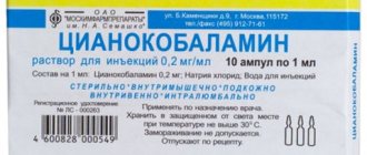
The drugs of choice for venous encephalopathy are venotonics, both for intravenous administration (L-lysine escitate, also known as horse chestnut salt, escin and amino acids), and for oral administration, these include Antistax, Troxevasin, Detralex, Phlebodia. The author of the site prefers to prescribe Detralex, although the choice of drug is the prerogative of the attending physician.
In addition, vasoactive drugs (Tanakan, Cavinton, Trental), metabolic agents (Mexidol), and specific diuretics that reduce the level of intracranial pressure (Diacarb, Mannitol) are used. Symptomatically, betahistine drugs, NSAIDs (especially for problems with the cervical spine), muscle relaxants (allow you to relieve spasm of the spinal muscles that impede venous outflow) can be used.
However, you should not bypass non-drug methods of treating the problem: exercise therapy, maintaining a correct lifestyle, limiting provoking factors (alcohol, bending work, etc.).
Venous encephalopathy, if diagnosed in a timely manner, responds quite well to treatment. Remember, your health is in your hands, and doctors can help restore it; it is only important to follow all the recommendations. Be healthy!
Causes
The following diseases can lead to venous encephalopathy:
- arterial hypertension (persistent increase in blood pressure);
- hypotension;
- vegetative-vascular dystonia;
- hyperthermia (increase in human body temperature due to various external factors);
- osteochondrosis of the cervical spine;
- diabetes ;
- atherosclerosis (high cholesterol);
- heart disease (coronary disease, arrhythmia, rheumatoid arthritis);
- brain tumor
- bacterial intoxication ;
- cirrhosis and necrosis (death of cells) of the liver;
- thrombophlebitis (blood thickening and formation of blood clots in the veins).
Not only serious diseases, but also traumatic brain injuries and many unfavorable factors can provoke the appearance of venous encephalopathy:
- psychological stress, stress, depression;
- abuse , taking drugs, antidepressants;
- taking hormonal medications without consulting a specialist;
- organism associated with work in hazardous industries;
- sedentary lifestyle, obesity;
- exposure of the body due to radiation, frequent x-rays or radiation therapy.
Anyone can be at risk.
results
Cephalgic syndrome.
At the start of treatment visit in the main group, the severity of headache according to VAS corresponded to the average level (43 mm). During therapy with L-lysine escinate, its intensity statistically significantly decreased by almost 2 times, amounting to 26.5 mm (p = 0.000; Fig. 2A). In the control group, at the start of treatment visit, the headache intensity index was slightly lower, 38 mm, but there was no statistically significant difference between the groups (p = 0.307). At visit 3, headache intensity decreased statistically insignificantly (p=0.909; Fig. 2B). Moreover, after the end of treatment, the severity of headache in the group of patients receiving L-lysine escinate was statistically significantly less than in the control group (p = 0.0000). When conducting a correlation analysis at visit 3, a highly significant correlation was revealed with a low level of headache in the group of patients who received L-lysine escinate (r=0.674; p=0.000), which was not observed in the control group.
Subjective neurological complaints.
As a rule, patients with chronic CVD present a number of complaints that are difficult to standardize; for this we used the SSNR [12]. In the main group of patients, at the start of treatment visit, the SSNR score (28 points) corresponded to a moderate intensity of subjective complaints; during therapy with L-lysine escinate, a moderate decrease in complaints was noted to 23.5 points according to the SSNR (p = 0.035; Fig. 3A). In the control group, the initial score on the SSNR was 37 points, which was not significantly higher than the indicator in the main group (p = 0.72). According to the results of treatment with vinpocetine, there was also a statistically significant improvement in the SSNR score to 26.5 points (p = 0.007; Fig. 3B) in the absence of differences from the indicator in the main group (p = 0.127).
Assessment of motor activity.
A characteristic symptom of cerebrovascular pathology is gait disturbance caused by various causes, including diseases of the musculoskeletal system, vestibular disorders, postural phobic instability and disorders of central origin. Often these reasons have a combined effect and it is almost impossible to single out the main one. However, the Tinetti scale is a useful tool for assessing gait impairment, regardless of cause.
In the group of patients receiving L-lysine aescinate, at the start of treatment visit, the total clinical score was 25 points, which corresponded to moderate walking impairment. During the therapy, a statistically significant improvement in walking was noted, the median score was 36 points (p = 0.000), indicating mild impairment (Fig. 4A). In patients in the control group, the initial total clinical score was 24 points, and during treatment it increased to 31.5 points (p=0.000; Fig. 4B). Intergroup analysis revealed that the groups did not differ statistically significantly at the screening visit, but during therapy in patients receiving L-lysine aescinate, disturbances in general motor activity became significantly less pronounced (p = 0.000). Also in the main group at the end of treatment visit, a negative correlation was revealed with the Tinetti scale score, which indicated a more favorable effect of therapy (r=-0.563; p=0.000).
Assessment of emotional disorders and asthenic disorders.
Patients in both groups at the screening visit, according to the HADS score of 5.5 (3.0; 6.5) and 6.0 (2.5; 7.0) points in the main and control groups, respectively, had no depression. At the same time, anxiety reached a subclinically expressed level - in the main group 9.5 (6.5; 10.0) points, in the control group - 9 (7.0; 10.5) points (p>0.05). After the end of therapy, the level of depression in the groups did not change, and the assessment of the severity of anxiety indicated its absence: in the main group 6 (4.0; 7.5) points, in the control group - 5.5 (3.5; 7.0) points (p>0.05).
The initial score on the modified asthenia scale MFI-20 at the screening visit in both groups corresponded to severe asthenia (Table 1).
After completion of the course of treatment in the main group, the median total clinical score on the MFI-20 scale was 49.0 (44.0; 56.0) points, and in the control group - 50.0 (46.0; 54.0) in the absence of statistical data significant differences between groups (p=0.564). A statistically significant decrease in the severity of asthenic syndrome was noted during treatment with both L-lysine escinate (p = 0.031) and vinpocetine (p = 0.038).
Assessment of ophthalmological parameters.
None of the patients had congestion in the fundus. In 2 patients of the main group and 1 in the control group, blurring of the optic discs was observed. It should be noted that these patients had a rather pronounced course of arterial hypertension, with frequent crises, high “working” blood pressure and a large number of antihypertensive drugs taken. Narrowing of the vessels of the arterial bed of the fundus was detected in 24 patients of the main group and in 22 of the control group. During therapy, a slight decrease in the number of patients with this symptom was noted in both groups, but the groups did not differ statistically significantly. At the same time, dilation of the veins detected before treatment in 26 patients of the main group and 28 patients of the control group remained at the end of treatment in 12 (p = 0.000) and 24 (p = 0.254) patients, respectively, which indicates a pronounced venotonic effect of L-lysine escinate .
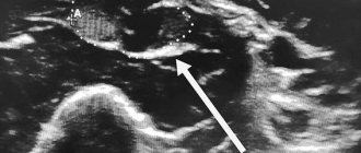
Assessment of ultrasound parameters of cerebral venous blood flow.
As a rule, when assessing cerebral venous hemodynamics, the following indicators are determined: the speed of blood flow through certain veins and their diameters [15, 16].
During treatment, both groups showed a statistically significant change in blood flow velocity in almost all veins studied. Thus, in the main group of patients receiving therapy with L-lysine escinate, the speed of blood flow through the veins of Rosenthal, the angular veins of the eye and the vertebral veins decreased statistically significantly. These changes indicate a redistribution of cerebral venous blood flow, possibly the inclusion of some additional pathways for the outflow of venous blood (Table 2). In the control group, there was also a slight decrease in the speed of blood flow through the veins of Rosenthal and the vertebral veins, less pronounced than in the main group. The speed through the bulbs of the jugular veins was quite high on both visits and did not change in any way during the therapy, which is understandable: as noted above, the main outflow in a horizontal position occurs precisely through the jugular veins, in addition, the bulbs are vessels of a fairly large diameter, and this affects the speed of blood flow. When conducting a correlation analysis, it was revealed that normalization of blood flow velocity in the veins of Rosenthal and in the angular veins of the eye was associated with the group of patients receiving L-lysine escinate (r=-0.340; p=0.008) and (r=-0.552; p=0.000) .
It should be noted that no significant adverse reactions were registered during this work: 4 patients of the main group had a local reaction in the form of burning and redness along the vein into which the infusion was carried out; In 6 patients in the control group, a moderate decrease in blood pressure was recorded, which did not require discontinuation of therapy.