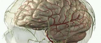
In medicine, the term sinus durae matris - sinuses of the dura mater, implies vascular collectors located between the plates of the dura mater. These are peculiar triangular ducts with endothelium on the surface, formed in the splits of the hard layer of the brain. They are supplied with blood from the internal and superficial vessels of the brain, and participate in the reabsorption of cerebrospinal fluid from the cavity between the arachnoid and non-solid medulla.
Functions of sines
There are specific tasks for the venous sinuses.
They perform the function of uninterrupted supply of blood and oxygen to the vessels of the brain. It is through them that blood directly flows from the head organ to several double veins located in the neck, which carry blood away from the upper part of the body. The sinuses of the dura mater perform the functions of blood vessels, and in addition take part in the metabolism of cerebrospinal fluid. The structure is very different from the cerebral vessels.
Successful drainage of blood from the cerebral vessels often saves from the occurrence of fatal pathologies. In cases where difficulties arise in the field of vascular blood circulation, it becomes possible to quickly eliminate it, thanks to the recanalization of blood vessels and the formation of collaterals.
Causes
The most common cause is infections that cause inflammation of the ear, tonsils, teeth, maxillary sinuses, facial tissues, eyes, as well as septic conditions due to viral, bacterial (including tuberculosis), and fungal infections.
In the absence of infectious diseases, sinus thrombosis occurs under the influence of the following factors:
- traumatic brain injury,
- operations on bones, skull tissues, internal organs,
- intracranial or other tumors,
- puncture of the spinal canal for diagnosis or pain relief,
- toxicosis of the second half of pregnancy with hypertension,
- caesarean section or natural birth,
- taking estrogen drugs (including contraceptives),
- extreme degree of dehydration,
- autoimmune diseases,
- increased blood clotting activity,
- circulatory disorders,
- diabetes.
The structure of the sinuses of the solid MO
The development of TMO collectors occurs due to their division into two sheets, which are similar to channels. These ducts are designed to distribute the venous flow of blood from the main human organ, which is subsequently sent to several double vessels that are located in the neck and transfer blood from the brain.
The dura mater plates that make up the sinus look like tightly stretched ropes that do not lose tension. This structure allows blood to flow freely from the head and neck, without in any way affecting the state of intracranial pressure.
The following types of dura mater reservoirs have been established in humans:
- Superior or inferior sagittal. The first is located longitudinally along the upper border of the falx bone and ends on the occipital fragment, and the next is longitudinally along the border of the falx below and flows into the straight sinus;
- Straight. It is located longitudinally of the fragment, where the falciform process passes into the cerebellar tentorium;
- Transverse (double). Formed on the transverse growth of the skull, located longitudinally along the posterior border of the cerebellar groove;
- Occipital. It is located in the cavity of the cerebellar arch, and then extends to the occipital junction;
- Sigmoid. Located in the division in the ventral fragment of the head bone tissue;
- Cavernous (double). It is located on the sides of the formation of a wedge-shaped bone in the body (sella turcica);
- Sphenoparietal sinus (double). Refers to a small wedge-shaped border of bone and ends in a cavernous reservoir.
Stony (double). Located close to both borders of the pyramidal bone of the temples.
The collectors of the medulla begin to collect anastomoses with venous vessels on the surface of the brain, through venous branches that connect the vascular sinuses of the dura mater with the external circulatory vessels of the head. These depressions begin to communicate with the diploic processes, which are typically located in the cranial vault and then pass into the vessels of the head. Then the blood tends to pass through the venous plexuses and then flows into the dura mater reservoirs.
Prevention, complications, prognosis
It is very important, if a person notices any changes or pain in his condition, to consult a doctor in time, because the sooner this disease is identified, the sooner and faster doctors will be able to help the sick patient. Therefore, there is no need to wait until the last moment, because waiting can often lead to a stroke or cerebral edema, or death. Other complications include blurred vision, deterioration in hormone production, possible formation of cysts in the lining of the brain and other cardiovascular diseases in humans
Other complications include blurred vision, deterioration in hormone production, possible formation of cysts in the lining of the brain and other cardiovascular diseases in humans.
Of course, the prognosis for recovery is influenced by the stage of the disease at which a person is; it all depends on how quickly the person turned to specialists. In any case, the mortality rate usually does not exceed twenty percent in advanced stages.
So, in order not to undergo such a difficult test, you need to adhere to preventive measures and check regularly with a doctor.
Types of dura sinuses
Nature very thoughtfully created humans, providing the dura mater with indentations to provide the main organ with oxygen and nutritional compounds.
Superior sagittal sinus
This cranial sinus is characterized by a large space with a complex structure. The sickle of the main human organ takes a significant part in its development. This is a crescent leaf. It is made of dura mater. The process originates from the top of the ethmoid bone, passes in the middle back, penetrating into the interhemispheric foramen, separating the brain regions from each other. The groove-like growth of the superior sagittal sinus is essentially the base of the falx bone.
This duct provides numerous lacunae on the sides. These are small cavities that are connected to a venous network of strong plates.
The superior sagittal reservoir has the following venous connections:
- the anterior parts belong to the vessels of the labial cavity (near the nose);
- the middle parts belong to the venous beds of the parietal fragments of the brain.
As a person grows, this collector of arteries and veins becomes larger and wider in terms of mass capacity. Its posterior fragment protrudes into the combined sinus drain.
Inferior sagittal sinus
This cistern of the structure of the cranium is presented in medical annals as sinus sagittalis inferior. It was so named because it is located in the lower location of the cerebral arch. Compared to the upper reservoir, it has a significantly small volume. Thanks to the large number of venous anastomoses, it is attached to the rectum.
Direct sine
This fragment of the skull is, in fact, the so-called continuation of the lower cistern from the rear side. It connects the rear sections of the superior tanks and the lower manifold. Along with the superior one, a large vessel is included in the anterior part of the nondual sinus. The posterior section of the cavity flows into the median fragment of the double descending duct, which developed due to the divergence of the dura mater of the skull, which is located in the groove of the hard tissue of the back of the head, extended laterally and towards the bottom, attached to the sinus. This fragment is called the sine drain.
Sigmoid venous sinus
This reservoir is the most significant and extensive. On the surface inside the scales of the occipital bone tissue, it is presented in a large groove. The venous reservoir then flows into the sigmoid sinus. Then it goes deeper into the mouth of the largest vessel, which carries out venous drainage from the head. Thus, the transverse sinus and sigmoid sinus are characterized as the main venous reservoirs. In addition, all other pockets go into the first one. Some vein sinuses are included in it directly, some through a smooth transition. On the temporal sides, the transverse recess continues with the sigmoid recess of the proper side. The place where the venous expansions of the sagittal, rectal and occipital sinuses are included in it is called the common drain.
Cavernous reservoir
It acquired this name because it has a large number of partitions. They provide the reservoir with an appropriate structure. The abducens, ophthalmic, trochlear nerve fibers that move the eyes, and in addition the carotid artery (which is inside) along with the sympathetic intertwining (autonomic nerves in the thoracolumbar region) are stretched through the cavernous sinus. Between the right and left localizations of space there are communicative connections. They are provided in the posterior and anterior intercavernous. Accordingly, a venous ring develops in the location of the sella turcica. The cavernous sinus (its flanking fragments) passes into the space of the sphenoid-parietal sinus, which lies on the border of the small branch of the bone in the form of a wedge.
Occipital venous sinus
The occipital cistern is located at the base of the arch and the upper part of the occipital region located inside. From above it refers to the transverse duct. At the bottom, this pocket is divided into two branches that encircle the joint at the back of the head. They are interconnected by sigmoid sinuses on both sides. The superficial veins of the main human organ and the veins and vessels of the spine are related to the occipital space.
Treatment
The development of cerebral and focal neurological symptoms in thrombosis of the cerebral veins and venous sinuses is based on changes in brain tissue and the development of intracranial hypertension. This combination is potentially dangerous and may be associated with an unfavorable outcome of the disease.
Therefore, it is necessary to carry out complex treatment, including pathogenetic (recanalization of venous sinuses) and symptomatic therapy (correction of intracranial hypertension, anti-inflammatory treatment).
The main goal of treatment for thrombosis of the cerebral veins and venous sinuses is to restore their patency. There are indications of the successful use of thrombolysis, but the number of hemorrhagic complications increases significantly. Currently, the drugs of choice are anticoagulants, in particular low molecular weight heparins.
According to various studies, the use of direct anticoagulants in the acute period of thrombosis of the cerebral veins and venous sinuses improves the outcome and reduces the risk of death and disability. According to ISCVT, 80 out of 624 patients with thrombosis of the cerebral veins and venous sinuses received low molecular weight heparins. 79% of these patients recovered, 8% had mild symptoms, 5% had significant neurological impairment, and 8% of patients died. These data indicate the effectiveness and safety of low molecular weight heparins in the acute period of thrombosis of the cerebral veins and venous sinuses.
Important Gamma-aminobutyric acid
In cases of infectious thrombosis of the venous sinuses, antibacterial therapy should be carried out using high doses of broad-spectrum antibiotics, such as cephalosporins (ceftriaxone 2 g/day IV), meropenem, ceftazidine (6 g/day IV), vancomycin (2 g /day i.v.).
The primary source of infection should be revised, and surgical methods should be used if necessary. It is not advisable to perform surgical treatment before prescribing drug therapy. First of all, you need to carry out antibiotic therapy. The decision to undergo surgical intervention on the ear or sinus is possible when control of the infection is achieved. There is no consensus on the feasibility and safety of anticoagulant therapy, although most authors adhere to the management of such patients using low molecular weight heparins.
At the end of the acute period of thrombosis of the cerebral veins and venous sinuses, it is recommended to prescribe indirect oral anticoagulants (acenocoumarol, warfarin) with maintaining the international normalized ratio (INR) within 2-3. In this case, direct anticoagulants are used until the INR reaches target values.
In case of thrombosis of the cerebral veins and venous sinuses during pregnancy, the administration of indirect anticoagulants should be avoided due to their teratogenic properties and the possibility of penetration through the placental barrier. In such cases, treatment with direct anticoagulants is recommended.
Currently, there are no studies clearly regulating the duration of use of oral anticoagulants. According to the EFNS recommendations (2006), indirect anticoagulants should be used for 3 months. with secondary thrombosis of the cerebral veins and venous sinuses, which developed in the presence of a so-called transient risk factor, within 6-12 months. in patients with idiopathic thrombosis and in “minor” thrombophilic conditions, such as deficiency of proteins C and S, heterozygous mutation of Leiden factor or mutations in the prothrombin gene (G20210A). Lifelong anticoagulant therapy is recommended for patients with recurrent venous sinus thrombosis, as well as for congenital thrombophilic conditions (homozygous Leiden factor mutation, antithrombin III deficiency).
In addition to basic therapy, measures should be taken to prevent complications such as seizures and intracranial hypertension. These conditions require the prescription of anticonvulsants, mechanical ventilation in hyperventilation mode with positive expiratory pressure, and the administration of osmotic diuretics. However, it should be remembered that excessive dehydration, in turn, worsens the rheological properties of the blood, thereby promoting further thrombus formation.
The effectiveness of glucocorticoids for cerebral edema resulting from thrombosis of the cerebral veins and venous sinuses has not been proven. In some cases, in severe forms of CVST, complicated by dislocation of brain structures, the issue of decompressive hemicraniotomy, which is a life-saving operation, may be considered.
Violations of structures
Pathologies of these choroid plexuses arise due to their blockage, which in turn is provoked by thrombosis, thrombophlebitis or compressive neoplasm of intracranial veins and arteries.
Inflammation of the structures of the main human organ can appear when infectious agents penetrate into the bloodstream (all kinds of unconnected vascular substrate, solid, liquid or vapor, circulating through the bloodstream, uncharacteristic in a normal state, capable of provoking blockage of an artery at a fairly large distance from the site of occurrence). The pathological agent can enter the meninges and the vascular beds of the head bone tissue on its surface . In this case, symptoms of peak manifestations of meningitis and other pathologies are likely to appear. In preschool children, a picture of neuropoisoning appears.
In some cases, neurosurgeons can determine damage to the base of the skull by seeing signs of intense exophthalmos. When a fracture occurs, the integrity of the internal carotid artery, which is in contact with the cavernous duct, is disrupted. The flow of venous blood, penetrating into the ophthalmic veins related to this reservoir, provokes pulsation, obvious hyperemia and protrusion of the apple of the optic organ. This deviation is otherwise called the carotid-cavernous anastomosis, and this is one of the extremely rare pathologies when listening to the skull with a phonendoscope makes it possible to hear the noise of blood flow in the area where the vessels join.
A comment
Sinus pericranii is a rare pathological condition. The work covers issues of its etiology, clinical manifestations and surgical treatment techniques. A very extensive literature is analyzed and various points of view on pathogenesis, morphology, classification and indications for surgery are given. The authors describe two of their own observations of surgical treatment of children with this pathology, and having successfully cured them, in one of them, it seems to them, they discovered a combination of two types of pathology: congenital Sinus pericranii and cavernous malformation. This may be true, but in reality, those shown in Fig. 4 microphotographs show only a small blood vessel (it is impossible to determine which one - arterial or venous - at the magnification used, about ×100), as well as 4 cavernous cavities that are not filled with blood. It is not clear how these cavities are embedded in the anomalous venous network and how they participate in blood flow through it.
In any case, the work illuminates a relatively rare and little-known problem and arouses interest.
S. Yakovlev, L. Shishkina, A. Melikyan
(Moscow)
Doctors' recommendations for pathologies
The main recommendation of doctors is a timely visit to a specialist to clarify the picture and nature of symptomatic manifestations. As well as preventing mechanical head injuries and protection from external factors, such as weather conditions.
Prevention of brain diseases is possible only if you visit a doctor and get rid of chronic diseases, in particular those that are associated with an increase in the viscosity of hemostasis or stratification of the walls of blood vessels. In addition, it is necessary to treat infectious pathologies in a timely manner; they are the ones that mostly cause deviations.
Nerves
The dura mater is innervated by various branches. In particular, branches of the vagus and trigeminal nerves approach it. In addition, innervation is provided by sympathetic fibers. They enter the hard shell in the thickness of the outer wall of blood vessels. In the area of the cranial anterior fossa, the dura mater receives processes from the optic nerve.
Its branch, the tentorial, provides innervation to the cerebellar tentorium and the falx cerebri. The cranial middle fossa is supplied by the meningeal process of the maxillary and part of the mandibular nerves. Most of the branches lie along the vessels of the membrane. In the tentorium cerebellum, however, the situation is somewhat different. There are few vessels there, and the branches of the nerves are located in it independently of them.
Important Norepinephrine and adrenaline
Sinus transversus
This sinus is the largest and widest. On the inside of the scales of the occipital bone, it corresponds to a wide groove. Next, the sinus transversus becomes the sigmoid sinus. Then it goes into the mouth of the internal jugular vessel. Sinus transversus and Sinus sigmoideus thus act as the main venous collectors. At the same time, all other sinuses flow into the first. Some venous sinuses enter it directly, some indirectly. On the right and left, the transverse sinus continues into the sinus sigmoideus of the corresponding side. The area where the venous sinuses sagittalis, rectus and occipitalis flow into it is called the drain.
General information
The brain contains 25 billion neurons that make up the gray matter. The weight of the organ varies depending on gender. For example, in men its weight is about 1375 g, in women - 1245 g. On average, its share in the total body weight is 2%. At the same time, scientists have found that the level of intellectual development is not related to brain mass. Mental abilities are affected by the number of connections created by the organ. Brain cells are neurons and glia. The former generate and transmit impulses, the latter perform additional functions. There are cavities inside the brain. They are called ventricles. The cranial nerves extend from the organ we are considering to different parts of the human body. They are paired. In total, 12 pairs of nerves depart from the brain. The brain is covered by three membranes: soft, hard and arachnoid. There are spaces between them. They circulate cerebrospinal fluid. It acts as an external hydrostatic environment for the central nervous system and also ensures the removal of metabolic products. The membranes of the brain differ in their structure and the number of vessels passing through them. However, they all provide protection for the contents of the upper part of the skull from mechanical damage.
Connection with elements of the cerebellum
In the anterior part, the sickle is fused with the cockscomb on the ethmoid bone. The posterior region of the process at the level of the occipital internal protrusion connects with the tentorium of the cerebellum. He, in turn, hangs over the cranial fossa like a gable tent. It contains the cerebellum. Its tentorium penetrates the transverse fissure in the cerebrum. Here it separates the cerebellar hemispheres from the occipital lobes. There are irregularities on the leading edge of the tentorium. Here a notch is formed, to which the brain stem adjoins in front. The lateral portions of the tentorium fuse with the edges of the groove in the posterior sections on the transverse sinus of the occipital bone and with the upper edges of the pyramids on the temporal bones. The connection extends to the posterior processes of the wedge-shaped element in the anterior parts on each side. The cerebellar falx is located in the sagittal plane. Its leading edge is free. It separates the cerebellar hemispheres. The posterior part of the falx is located along the occipital internal crest. It extends to the edge of the large hole and covers it with two legs on both sides. At the base of the falx there is the occipital sinus.