- What is duplex scanning of head and neck vessels?
- Indications for the study
- Methods of ultrasound diagnostics of blood vessels
- Preparation
- How is ultrasound scanning of the vessels of the head and neck performed?
- Information content of duplex scanning
- Popular questions
Duplex scanning of head and neck vessels
is a non-invasive diagnostic study based on the properties of an ultrasound wave reflected from moving red blood cells, forming an image of the vessel. This procedure, which allows you to assess the condition of the carotid, subclavian and vertebral arteries, helps to identify and prevent the progression of symptoms of vascular diseases and cerebrovascular accidents.
Using duplex scanning, you can obtain information about the anatomical course of the arteries, their diameter, the state of the lumen and walls, and assess blood flow. Ultrasound scanning is also widely used as a non-invasive method for detecting atherosclerotic plaques and assessing their condition. The technique makes it possible to determine hemodynamic disturbances caused by the presence of a plaque Source: Tyulyakova S.Sh. Condition of the great vessels of the neck and head according to ultrasound duplex scanning in acute ischemic stroke / S.Sh. Tyulyakova [and others] // Medical Bulletin of Bashkortostan. - 2011. - pp. 294-296. , assess the structure of the plaque, determine its thrombogenicity, determine the advisability of surgical treatment Source: Lagoda O.V. Duplex scanning in the diagnosis of cerebral vascular pathology / O.V. Lagoda, A.O. Chechetkin // Nervous diseases. - 2004. - No. 3. - P. 19-24. .
Indications for ultrasound of the vessels of the head and neck
- frequent dizziness, headaches;
- fainting;
- high blood cholesterol levels;
- arrhythmia;
- hypertension;
- osteochondrosis of the cervical spine, neck pain of another etiology;
- heart disease;
- sleep disorders;
- traumatic brain injuries and their consequences;
- complaints indicating the presence of pathology of the vessels of the neck (ringing in the ears, visual and auditory disturbances, unsteadiness of gait, decreased memory, weakened attention);
- preparation for surgery on the heart or blood vessels;
- suspected brain tumor.
Ultrasound Doppler ultrasound (Doppler ultrasound) of the vessels of the head and neck can be performed for preventive purposes to minimize the risk of cerebral stroke.
Bogomolov Sergey Nikolaevich
cardiologist
Duplex (ultrasound) scanning of the vessels of the head and neck is performed to assess the degree of disruption of blood flow in the main blood vessels. The study has the greatest diagnostic value in patients with chronic cerebrovascular accidents, dizziness, headaches, high cholesterol, stroke, as well as in young patients with unfavorable heredity for heart and vascular diseases and in patients with vegetative-vascular dystonia.
Vascular Dopplerography (USDG)
Ultrasound ultrasound cost
Dopplerography of scrotal vessels (USDG)
RUB 2844
Duplex scanning of the great vessels of internal organs
3872 RUR
Doppler ultrasound of the carotid and vertebral arteries, brachiocephalic (great vessels of the head and neck)
3449 RUR
Dopplerography in duplex and color modes (USDG) of paired arteries of the upper extremities
3449 RUR
Dopplerography in duplex and color modes (USDG) of paired arteries of the lower extremities
3449 RUR
Dopplerography in duplex and color modes (USDG) of paired veins of the upper extremities (2 extremities)
3449 RUR
Doppler ultrasound of the vessels of the head, neck, upper and lower extremities is a modern, highly informative method for diagnosing the state of the vascular bed and the main characteristics of blood flow. During the examination, the specialist uses an ultrasound diagnostic device together with Doppler sensors.
Ultrasonic beams sent by sensors are reflected from moving blood particles and vessel walls. This allows you to obtain accurate data regarding the thickness of the vascular walls, the presence of atherosclerotic plaques, narrowing of the vascular bed and blood flow speed.
When is it necessary to do an ultrasound scan?
A neurologist or therapist will refer you for an ultrasound scan of the vessels of the brain and neck if persistent headaches, speech and vision disturbances, partial paralysis of the limbs, or periodic loss of consciousness occur.
Ultrasound diagnostics of the vessels of the neck and head is prescribed if the following pathological conditions are suspected:
- atherosclerotic and inflammatory lesions of blood vessels (arteries);
- any disturbances of venous blood flow;
- spasms and narrowing of the lumen of blood vessels, which impairs their patency and affects the speed of blood flow;
- age-related and other changes in the elasticity of vascular walls;
- congenital anomalies of vascular anatomy (arterial hypoplasia, pathological tortuosity, etc.);
- hypertension;
- increased intracranial pressure and vasospasm;
- speech development delay.
You should sign up for an ultrasound scan of the lower extremities on the recommendation of a therapist or phlebologist if you suspect atherosclerotic and inflammatory lesions of the arteries of the legs and varicose veins. The patient himself can notice the symptoms of these diseases when pain, chilliness and numbness of the lower extremities occur.
How is Doppler ultrasound of blood vessels performed?
An important advantage of this diagnostic method is that there is no need for special preparation. Ultrasound of the vessels of the neck, head and extremities is absolutely safe and painless for patients, which allows the procedure to be used even for children.
The entire procedure takes 20-40 minutes, during which the patient lies or sits on the couch. The specialist applies a special gel to the skin to facilitate the sliding of the sensor and conducts the examination using special equipment.
Ultrasound beams emitted by the sensor are reflected from the structures of the vascular bed, converted and displayed on a computer monitor. As a result, the doctor can detect any problems associated with morphological pathologies of blood vessels and functional disorders of blood flow.
“Capital Medical Clinic” offers its patients Dopplerography of blood vessels at the most affordable prices in Moscow using innovative high-tech ultrasound equipment. Diagnostics and interpretation of results in our clinic are carried out by highly qualified specialists with extensive experience in this field.
Share on social media networks:
DOCTORS OF THE DEPARTMENT Vascular Dopplerography (USDG)
Gorbunova Alena Vladimirovna
Ultrasound diagnostic doctor
Sign up More details
Preparation for the procedure
Duplex scanning of head and neck vessels does not require special preparation, but has several recommendations.
- On the appointed day, the patient is not recommended to drink coffee, strong tea or alcohol.
- For two hours before the test, you should refrain from eating and smoking, as this may affect the information content of the results.
- If you need to take medications on a regular basis, you should inform your doctor.
Features of ultrasound examination of head vessels
Before starting diagnostic procedures, the diagnostician will ask the patient to remove any jewelry from the neck and take a supine position on the medical couch. Medical gel is applied to the areas of the skin located above the passage of the diagnosed vessels, which will ensure a tight fit of the ultrasound scanner sensor.
By moving it over the area under study and positioning it at different angles, the doctor receives an image on the screen that allows him to obtain the necessary information.
If necessary, functional tests can be performed, during which the diagnostician asks the patient, for example, to hold his breath.
What does duplex scanning show?
When performing duplex scanning, the structure of the main vessel is studied, the diameter of its lumen is determined, possible obstacles are identified (atherosclerotic plaques, blood clots, foci of inflammation, solid, liquid or gaseous intravascular substrates, etc.). Also during the procedure, signs of arterial aneurysm, vascular spasm, impaired venous outflow and other pathological conditions may be detected. This technique allows us to assess the reserve capabilities of blood circulation and regulation of vascular tone.
Doppler ultrasound: what is it?
The abbreviation “UZDG” stands for “ultrasound triplex Dopplerography”.
This is one of the ultrasound diagnostic techniques based on the ability of moving blood cells to reflect acoustic ultrasound waves. Thanks to it, it is possible to determine the parameters of cerebral circulation and identify those areas where it is absent or its speed changes. The procedure is carried out in real time and is reliable. In the process, the ultrasound scanner sensor emits waves, and then picks up their modified reflections - and after they are converted, a corresponding image is displayed on the scanner screen.
Where to do ultrasound duplex scanning of blood vessels
If you need to do an ultrasound scan and duplex scanning of the vessels of the head and neck in St. Petersburg, contact the medical office. Our specialists have at their disposal the latest Italian expert-class ultrasound equipment, which allows them to make a diagnosis as quickly and accurately as possible.
You can find out the price of a diagnostic test, make an appointment and find out other questions by telephone or by filling out an electronic form.
| Name of service (price list incomplete) | Price, rub.) | In installments* |
| Transcranial duplex scanning (TCDS) of cerebral vessels | 3 600 | — |
| Ultrasound scanning of brachiocephalic vessels and vertebral arteries | 3 200 | — |
| Transcranial ultrasound scanning (TCDS) of arteries and veins, brachiocephalic vessels and vertebral arteries | 6 700 | — |
* You can read more about the conditions here - Treatment on credit or in installments.
How is the examination carried out?
When examining the head and neck, ultrasound examination is performed in a sitting position.
All other types of examination are performed lying down. In some cases, when examining the veins of the lower extremities, the patient will be asked to stand or place the leg in a certain position. A gel is applied to the area under study to facilitate better penetration of ultrasound into the vessels. Next, a sensor is passed over this place several times in different directions, which transmits data to the monitor.
Based on the data obtained, a conclusion is formed. Based on it, a neurologist, phlebologist, cardiologist or other specialized specialist makes a diagnosis and prescribes treatment
Advantages and disadvantages of ultrasound of the head and neck vessels
Ultrasound of the vessels of the brain and neck has many advantages:
- The method is highly informative and allows you to evaluate not only the structure and anatomy of blood vessels, but also their functionality.
- Absolute safety. Ultrasound can be performed on patients of all ages as many times as their condition requires. There is no radiation exposure.
- The procedure is tolerated comfortably by patients. It is painless and does not require special preparation.
- No contraindications. More precisely, there is one contraindication - an open wound in the area of the study site. In this case, the ultrasound is postponed until it heals.
Disadvantages include the inaccessibility of some vessels for imaging.
How is an ultrasound scan of the neck and head vessels performed?
During an ultrasound scan of the vessels of the brain and neck, the basilar, vertebral and carotid arteries, as well as the great vessels of the brain, are scanned. The following indicators are normally determined:
- The wall thickness should be in the range of 0.9-1.1 mm.
- The diameter of the vertebral artery does not exceed 2 mm.
- The vessels are passable, their lumen is free.
- There is no turbulence of blood flow and pathological branching of blood vessels.
During the scan, the following pathologies may be detected:
- Atherosclerosis. With atherosclerosis, there is a thickening of the walls of blood vessels, a violation of their echogenic structure. In some cases, there is a narrowing of the intravascular lumen, disruption of the course of blood vessels, and the appearance of pathological tortuosity and kinks.
- Vascular stenosis - manifested by a narrowing of the diameter or lumen of veins and/or arteries. The main causes of stenosis are atherosclerotic plaques and blood clots. With incomplete stenosis, the lumen of the vessel is narrowed, and with complete stenosis, it is completely absent. Ultrasound allows you to determine the degree of narrowing.
- Pathologies of vascular development - pathological tortuosity, aneurysms, abundant blood supply in pathological foci.
- Vasculitis is inflammation of blood vessels . It manifests itself as a violation of the echogenicity of the layers of the vascular wall.
After the examination is completed, the patient is given a conclusion.
It is interpreted by the attending physician, for example, a neurologist, taking into account clinical manifestations. As a rule, ultrasound of the cerebral vessels alone is not enough to make a diagnosis. Therefore, other instrumental research methods are performed, for example, head CT, EEG. Book a consultation around the clock +7+7+78
After research
After the examination, the doctor gives you a napkin to wipe off the gel. During this time, a diagnostic protocol is drawn up. If necessary, the protocol can be supplemented by printing an ultrasound image on a printer, although most often a conclusion from an experienced specialist is sufficient. Duplex ultrasound scanning of the neck arteries is a safe and non-invasive method for diagnosing the pathology of the carotid and vertebral arteries and allows us to identify many dangerous diseases. Duplex MAG scanning should be performed in all patients at risk of ischemic stroke or before complex vascular interventions.
Ultrasound of cerebral vessels
Ultrasound of blood vessels in the neck makes it possible to determine the condition of the vessels supplying the brain.
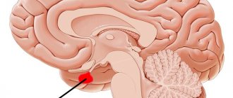
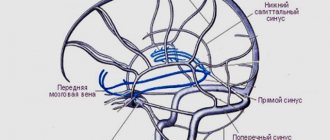
All cerebral arteries are interconnected and originate from the cervical arteries. Before examining the vascular bed of the brain, an ultrasound of the neck vessels is performed. This will provide information about the condition of the vertebral arteries, carotid artery, aorta, subclavian and other vessels of the neck. Having excluded pathologies at this stage, it is possible to perform an ultrasound of the vessels of the head, assessing the functioning of the main arteries of the brain. For this, there are three access zones: orbital, transtemporal (a thin section of the temporal bone) and occipital (through the foramen magnum). By examining the vessels through the orbital approach, the state of blood flow in the ophthalmic arteries is assessed. This area provides information about the presence of disturbances in the blood supply to the brain due to blockage of intracerebral vessels. Assess the condition of the vessels adjacent to this zone in the cranial cavity.
The transtemporal access zone makes it possible to study the state of the vessels of the brain. The temporal bone in this area absorbs little ultrasonic waves, making it possible to study the blood flow in the anterior, posterior and middle cerebral arteries. This procedure is informative in identifying intracranial hematomas. With an ultrasound of the head, you can examine the substance of the brain that is visible in this area and examine large vessels in the cranial cavity.
The occipital examination area evaluates the condition of the vertebral veins and vessels in the brain, and identifies disorders in the brain itself.
An ultrasound scan of the brain will help identify existing but not yet manifested aneurysms, narrowing and abnormal placement of cerebral vessels, both congenital and after trauma. By assessing the speed of arterial blood flow, it is possible to determine the types of circulatory disorders. By measuring the speed of venous blood flow, we can draw conclusions about the presence of increased intracranial pressure. Finding out the cause of pathological conditions using vascular ultrasound will make it possible to prescribe comprehensive treatment for many diseases.
1. Doppler ultrasound of the main arteries of the head (USDG)
The study is carried out using a special ultrasound sensor, which can assess blood flow disturbances and identify changes in the vascular walls. It is based on a change in the frequency of ultrasonic waves that are reflected from moving blood particles (red blood cells). The vessels as such are not visible, so only vascular patency can be assessed.
2. Duplex scanning of the main arteries of the head (DS MAG)
Transcranial duplex examination is performed only after ultrasound of the neck vessels. This method examines both the speed and direction of blood flow and the anatomical state of the vessel itself. A specialized scanner that emits ultrasonic waves works on the principle of an echolocator. A two-dimensional color image is created on the device’s monitor, showing the vascular wall and the lumen inside the vessel. The vascular lumen is examined in the longitudinal and transverse directions. Scanning can be performed in color mode (provides information about the qualitative state of blood flow) and spectral mode (quantitative state of blood flow). This type of study is carried out through the transtemporal access zone and the suboccipital (occipital) zone. Scanning is possible through transoccipital (above the occipital protuberance) and transorbital (orbital) access.
3. Triplex scanning
This study combines duplex scanning methods and color Doppler functions. The monitor of the device displays the blood flow in the vascular cavity in the form of a color picture. It is possible to determine the qualitative condition of the vascular wall, its anatomy and lumen diameter, the direction and speed of blood flow, the presence of atherosclerotic plaques, calcareous deposits, aneurysms, small blood clots and disturbances in the patency of the vascular bed.
Indications for ultrasound of the vessels of the neck and brain.
All conditions in which it appears:
- noise in the head
- impaired coordination and balance,
- dizziness, including when turning the head,
- incomprehensible and sudden loss of consciousness,
- transient ischemic attacks,
- floaters before eyes,
- noise in ears,
- migraine conditions,
- frequent incoming numbness and weakness in the limbs,
- speech disorders,
- in the presence of cerebrovascular disease and a history of vertebrobasilar insufficiency.
Ultrasound is also indicated for:
- vegetative-vascular dystonia,
- diabetes mellitus,
- suspected myocardial infarction, stroke or after it,
- angina pectoris
- in the treatment of headaches of various etiologies,
- hypertension,
- obesity,
- high cholesterol levels,
- bleeding disorders and other cases.
Patients over forty years of age should have their cerebral vessels examined as routine medical examinations, as they are a risk group.