Human anatomy is undoubtedly a major core subject for study in medical schools. Despite the fact that normal human anatomy is a discipline that stood at the origins of the development of medicine, a large number of scientific works still appear that make adjustments to modern anatomical atlases.
It would seem that human anatomy cannot change so quickly with the course of evolution, but our understanding of it is constantly improving as new research methods appear - evidence of this is provided by ever new versions of the anatomy atlas.
Atlas of anatomy Sinelnikova R.D. in 4 volumes, this is perhaps the most authoritative and time-tested source of knowledge on this topic. It is constantly republished, delighting us with its visual illustrations and text accessible to everyone. Many students tried to download Sinelnikov’s atlas for study, but the links either did not work, or there was a virus in the folder... We solved this problem by making a website dedicated to this source.
The main goal of studying human anatomy is to create a fundamental knowledge base for students for further study of other medical disciplines. It is difficult to imagine mastering a curriculum in physiology, pathological physiology, pathological and topographic anatomy, operative surgery, and a whole range of clinical disciplines without a thorough study of normal human anatomy.
It is very important for a student to have a visual image of the material studied; for this it is necessary to study human anatomy in pictures. The main feature of this science. of course, is the structuring of its sections and subsections, as well as a clear systematization of the entire nomenclature.
Thus, we can distinguish the following directions that correspond to each system:
- osteology (section on the bones of the human skeleton). Studies the skeleton, both as a whole mechanism and as individual bones. There is also a study of age-related changes in bones.
- syndesmology (joints, ligaments). An extremely important section for future orthopedists and traumatologists.
- myology (muscular system). He studies not only the structure, but also the development and physiology.
- splanchnology (internal organs). Includes the anatomy of the endocrine, digestive, respiratory, excretory and genitourinary systems.
- angiology (vessels and their derivatives). Information is presented on the structure of blood and lymphatic vessels.
- neurology (central and peripheral nervous system). An extremely important section for successful diagnosis of diseases and perhaps the most difficult.
- aesthesiology (the science of the sense organs). Everything about vision and hearing. And also about taste, olfactory and tactile sensitivity. Closely related to neurology.
IV ventricle
The fourth ventricle (ventriculus quartus) (Fig. 253) is a cavity, the base of which is a rhomboid fossa. With its tent-shaped roof, the fourth ventricle projects into the cerebellum.
The rhomboid fossa (fossa rhomboidea) is bounded above by the superior cerebellar peduncles, and below by the inferior. It can only be seen after the cerebellum is removed. The upper corner of the rhomboid fossa communicates with the cerebral aqueduct, the lower and lateral corners with the subarachnoid space. The inferior angle is also the link between the fourth ventricle and the central canal of the spinal cord.
The rhomboid fossa is divided into superior and inferior triangles. The border is the medullary stripes, which are auditory fibers and pass between the lateral angles in which the auditory nuclei are located (VIII pair). A median groove (sulcus medianus) (Fig. 263) runs through the center of the rhomboid fossa, on both sides of which there are medial elevations (eminentiae mediales) (Fig. 263). On the back of the eminence there is a facial tubercle (colliculus facialis) (Fig. 263). In general, the bottom of the IV stomach is the site of projection of the nuclei of cranial nerves from the V to the XII pairs.
| Rice. 263. IV ventricle 1 - roof of the midbrain; 2 - median groove; 3 - medial eminence; 4 - superior cerebellar peduncle; 5 - middle cerebellar peduncle; 6 - facial tubercle; 7 - inferior cerebellar peduncle; 8 - wedge-shaped tubercle of the medulla oblongata; 9 - thin tubercle of the medulla oblongata; 10 - wedge-shaped fascicle of the medulla oblongata; 11 - thin bundle of the medulla oblongata |
In the upper triangle, in the area of the median sulcus, there are the nuclei of the trigeminal nerve (V pair), the nucleus of the abducens nerve (VI pair) and the slightly deeper nucleus of the facial nerve (VII pair). Rounding the nucleus of the abducens nerve, the processes of the bodies of the nucleus of the facial nerve form an elevation of the rhomboid fossa, called the facial tubercle. In the lower triangle are located the nuclei of the vestibular-cochlear nerve (VIII pair), which are called vestibular. The nuclei of the glossopharyngeal (IX pair) and vagus nerves (X pair) are projected into the triangle of the vagus nerve. They have a common motor nucleus, the cell processes of which form the fibers of the accessory nerve (XI pair). The nucleus of the hypoglossal nerve (XII pair) projects into the triangle of the hypoglossal nerve on the sides of the posterior median sulcus of the medulla oblongata.
Colloid cyst of the 3rd ventricle
Colloid cyst of the 3rd ventricle is a disease common among adults 20-40 years old. It is characterized by the appearance of a benign round formation in the cavity of the 3rd ventricle, which is not prone to rapid growth and metastasis.
The colloid cyst itself does not pose any danger to human health. Problems begin if it reaches a large size and interferes with the outflow of cerebrospinal fluid. In this case, the patient experiences neurological symptoms associated with increased intracranial pressure:
- severe headache;
- vomit;
- visual impairment;
- convulsions.
Diagnosis and treatment of colloid cyst of the third ventricle is carried out jointly by a neurologist and a neurosurgeon. If the size of the formation is pronounced, determined on CT or MRI, surgical treatment of the cyst is prescribed. After the operation, the normal flow of cerebrospinal fluid is quickly restored, and all symptoms of the disease disappear.
Which doctors should you contact when diagnosing ICH?
If the 3rd ventricle of the baby’s brain is slightly dilated and the mother has no serious complaints, regular monitoring by a local pediatrician is sufficient. Consultation with a neurologist and neurosurgeon is necessary if there is significant dilatation of the ventricles on ultrasound or symptoms of ICH:
- the child began to suck the breast worse;
- the fontanel is tense, protruding above the surface of the skull;
- the saphenous veins of the scalp are dilated;
- Graefe's symptom - a section of white sclera between the iris and eyelid when looking down;
- loud, sharp cry;
- vomit;
- divergence of the sutures of the skull;
- rapid increase in head size.
Doctors determine further tactics for treating a baby with hydrocephalus: conservative means prescribing vascular drugs, massage, physiotherapy; surgical – performing an operation. After therapy, children quickly recover, the activity of the nervous system is restored.
Liquor features of this liquid
The rate of production of this type of liquid in a person per day is about 500 ml. Complete renewal of the cerebrospinal fluid occurs within a period of 4 to 7 hours. If the cerebrospinal fluid is poorly absorbed or its outflow is disrupted, the brain is severely compressed. If everything is in order with the cerebrospinal fluid, its presence protects the gray and white matter from damage of any type, especially mechanical. CSF transports substances important to the central nervous system, while simultaneously removing unnecessary substances. This is possible because the central nervous system is completely immersed in a fluid called cerebrospinal fluid. It contains:
- vitamins;
- hormones;
- compounds of organic and inorganic types;
- chlorine;
- glucose;
- proteins;
- oxygen.
Treatment
Treatment for tumors, hematomas and parasites of g. g. m. is only surgical. To remove the intraventricular focus, ventriculotomy is performed (see) - dissection of the wall of the ventricle with opening of its lumen.
In inflammatory processes, surgical intervention is resorted to in cases of development of occlusive phenomena (see Hydrocephalus). Ventriculopuncture is used as a temporary measure for acute occlusions of the cerebrospinal fluid outflow tract to reduce intraventricular pressure (see).
In cases where the occlusion cannot be surgically eliminated, palliative operations are performed aimed at creating a roundabout path for the outflow of cerebrospinal fluid from the ventricles (ventriculostomy operations, perforation of the terminal plate, ventriculosubdural anastomosis, ventriculocisternostomy).
Among the conservative methods of treating ventriculitis, dehydration is used to reduce intracranial pressure and reduce hypertensive syndrome (see Dehydration therapy). For acute and chronic infectious ventriculitis, anti-inflammatory treatment is carried out.
The prognosis for tumors of the gastrointestinal tract is always serious and is determined by their good quality, localization within the ventricular system, and relationship to adjacent brain formations. With hemorrhages in the gastrointestinal tract, in most cases the prognosis is unfavorable. In inflammatory processes, the prognosis largely depends on concomitant disturbances in the circulation of cerebrospinal fluid. The prognosis for parasitic lesions is determined by the multiplicity of lesions and the degree of concomitant inflammatory and adhesive processes in the ventricles and membranes of the brain.
See also Intracranial pressure, Hypertensive syndrome.
Bibliography:
Multi-volume guide to neurology, ed. S. N. Davidenkova, vol. 5, M., 1961; Multi-volume guide to surgery, ed. B.V. Petrovsky, vol. 3, book. 2, M., 1968; Patten B. M. Human embryology, trans. from English, M., 1959; Shelia R. N. Tumors of the ventricular system of the brain, L., 1973; G 1 a g a M. Das Nerven-system des Menschen, Lpz., 1953; G o r-rales M. a. T orre a 1 ba G. The third ventricle, Normal anatomy and changes in some pathological conditions, Neuroradiology, v. 11, p. 271, 1976, bibliogr.; Messert B., Wanna-maker BB a. Dudley AW Reevaluation of the size of the laterol ventricles of the brain, Postmortem study of an adult population, Neurology (Min-neap.), v. 22, p. 941, 1972.
Causes of ventriculomegaly
Dilatation, or an increase in the size of the ventricles of the brain, is called ventriculomegaly. It must be said right away that, whatever the reason led to this phenomenon, their asymmetry should cause the greatest concern. If ventriculomegaly is symmetrical, then it can be either a normal variant or a sign of hydrocephalus, or develop for a number of other reasons. But if their asymmetry is revealed and one ventricle is larger than the other, or they are disproportionate, then, most likely, we are talking about a volumetric formation of the brain, or the consequences of an injury; The newborn should have an urgent consultation with a neurosurgeon, otherwise the consequences may be severe and unpredictable.
But mild ventricular asymmetry may be normal. If the dimensions of the right and left ventricles at the level of the foramen of Monroe differ by up to 2 mm, we are not talking about pathology. You just need to monitor it in a timely manner so that this difference does not increase.
As a rule, pathological dilatation of the ventricles begins with their occipital horns. The screening method for examining them is the baby, carried out through the large fontanel. If the ventricles are poorly visible, this does not mean that they are enlarged. In order to make this diagnosis, you need to see them clearly.
In relation to newborns, we can talk about dilatation of the lateral ventricles of the brain only when the size of the anterior horns in diagonal sections at the level of the foramen of Monro, according to neurosonography, exceeds 5 mm and the concavity of the fundus contour disappears.
The causes of ventriculomegaly can be either congenital or acquired. Congenital causes include:
- Pathological course of pregnancy and complications during childbirth.
- Phenomena of acute intrauterine fetal hypoxia.
- Malformations of the central nervous system and other developmental defects.
- Premature birth.
- Perinatal trauma.
Particular attention should be paid to hemorrhages, such as subdural and subarachnoid. It is in these cases that significant asymmetry of the ventricles occurs, which occurs due to the appearance of blood volume, which causes compression of one of the ventricles of the brain.
- Intrauterine infections, septic complications in mother and child.
- Delayed expulsion period (long interval between the breaking of water and the birth of the baby).
- Some extragenital pathology of the mother (diabetes, heart defects, etc.).
In addition to these congenital reasons, there are also acquired reasons why the size of the lateral ventricles may be increased:
- space-occupying formations – ranging from cysts and hemangiomas to brain tumors;
- hydrocephalus.
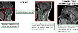
Special mention should be made about hydrocephalus in newborns. With this disease, an excess amount of cerebrospinal fluid accumulates in the brain structures, which causes general cerebral symptoms and can also cause acute conditions.
Hydrocephalus does not immediately cause an increase in the cerebrospinal fluid cavities of the brain. Their size can remain unchanged for quite a long time, and only after decompensation caused by a sharp increase in intracranial pressure, expansion of primarily the lateral ventricles occurs. Since they are not central structures communicating with the vast cerebrospinal fluid spaces of the spinal cord and the main tank, they experience the greatest pressure.
Symptoms of hydrocephalus
This disease is manifested by certain symptoms - signs of deterioration in health, which should definitely be paid attention to. They directly depend on the age group to which the patient belongs, as well as on the degree of progression of the disease
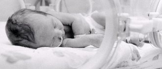
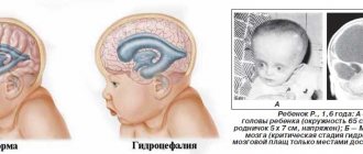
For example, hydrocephalus of the brain in newborns is accompanied by a number of features. First of all, those children who have been diagnosed with this disease have an excessively large head circumference, which continues to increase in the future. At the same time, a convex fontanel is noticeable on the parietal part of the child’s head. Other manifestations of this disease in young children include vomiting, poor sleep, irritability, eye rolling and convulsions. Most often, the development of such children is delayed, complicated by poor perception of information, slow thinking, learning difficulties, etc.
Hydrocephalus of the brain in children can appear during the mother’s pregnancy. This type of this disease is called congenital. Its occurrence is caused by intrauterine infections, fetal malformations, and hemorrhages in the ventricles of the brain in an unborn child. Another type of this disease is acquired hydrocephalus. It develops after the baby is born. Its causes may be traumatic brain injuries received during childbirth, as well as various infectious diseases.
All of the above types of hydrocephalus are in an active progressive form, in which intracranial pressure increases, brain tissue atrophies and the ventricles of the brain expand. But the expansion of the ventricles of the brain can be passive, this form is called moderate external hydrocephalus. Doctors believe that moderate external hydrocephalus is a rather dangerous disease, since in most cases there are no symptoms characteristic of hydrocephalus. It is worth noting that moderate external hydrocephalus leads to impaired blood circulation in the brain and the patient begins to suffer from nervous system disorders, lethargy, and migraines.
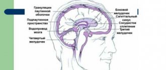
What is a cerebral ventricle
The ventricular system of the brain is presented in the form of reservoirs in which the secreted secretion - cerebrospinal fluid - accumulates. Continuous circulation of fluid in the subarachnoid space, ventricles and spinal canal is achieved through openings (anastomosis), which are necessary for the process of cerebrospinal fluid circulation.
The choroid plexuses of the ventricles, namely glandular cells, produce cerebrospinal fluid, the amount of which is determined by a person’s age. The size of the ventricles also increases as the central nervous system matures; normally, the lateral cisterns are larger.
Type of ventricles GM
In a newborn child, the total volume of fluid is about 10 milliliters, and in an adult it is from 150 to 250. But regardless of age, the cerebrospinal fluid is renewed up to 6 times per day. How much secretion glandular cells produce per day depends on the size of the ventricles and the influence of various factors (psycho-emotional background, blood pressure, central nervous system diseases).
The cerebral ventricles are lined with a layer of epithelium, each cell in it has cilia that move the accumulated fluid. Thus, the cerebrospinal fluid moves passively through an open system with the help of vibration from large cerebral vessels and the villous layer. These cells (tanycytes) have processes that penetrate into the diencephalon.
Their anatomical structure is very similar to nerve fibers, so ependyma is involved in the transmission of nerve signals, but there are also unique features in the structure that affect the overall functionality of the cells.
Summarizing all of the above, we can answer the question - what are the ventricles responsible for? The main task of the cavities is the production of secretion, which in turn is an important shock-absorbing system and supports the trophism of brain tissue.
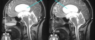
The ventricles increase in proportion to the growth of the skull. But there are deviations in which the cavities are significantly expanded and this leads to neuralgic symptoms. It is possible to assess the liquor flow and the size of cavities using non-invasive research methods.
In children under 1 - 1.5 years of age, diagnosis is carried out through the central, non-overgrown fontanel using ultrasound (neurosonography). This method is not suitable for adults, since the ossified areas of the arch do not conduct the ultrasonic wave. MRI or CT is used for diagnosis.
The table shows the normative values of the structures of the ventricular system in an adult:
| Structural unit | Average standard values in millimeters |
| Anterior horns of large cavities (lateral). | From 2 to 5 |
| Sylvian fissure. | From 3 to 5 |
| 3 cerebral ventricle | From 2.2 to 4.7 |
| 4 cerebral ventricle | From 12 to 14 |
The side tanks normally have no asymmetry and are identical in size.
For a general assessment of the ventricular system, the index is also calculated:
- Evans - the distance between the anterior horns of the lateral ventricles and the largest bitemporal diameter of the skull (from 24 to 30%);
- Schlatenbrandt-Nurenberger – width of the 3rd ventricle and transverse diameter of the skull (from 30 to 50 mm);
- Akimova-Komissarenko - the width of the 4th ventricle in relation to the diameter of the cranial fossa (from 11.5 to 14 mm.). The overall coefficient is calculated, where a value of 5.1 is the norm.
With significant asymmetry of the lateral cisterns, the reason always lies in organic pathology (inflammatory processes, neoplasms).