Congenital malformations of the fetus are not as common as we have already said - approximately every 30 pregnancies have clinically significant changes in the fetus, although small deviations or features in the development of one or another organ in the fetus are much more common.
It is the fact of the relative rarity of fetal pathology that causes carelessness among expectant mothers, and their motivation is the simplest - my husband and I are healthy, and, therefore, our children are also healthy! Alas, not everything is so rosy and simple!
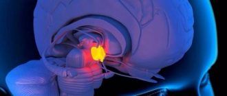
A fairly common pathology (5 per 1000 births), but rarely diagnosed during pregnancy, and almost never diagnosed before the 20th week of pregnancy, is agenesis of the corpus callosum (CAC) . Among all fetal malformations, this pathology accounts for 2-3%.
— What is the corpus callosum, and why did it appear in the brain, where could the calluses come from? – you ask indignantly.
- This is a small but important part of the brain that ensures the exchange of information between the right and left hemispheres of the brain. Without it, the right and left hemispheres live on their own, and the two halves of the brain have independent capabilities to participate in the processes of consciousness, memory accumulation, communication and regulation of motor activity.
A precedent that has become a parable is created when “the right hand does not know what the left hand is doing” - and, therefore, with agenesis of the corpus callosum, a significant intellectual deficit is expected in a person’s neurological development. In addition, seizures, large head sizes, giant brain cysts and cerebral palsy (up to 30%) are also consequences of AMT.
General information
Agenesis of the corpus callosum (ACC) is a rare disorder that is present at birth (congenital).
It is characterized by partial or complete absence (agenesis) of the corpus callosum. The corpus callosum consists of transverse fibers. The cause of agenesis of the corpus callosum is usually unknown, but it can be hereditary in either an autosomal recessive or X-linked dominant pattern. In addition, agenesis can also be caused by infection or injury during weeks 12 to 22 of pregnancy (intrauterine life), resulting in impaired fetal brain development. Prenatal alcohol exposure can also lead to AMT. In some cases, with AMT, mental retardation may occur, but intelligence is mildly impaired, and subtle psychosocial symptoms may also be present.
Partial agenesis of the corpus callosum
Fig.1
Complete agenesis of the corpus callosum
Fig.2
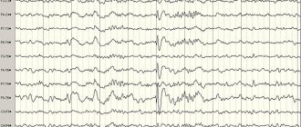
AMT is often diagnosed during the first 2 years of life. Seizures of epilepsy may be the first symptom indicating that a child should be tested for brain dysfunction. The disorder can also remain without visible symptoms in the mildest cases for many years.
Functions of the organ
The splenium of the corpus callosum is a thickening in the posterior part. The middle section is the trunk. The main part is 2/3 of the entire length of the solder.
The front one is a knee that ends in a thin beak.
The structure of the MT is such that it lies deep in the longitudinal sulcus and connects only the hemispheres, without affecting the rest of the brain.
For a long time after its discovery, scientists could not understand what specific functions the corpus callosum performs. It was initially assumed that it contained a focus of epileptiform activity.
For patients with epilepsy, surgeons cut it open to eliminate seizures. The seizures passed, but people's behavior changed. Thus, a patient with a dissected corpus callosum hugged his wife with his right hand and pushed her away from him with his left.
And only experiments on animals, and subsequently with the participation of a volunteer, made it clear:
- The obvious function is to physically connect the right and left hemispheres, since the corpus callosum is located deep in the longitudinal fissure between the hemispheres. Nerve fibers connect both symmetrical and asymmetrical sections of different hemispheres, as well as different sections of the same hemisphere.
- Information communication between the hemispheres. Neuronal fibers (white matter) fan out in all directions, transmitting information received by different lobes of the hemispheres.
- Coordination of the hemispheres. MT ensures not only the exchange of received information between the left and right hemispheres, it is also responsible for analyzing this information and responding adequately.
The corpus callosum is covered on top by a small layer of cerebral gray matter, which explains, accordingly, the gray covering on it. Upon visual examination, 3 main sections can be distinguished:
- trunk (or midbrain);
- knee (part of the brain located in the front);
- beak or splenium of the corpus callosum (posterior section).
The middle section, when viewed, looks like a bulge, which is also the longest part of the entire brain. The posterior section is visually visible as a thickening relative to other sections and zones, which is freely located above neighboring areas of the brain. The gray matter is represented by stripes and is located on top.
Functions provided by the corpus callosum:
transfer of information (impulses) important for the functioning of the body from one hemisphere to the other; formation of the main characteristics that define personality and its characteristics; basic (basic, defining) skills and the possibility of their application throughout a person’s life; work on the formation of the emotional and personal sphere
Morphology
Classic neuroradiological signs of agenesis of the corpus callosum:
- The anterior horns and bodies of the lateral ventricles are widely spaced and parallel (not curved). The anterior horns are narrow and acute-angled. The posterior horns are often disproportionately enlarged (colpocephaly). The concave medial borders of the lateral ventricles are caused by protrusion of the longitudinal fasciculi.
- The third ventricle is usually dilated and elevated with varying degrees of dorsal expansion and mixing between the lateral ventricles. The interventricular foramina are often elongated.
- The interhemispheric groove appears to be a continuation of the anterior section of the third ventricle, since the knee is absent. In the coronal projection, the interhemispheric groove expands downwards between the lateral ventricles towards the roof of the third ventricle. In the sagittal plane, the usual cingulate gyrus is absent, and the middle sulci have a radial or spoke-shaped configuration. Interhemispheric cysts are often visible around the third ventricle. As they increase in size, these cysts can acquire an abnormal configuration and hide underlying defects.
- Absence of the corpus callosum and septum pellucidum.
- Angulation of the anterior horns of the lateral ventricles and their depression along the medial surface by Probst's bundles.
- Radial pattern of grooves and convolutions extending from the roof of the third ventricle.
In dry but important remainder
Thus, the corpus callosum of the brain, despite its tiny size, has a great influence on a person’s life. It allows the formation of personality, is responsible for the emergence of habits, conscious actions, the ability to communicate and distinguish between objects.
That is why it is extremely important to take care of your health during pregnancy, since the main metabolic disorders are formed during this period.
We should not forget that the corpus callosum forms the intellect and makes a person a personality. Despite all attempts to study this structure, scientists have not yet been able to uncover all its secrets, and therefore very few methods for treating disorders, if any, have been developed.
The main ones are drug therapy and a special set of exercises - exercise therapy, which allows you to maintain optimal indicators of physical development. Measures to eliminate the symptoms of disorders should be taken immediately, otherwise the desired improvements may not occur.
Signs and symptoms
Agenesis of the corpus callosum (ACC) may initially present with the onset of epileptic seizures in the first weeks of life or during the first two years. However, not all people with AMT have seizures. Other symptoms that may begin early in life are problems with feeding and holding the head up. Sitting, standing and walking may also be affected. Impaired mental and physical development and/or hydrocephalus are also early-onset symptoms of this disorder.
Non-progressive mental retardation, impaired hand-eye coordination, and visual or auditory memory can be diagnosed through neurological testing in patients with AMT. In some mild cases, symptoms may not appear for many years. In mild cases, AMT may be missed due to the absence of obvious symptoms in childhood.
Some patients may have deep-set eyes and a bulging forehead. There may be an abnormally small head (microcephaly) or an unusually large head (macrocephaly). Folds of skin in front of the ears, one or more bent fingers (camptodactyly), and slow growth may also be associated with some cases of agenesis of the corpus callosum. In other cases, wide eyes, a small nose with inverted nostrils, abnormally shaped ears, an excessively long neck, short arms, decreased muscle tone (hypotonia), laryngeal abnormalities, heart defects, and symptoms of Pierre-Robin syndrome may be present.
Aicardi syndrome , considered to be inherited in an X-linked dominant pattern, consists of agenesis of the corpus callosum, infantile spasms, and abnormal ocular structure. This syndrome is an extremely rare congenital disorder that affects the middle layer of the eyes (choroid) and retina, as well as the absence of the corpus callosum, accompanied by severe mental retardation. Only women suffer from this syndrome.
Anderman syndrome is a genetic disease characterized by a combination of agenesis of the corpus callosum, mental retardation and progressive sensorimotor disorders of the nervous system (neuropathy). All known cases of this disorder come from the Charlevoux district and the Saguenay-Lac Saint-Jean region in Quebec, Canada. The gene causing this rare form of agenesis of the corpus callosum has recently been identified and is currently being tested (SLC12A6).
XLAG (X-linked lissencephaly with true hermaphroditism) is a rare genetic disorder in which males have a small and smooth brain (lissencephaly), a small penis, severe mental retardation and intractable epilepsy. It is caused by mutations in the ARX gene. In women, these same mutations can cause agenesis of the corpus callosum on their own, while less severe mutations in men can cause mental retardation. Diagnosis of this disorder is available in clinical use.
Etiology
The causes of abnormalities in the development of the corpus callosum can be genetic, infectious, vascular or toxic.
Genetic causes
There are more than 60 genetic and chromosomal abnormalities in which corpus callosum pathology is a nonspecific feature.
The genetics of AMT in humans are diverse and reflect the complexity of corpus callosum development. A combination of genetic mechanisms, including Mendelian single-gene mutation, sporadic single-gene mutation, and complex genetics (which may be a combination of inherited and sporadic mutations) may play a role in the etiology of AMT. The causes of 30-45% of AMT cases can be identified. 10% have chromosomal abnormalities, and the remaining 20-35% have identifiable genetic syndromes. Most individuals with AMT have no obvious hereditary cause or genetic syndrome, suggesting that AMT may be caused by random new ( de novo)
) genetic mutations.
Related disorders
Agenesis of the corpus callosum can be combined with spina bifida. Some of the contents of the spine may protrude through the abnormal opening, leading to the formation of a meningocele or meningomyelocele.
- hydrocephalus 30%
- arachnoid cysts
- intracranial lipoma 10%
- Dandy-Walker malformation 11%
- Interhemispheric cyst
- Arnold-Chiari malformation 7%
- Median encephalocele
- Porencephaly
- Holoprosencephaly
- Polymicrogyria
- Heterotopia
The big commission is under attack...
Disorders of the corpus callosum are a rare phenomenon, occurring in 2% of all cases of diseases of the brain and central nervous system. In case of diseases of the corpus callosum, the following are observed:
- disorders of various nature and intensity, manifested in the emotional, personal and cognitive spheres;
- physiological problems in the functioning of the limbs;
- problems with the eyeballs and vision in general.
Corresponding diseases develop - agenesis, hypoplasia and dysplasia (dysgenesis) of the corpus callosum of the brain.
Treatment
Treatment is symptomatic and supportive depending on the severity of symptoms. If hydrocephalus is present, it is treated with surgical shunting to reduce the elevated ICP. Genetic counseling may also benefit families with this disorder.
Author: radiologist, Ph.D. Vlasov Evgeniy Alexandrovich
Full or partial reprint of this article is permitted by installing an active hyperlink to the source
If you still have doubts about the conclusions of your MRI, you can order a review of your study with a detailed transcript here: