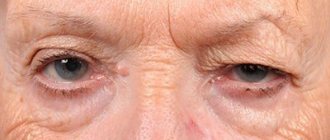
Surely you have encountered the phenomenon of floating spots in front of your eyes and glare. These are tiny shapes, lines, shadows or specks that feel like they are drifting across the surface of the eye. You may have wondered what this is and whether it is a dangerous symptom. Most often, the appearance of floating spots is normal and does not indicate problems with vision or eye condition. However, when this symptom becomes too frequent and is accompanied by glare, it may indicate a more serious problem.
Flares are flashes of light in the form of threads or stars. This can be either one flash in one visual zone, or multiple flashes in a wider area. Sometimes flashes may not be noticed because they most often appear on the side or in the periphery.
If you suddenly feel this unpleasant symptom, or the flashes and flickers become very frequent, you need to urgently make an appointment with an ophthalmologist to rule out any serious eye diseases.
What causes the floating fly effect?
The vitreous humor of the eye is a clear gel that fills most of the eyeball and resembles raw egg white. Inside the vitreous are small clumps of protein that shift and move with your eyes. When these tiny clumps of protein cast shadows on the retina, the light-sensitive surface at the back of the eye, you see floating shadows.
As we age, the vitreous shrinks, producing more proteins. This is why, over time, it becomes more and more common to feel like you are seeing floating shadows. Opacities are more common in nearsighted people and diabetics and are more likely to occur after cataract surgery or eye injury.
If you get tired of the effect of floating shadows, try moving your eyes up and down or side to side to gently move them to the periphery of your vision.
Symptoms
If the patient experiences a complete detachment, not only a sensation of cobwebs in the eye is formed, but also additional symptoms:
- lightning and flashes before the eyes, intensifying when the eyeballs move;
- floating dots and stripes, larger geometric shapes.
If the condition is less severe, other than floating webs, other symptoms may not occur. In the first stages, vision remains normal. But if timely therapy is not carried out, complications arise. It is they that lead to negative symptoms that patients discover on their own when they present complaints to the doctor.
What causes glare?
Glare occurs as a result of movement or tension of the retinal nerve cells. As the vitreous shrinks over time, it can pull on the retina, causing you to “see stars” or flashes of light. The process of separation of the vitreous from the retina is called “posterior vitreous detachment.” This is not a health hazard.
In about 16% of cases, the detachment process creates tiny tears in the retina, which can lead to retinal detachment. This is already a dangerous condition; if it is not treated, blindness is possible.
Other possible reasons why you are seeing strange flashes are an eye injury or a migraine.
Treatment
Usually, “floaters” in the eyes either go away on their own, or the person adapts and simply stops noticing them. At the moment, there is no effective method of drug treatment that can completely get rid of “annoying flies.”
For significant opacities, resorption therapy is used. Drugs are used to regulate metabolic processes in the eyes. For this purpose, Wobenzym tablets and Emoxipin eye drops are used. Take measures to improve immunity using vitamin and mineral preparations with lutein.
“Floaters” in the eyes can only be effectively eliminated surgically. Surgical treatment is carried out using two methods: vitreolysis and vitrectomy. In the first case, the cloudy fragments are broken into small sizes using a laser method, which are no longer perceived by vision as an image defect. During a vitrectomy, the vitreous humor is removed. But such an operation should be performed only if there are serious indications for it.
To prevent the development of eye floater syndrome, you need to reconsider your lifestyle. Perhaps a more flexible or, conversely, gentle mechanism of physiological mobility is needed. It is also recommended to balance meals according to age categories and taking into account health status. To maintain eye health, if you already have visual impairments, you must use glasses or contact lenses. This will prevent eyes from straining and will prevent the appearance of “floaters” in the eye.
Team of doctors Ochkov.Net
When to see a doctor about floaters
If you experience any of the following symptoms, contact your eye doctor immediately for emergency eye care.
Symptoms that should not be ignored:
- The sudden appearance of floaters accompanied by flashes (which can be of any shape or size).
- An increase in floating particles, accompanied by a darkening of one side of the visual field.
- Shadows in peripheral vision
- Do you see glare all the time?
In many cases, the appearance of floaters before the eyes is not a cause for concern; however, the above symptoms may indicate a retinal detachment, and if left untreated, it can cause permanent vision loss or even blindness.
If you experience the above symptoms, you should see a doctor within 24 hours. You will have to endure the discomfort of having your pupils dilated during an eye exam so that the ophthalmologist can get a good look at the peripheral retina and diagnose or rule out a retinal tear or other serious condition. It is worth finding out whether there is a threat to vision. If it's just an uncomplicated posterior vitreous detachment, which is quite common, or an ocular migraine, then you can breathe a sigh of relief.
Diagnostics
During the initial examination by an ophthalmologist, it is necessary to establish under what circumstances symptoms appear (bright lighting, work at close range, at the height of a hypertensive crisis) and when they disappear. It is important to clarify the duration of the process. Basic diagnostic methods:
- Visometry.
Vitreous opacities of small size and peripheral localization do not affect visual acuity. With a pronounced degree of impairment, visual dysfunction can be observed. - Biomicroscopy of the eye.
With asteroid hyalosis, a large number of small yellowish or yellowish-white shiny formations are observed in the projection of the light beam. The effect of reflection is characteristic. However, the technique does not allow assessment of peripheral structures. - Ultrasound of the eye.
In gray scale B-mode, individuals with asteroid hyalosis show multiple small discrete echogenic inclusions with varying acoustic density. In A-mode, repeating high-amplitude complexes are recorded in the projection of the entire anechoic space of the vitreous cavity. - Spectral optical coherence tomography ( SD - OCT )
. OCT makes it possible to clearly visualize vitreal lesions located preretinal. With more anterior localization, they are indirectly detected as darkening of the OCT image. - Scanning laser ophthalmoscopy
. Depending on the density of the opacities, they look like white-gray shadows or penumbra. Using a series of photographs, you can estimate the size and approximate density. A Weiss ring is often detected in the fundus. - Dynamic Light Scattering ( DLS )
. The laser nanodetector system is used to image particles ranging in size from 3 nanometers to 3 microns. An ophthalmologist can evaluate collagen fiber aggregation in myopic or diabetic vitreopathy. - Determination of contrast sensitivity
. In the presence of primary compactions in the vitreous body, contrast sensitivity (CS) decreases. This increases the degree of light scattering. Studies are carried out before and after surgery. Asteroid hyalosis does not affect CP indicators.
Ophthalmological examination
How to help yourself with a headache
If a headache radiates to the eyes after hard work, you should rest well, take a walk in the fresh air, and give yourself an acupressure massage. You can take a warm bath with chamomile infusion added to the water. Take a break from working at the computer or watching TV
Give yourself a head massage. Relax, drink warm milk with honey, tea with lemon balm. Do simple exercises to relieve headaches:
- sit on a chair, keep your back straight and your head free;
- without effort, just under the influence of gravity, tilt it towards your chest;
- stay in this position for twenty seconds; take a break for 30 seconds;
- Bend again for 20 seconds.
Repeat the exercise 15-16 times.
The second exercise is done in the following sequence:
- sitting or standing, raise your hands to your head;
- the thumbs of each hand are pressed to the zygomatic arches, with the remaining fingers clasping the back of the head;
- look up;
- while inhaling, try to throw your head back for 10 seconds, while holding it with your hands;
- while exhaling, look down for 6-8 seconds;
- tilting your head to your chest as much as possible, stretch but do not strain the neck muscles.
Repeat the inhale-exhale cycle 5-6 times.
To relieve headaches coming from the cervical spine, rehabilitation experts recommend performing the following exercise:
- sitting on a chair, with one hand clasp your head from above on the side in which the pain is felt more strongly;
- Place the index finger at the level of the beginning of the ear;
- With a little hand effort, turn your head to the “healthy” side;
- Press your free palm from below to your chin and cheek;
- while inhaling for 10 seconds, looking down, press your chin to your lower palm against its resistance;
- As you exhale, relax for 6-8 seconds and look up.
- repeat the exercise 5-6 times, slightly changing the turn of the head.
Consequences of untimely therapy
If a patient does not see an ophthalmologist for a long time with a black cobweb before his eyes, complications gradually develop:
- complete loss of vision;
- disability;
- rhegmatogenous retinal detachment;
- macular hole;
- serious defects of the retina.
If a person has cobwebs floating before his eyes even after thoroughly washing and removing lenses, it is better to consult an ophthalmologist in a timely manner. The earlier the defect is detected, the higher the chance of complete preservation of vision.
Author's rating
Author of the article
Alexandrova O.M.
Articles written
2100
about the author
Was the article helpful?
Rate the material on a five-point scale!
( 2 ratings, average: 5.00 out of 5)
If you have any questions or want to share your opinion or experience, write a comment below.
Causes of flashes before the eyes
Sometimes such complaints are a benign condition associated with excessive visual stress or age-related changes in the eye. However, more often than not, the cause of outbreaks is a serious problem that requires immediate medical attention. The causes of the condition may be:
- Posterior vitreous detachment. The gel-like substance that fills the central part of the eye and is attached to the retina is called the vitreous body. With age, it gradually shrinks, peeling off from its original place. This process retracts the retina, causing flashes to appear in the eyes. Similar symptoms are noticeable when moving the eye. There is no specific treatment for this condition. However, patients with this diagnosis must undergo regular ophthalmological examinations. Sometimes, such symptoms hide a more dangerous pathology - retinal detachment, which can cause irreversible vision loss - blindness.
- Retinal detachment or tears. Very often, flashes of light are a symptom of a violation of the integrity of the retina and the beginning of its detachment. It is necessary to pay special attention to the situation if such a condition appears after physical activity (heavy lifting) or serious nervous stress. In this case, flashes of light are often accompanied by the appearance of a veil in front of the affected eye and a sudden decrease in visual acuity. In case of retinal detachment, urgent medical attention is required - surgery.
- Migraine. Similar visual symptoms may precede headache attacks. In this case, flashes of light take the form of bright white zigzags, sparks or geometric shapes. As a rule, they are localized in the periphery of the visual field in one eye or two at the same time. Light flashes of light in the eyes occur without headache attacks. This phenomenon is called ocular migraine. They do not fall within the competence of an ophthalmologist; they are dealt with by neurologists.
- Vascular diseases. Such pathologies include: hypertension, atherosclerosis, diabetes mellitus, etc. Patients note the periodic occurrence of flashes before closed eyes or in the dark. The condition is caused by short-term vascular spasms with impaired blood circulation in the retinal tissue. As a rule, in this case, manifestations of angiopathy or retinopathy are visible in the fundus.
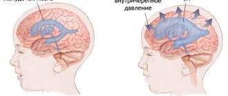
- Cervical osteochondrosis. When the vertebral arteries (pass in the bone canals of the cervical vertebrae) are compressed, the blood supply to the brain (including the cerebellum) is disrupted, so not only visual disturbances appear, but also dizziness. Often, vertebral artery syndrome (VAS) occurs in people who work at a computer for a long time and lead a sedentary lifestyle.
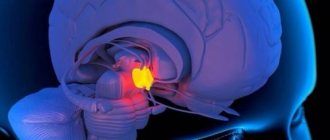
- Intracranial tumors of the occipital lobe of the brain. In this case, visible phenomena differ in shape and color and have a constant nature of occurrence.
- Inflammatory processes in the retina and choroid of the eye. The diseases that cause them are called retinitis and choroiditis and always occur against the background of accompanying signs of inflammation: blurred vision, decreased visual acuity.