How to find out what is happening inside the human body during illness? For centuries, the only realistic answer was to “cut and see” - not a very happy prospect for the patient! At the very end of the century, fluoroscopy was invented, and the capabilities of doctors fundamentally expanded. This method is excellent for obtaining images of dense tissues, primarily bones. But it was impossible to obtain a clear image of, for example, the brain with its help. That is why physicists and doctors continued to actively look for other methods of “candling” our body.
High tech
In the first years of its use, magnetic resonance imaging was called differently: NMR imaging, from the words “nuclear magnetic resonance.” It was renamed after 1986, when, in connection with the accident at the Chernobyl nuclear power plant, people began to fear anything containing the word “nuclear” in the name.
The principle, however, remains the same. It has nothing to do with radioactivity. The electromagnetic field that the tomograph generates affects the nuclei of hydrogen atoms present in any tissue of the human body. They generate a response signal, which is recorded by the device.
The nuclear magnetic resonance technique is used not only in medicine - it can be used to study the structure of almost any substance. But it was for its adaptation to the study of living organisms that American scientists Paul Lauterbur and Peter Mansfield received the Nobel Prize in 2003. The research itself that led to the creation of the tomograph was carried out in the late 1960s and early 1970s.
In science, it often happens that some idea is “in the air.” The use of nuclear magnetic resonance in medicine was explored at that time not only by Lauterbur and Mansfield. Back in 1960, the first prototype of a magnetic resonance imaging scanner was developed by Vladislav Aleksandrovich Ivanov, a graduate of the Leningrad Air Force Engineering Academy named after A.F. Mozhaisky. Unfortunately, the finalization of the device and the subsequent receipt of a patent took more than ten years, so Western scientists received an advantage in time - and therefore the laurels of discoverers.
How it's done?
Magnetic resonance imaging is often compared to computed tomography (CT). Both methods provide layer-by-layer images of internal organs, but computed tomography uses X-rays, so the method allows you to look mainly at bones and dense tissue. MRI uses a strong magnetic field and is better suited for imaging soft tissue.
Carrying out an MRI requires quite a lot of time - 30-40 minutes, and it is highly advisable to remain motionless during the entire scanning period. Computed tomography is more convenient in this sense: it takes no more than 10 minutes. But magnetic resonance imaging is allowed to be done frequently, since it is not accompanied by the risk of radiation. Unlike CT, magnetic resonance imaging appears to be safe during pregnancy, according to studies. In Russia, it is not recommended to do it during the first trimester, but this is most likely a precaution.
Preparing for the examination
An MRI examination of the heart and coronary vessels does not require any special preparation. Women should ensure that they are not pregnant, and those with kidney disease should ensure that they do not have kidney failure. Immediately before starting the procedure, it is necessary to get rid of metal objects, jewelry, excess clothing, and removable dentures. Clothing should be comfortable and loose.
If you do an MRI of the heart with contrast, you should not drink 5-6 hours before the examination. Is it possible to do a cardiac MRI if there is an increased level of anxiety? Yes, you just need to start taking sedatives the day before. Is it possible to do a cardiac MRI if there is pain? Yes, you can, but you just need to take an antispasmodic before the procedure.
Carrying out MRI of the heart requires calm and measured work of the heart muscle. Because a rapid heartbeat creates additional movements of the chest and unnecessary noise in the image.
Varieties
Magnetic resonance spectroscopy
Why: Allows you to determine the level of metabolism in a specific organ or tissue and reflects changes in metabolism, if any.
Bonus: The technique allows you to detect a violation of the blood supply to an organ or an inflammatory process even before pronounced symptoms appear, and stop the development of the disease at the very beginning.
Magnetic resonance angiography
Why: Used to study blood vessels, detects aneurysms, stenoses, atherosclerosis and even allows you to estimate the speed of blood flow.
Bonus: One of the options, 4D angiography, tracks the movement of blood throughout the body over time, and even “distinguishes” between arterial and venous blood flow.
Functional MRI
Why: Shows in real time how blood flow to certain parts of the organ changes. It is actively used for brain research: those nerve cells that are currently involved in work are most actively supplied with blood.
Bonus: A lot of psychological and neuroscience research is being done using fMRI. For example, in 2015, scientists from Oklahoma found that people who prefer to buy expensive organic products have increased activity in the insular cortex (responsible, among other things, for the feeling of disgust).
MRI in upright position
Why: Pathologies of the intervertebral discs, unless they have reached extreme stages, are hardly noticeable when the weight of our body does not press on the spine. Therefore, sometimes, to identify problems with the back, the patient, after a “regular” tomography, is turned 90 degrees in a supine position and placed vertically. A load appears on the spine - the same as that of a standing person. Protrusions and other problems manifest themselves, although they could be hidden in a horizontal position.
Bonus: During an upright MRI, the patient may be asked to flex or straighten their neck. Scanning in two positions further increases the information content of the study.
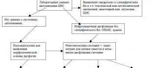
Diagnosis by neuromelanin
Why: Neuromelanin is a substance found in the cells of the substantia nigra of the brain. This area serves as the source of the neurotransmitter dopamine. Disruption of the substantia nigra is the main cause of parkinsonism.
Bonus: Neuromelanin has paramagnetic properties, which means that the places where it accumulates the most are visible on MRI. The amount of neuromelanin is an indicator of the activity of the cells of the substantia nigra, and if this activity is low, it is worth considering whether the patient is in the early stages of parkinsonism. Bonus: Most likely, schizophrenia can be diagnosed using the same method. Neuromelanin is also found in the locus coeruleus, the area of the brain that produces serotonin. It is assumed that problems in the system of neurons that produce and perceive serotonin are an important factor in the development of schizophrenia.
Virtopsy
Why: Used in forensics to assess the condition of the victim’s body, find injuries, and also find out what kind of weapon they could – or could not – have been caused. It is performed like a regular MRI. The results of a virtopsy (virtual autopsy) are easier to evaluate objectively than with a traditional autopsy, since it is not just a set of images, but also a lot of quantitative data.
Bonus: Virtopsy can be performed using a robot programmed to “illuminate” certain areas of the body.
MRI thermometer
Why: Sometimes they try to increase the effectiveness of radiation and drug therapy for malignant tumors by local heating of the affected tissue. To check whether the tumor tissue has warmed up to the desired temperature, and to avoid damage to healthy cells, a sensor can be installed in the tumor. However, in 2008, a much less painful method was tested on mice - MRI thermometry. It is based on obtaining resonance from hydrogen atoms from water and fat molecules contained in the tissues of the object under study. The frequency of radio waves emitted by hydrogen changes with the temperature of the tissues being examined, and the difference in these resonant frequencies allows one to accurately determine this temperature.
Bonus: By 2014, the technique had already been tested on living people, so it will soon be possible to see it in action in clinics.
WHEN SYMPTOMS APPEAR
People suffering from claustrophobia begin to panic if they need to get into an elevator or go down to the basement. When a room has a low ceiling or no windows, a small space - all this provokes attacks of fear. Often, an anxious state begins when a man has a tight tie or clothes that put pressure on his neck. People with this disease are afraid to stand in line for a long time, sit in the dentist's chair, and are afraid of crowds. On an airplane, in a tunnel, sometimes even in a car, these symptoms can also appear. Fear arises while staying in a shower stall, in a solarium, in a cave, in the subway.
MRI and metal
The tomograph generates a powerful magnetic field that affects any electrical conductors present in the body: metal pins and endoprostheses, pacemakers, cochlear implants. The presence of complex electronic devices, as a rule, serves as an absolute contraindication to tomography, but this study can sometimes still be combined with prosthetic joints and similar mechanical devices.
Contrary to popular belief, the magnetic field of a tomograph is hardly capable of moving an endoprosthesis, much less pulling it out of the body. However, it may interfere with the localization of loosely attached objects, such as a clip on a blood vessel. The magnetic field can also heat the implants, sometimes noticeably to the patient. However, the main problem is different: foreign bodies create strong interference and sharply degrade image quality. An MRI with a metal prosthesis is not necessarily dangerous, but it is almost certainly useless - at least if you want to take a picture of the exact area in which the implant is installed.
The good news is that many modern implants are made from materials that cannot be magnetized and therefore do not interfere with MRI scanning. If you have any implant, be sure to find out what substance it is made of and ask your doctor to give you a certificate about whether a tomography is possible in your case.
Dentures and braces are most often made from alloys that cannot be magnetized, and therefore do not interfere with MRI, however, in this case, you need to be on the safe side: check with the dentist what materials he used, and convey this information to the doctor, who will perform MRI.
The list of contraindications for MRI also mentions the presence of tattoos made with metal-containing dyes; the reason is the same: the metal particles will heat up, shift and cause discomfort. However, it should be noted that modern dyes do not contain any metal particles. You should only worry if you got a tattoo forty years ago in the yard behind the garages, with a rusty needle, using a dye made by a folk craftsman from completely unknown ingredients.
Other contraindications to MRI
- Claustrophobia.
The tomograph is cramped, dark and noisy; you must be sure that this will not prevent you from remaining calm and still.
- First trimester of pregnancy
There is no proven harm to the child, but doctors prefer to play it safe.
- Weight more than 120 kilograms
In this case, the table may not support the patient, and he will be desperately cramped inside the pipe. However, a solution has already been created: an open tomograph. During the procedure, the patient is not in a cylinder, but in a semicircular groove, and the table of such a tomograph can withstand up to 200 kg. A similar option can be offered to those suffering from claustrophobia. True, open tomographs of sufficient power are just entering the market and are still quite rare.
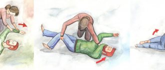
Claustrophobia
People experience their first attacks of claustrophobia between the ages of 25 and 45. Rarely, the pathology is detected in acute form in children. A phobia can manifest itself even if there is currently no stimulus, but its appearance is expected. For example, if you have to walk through a hall full of people. The increase in the feeling of fear occurs gradually, and is accompanied by common symptoms: rapid heartbeat, sweating, etc. The form of pathology influences how a person behaves when an attack of claustrophobia begins.
If the disorder is neurotic in nature, then, despite the horror experienced, the person retains the ability to respond adequately. He can remember tips for dealing with an attack, try to distract himself, and calmly leave the room. In the psychotic form of pathology, during an attack the patient loses control over himself and tries to escape by any means, even harming himself. For example, if a person sees that the door to the office is closed, he begins to behave inappropriately: trying to climb through the window he broke, loudly calling for help, not paying attention to those around him.
How much to hang in a Tesla?
The magnetic field induction of the tomograph is measured in Tesla. The unit is named after the inventor Nikola Tesla, who clarified much about the nature of electricity and magnetism. The more Tesla markings a tomograph shows, the more powerful it is and the more detailed images it can provide. Typically in medicine, tomographs with induction from 0.2 to 3 Tesla are used; high power is not required. Still would! The magnetic field induction of sunspots is only 3 times higher.
- 0.23-0.35 Tesla (low-floor) - preliminary diagnostics only. For example, such a tomograph will be able to determine whether there is a tumor or not, but will provide almost no information about its structure;
- 1 Tesla (mid-field) - such a tomograph will not allow you to examine the smallest details, but it is quite enough to get a general idea of the location and severity of the damage.
- 1.5-3 Tesla (high-field) – visualization of small areas of tissue. Such a tomograph will help to find metastases and determine whether there is a pathological change in the tissue around the joint capsule or inside the mammary glands after the installation of implants.
Author: Svetlana Yastrebova Published: October 19, 2021
Contraindications
It is not recommended to perform MRI of the heart and blood vessels in the following categories of patients:
- in the presence of foreign bodies inside the body, for example, shunts, prostheses, neuro- and cardiac pacemakers, bullets, piercings;
- in the presence of implants in the form of magnetic and electronic devices, for example, hearing aids;
- in the early stages of pregnancy, up to about three months, there is a danger of exposure to the fetus;
- in the presence of claustrophobia or convulsive syndrome;
- presence of stented vessels;
- presence of excess weight, more precisely, if the patient’s weight exceeds 150 kg.
Cardiac MRI with contrast is not performed:
- with individual intolerance to gadolinium;
- heart failure;
- renal failure.
MRI of cardiac vessels after bypass surgery can be performed no earlier than six months after surgery.