Paresthesia is numbness of the limbs or other areas, which is accompanied by subjective symptoms such as burning or tingling. In many cases, the disease is curable - medications and physiotherapeutic procedures are used for therapy. If necessary, surgery may be prescribed.
Paresthesia: what is it?
This term refers to a sensitivity disorder in which a person feels a burning, tingling or “crawling” sensation. These are subjective sensations associated with objective reasons. As a rule, they are associated with irritation of a nerve that lies close to the surface of the skin. This may be the result of a shock, mechanical pressure, or due to a temporary interruption of the blood supply.
The main factors leading to paresthesia:
- prolonged compression, for example, of a limb;
- osteochondrosis of any part of the spine;
- tumors of different nature (benign, oncological);
- tourniquets that are applied to stop bleeding;
- nerve ending injuries;
- burns, exposure to high temperature (this may cause problems with the sensitivity of the tongue and lips);
- inflammation in the vascular tissues that supply nerve cells with blood;
- diabetes mellitus and other hormonal disorders;
- poisoning with chemical poisons, biological toxic substances;
- deficiency of certain B vitamins;
- Iron-deficiency anemia;
- intermittent claudication;
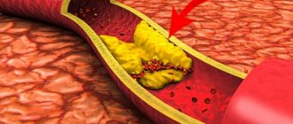
- atherosclerosis;
- incorrect position at night (during sleep);
- mechanical shocks.
Facial paresthesia secondary to endodontic treatment
Paresthesia is a disorder of neurosensitivity caused by damage to the nervous tissue, which is characterized by subjective sensations of burning, numbness or attacks of pain, as well as partial loss of sensitivity. It is important to differentiate between paresthesia, dysesthesia and anesthesia. Dysesthesia manifests itself in the form of unpleasant abnormal sensations that occur spontaneously or as a result of the action of a provoking factor. Special cases of dysesthesia include hyperalgesia and allodynia. Dysesthesia is always accompanied by unpleasant sensations, unlike paresthesia. From a practical point of view, paresthesia refers to a case of abnormal sensation and, therefore, can be classified as a separate variant of dysesthesia. Anesthesia is characterized by sensory loss of perception caused by drug exposure or neural dysfunction.
Endodontically provoked paresthesias require careful examination due to the close anatomical proximity of the root apex and the neurovascular bundle of the jaw. In addition, if the damage is severe, irreversible nerve pathologies may occur. The purpose of our study was to analyze the literature data concerning paresthesia as a complication of endodontic treatment, as well as its causes, diagnostic features, the most severe complications and treatment options.
Paresthesia as a complication of endodontic treatment
Paresthesias associated with endodontic treatment are most characteristic of the inferior alveolar nerve (IN) and its mental branch. However, the prevalence of paresthesia associated with specific endodontic problems in individual teeth has not yet been established. A retrospective study of paresthesia cases associated with treatment of mandibular premolars found the incidence to be 0.96% (8/832). However, in many cases, paresthesia may go unnoticed by the endodontist and, therefore, such cases are not reported.
Some studies examining the relationship between paresthesia and endodontic infection have found both problems predominantly in teeth with extensive periradicular lesions (Figure 1). This type of nervous disorder is mainly of a compressive nature, but the possible migration of inflammatory mediators and bacterial products from the affected area to the nerve formations also remains an equally important etiological factor. In particular, inflammatory mediators such as interleukin 1, tumor necrosis factor and nitric oxide have inherent neurotoxic activity, and bacterial endotoxins such as lipopolysaccharides can also cause damage to nerve tissue.
Photo 1. Periapical radiograph of the mandibular second molar: visualizes the anatomical proximity between the root apex and the mandibular canal.
Regarding endodontic treatment as a possible cause of paresthesia, Rowe suggested that the nerve may be damaged directly during root canal treatment due to overtreatment (Figure 1). However, Rowe also argued that mechanical damage to the nerve from an endodontic instrument is easily repaired, so this form of paresthesia is temporary.
The possibility of paresthesia occurring as a result of direct contamination by microorganisms requires detailed consideration, taking into account the possibilities of their biological aggression, since this particular pathogenetic mechanism has not yet been studied in detail in the literature. Cytotoxicity and mechanical pressure of sealers in areas close to the mandibular canal are also potential mechanisms for possible nerve injury due to endodontic manipulation. In this case, the already mentioned inferior alveolar nerve and its mental branch are the most frequently damaged structures. Materials that can initiate such complications contain paraformaldehyde.
For this systematic review, the PubMed database was searched using the keyword combination “endodontic and paresthesia” to select all case reports of paresthesia associated with endodontic complications published within the last 10 years (January 2002 to December 2012). A total of 40 cases were found. Some clinical cases published earlier than this period will also be discussed in this article.
Several cases of paresthesia associated with endodontic complications in the maxilla have also been found in the literature. Orr described 2 cases of paresthesia of the maxillary nerve caused by apical extension of N2 paste (Indrag-Agsa, Bologna, Italy). Orr emphasized that N2 paste contains paraformaldehyde, which fully explains the pathology. Ree and Messer described paresthesia that occurred during retreatment of the maxillary central incisor. Perforation of the root apex resulted in transudation of sodium hypochlorite (NaOCl) into the periradicular tissues. The consequences of this incident were not long in coming, so pain, swelling and paresthesia immediately appeared in the areas of the lower orbit and nose. Pelka and Petschelt presented another case of paresthesia caused by accidental passage of NaOCl beyond the apical foramen in the upper left lateral incisor. This resulted in paresthesia of the left facial and infraorbital nerves. During in vitro studies, Schwarze et al observed that sealers/pastes containing paraformaldehyde, such as Endomethasone (Endomethasone, Septodont, Saint-Maur, France) and N2, were more toxic and mutagenic than sealers containing calcium hydroxide.
Overpreparation of the root canal often results in widening of the apical foramen and leveling of the apical constriction, which promotes the removal of irrigation solutions or filling material beyond the root apex. This, in turn, can cause chemical or mechanical damage to the nerve bundle. The diameter of bone lesions of endodontic origin may also influence the onset of paresthesia, especially in cases of premolars and mandibular molars.
Anatomical relationship of the inferior alveolar nerve and root apexes
Tilotta-Yasukawa et al determined the proximity of the apices of premolars and molars in relation to the mandibular canal, as well as the relationship between the inferior alveolar nerve and its corresponding artery, to understand how endodontic material spreads through bone before entering the mandibular canal. They noted that the distance between the root apex and the mandibular canal was more variable (and generally larger) for the first molar compared to the second and third molars (1-4 mm and less than 1 mm, respectively, in 35 of 40 cases studied on the mandibular jaws). The authors concluded that in its posterior region, the mandible is less dense and has a greater amount of cancellous bone. It should be noted that the inferior alveolar artery itself may be associated with the occurrence of paresthesia, as it can act as a router for the distribution of materials, microorganisms and irrigation solutions to the nerve structures.
Littner et al studied 22 jaws and found that the superior portion of the mandibular canal may be located 3.5 to 5.4 mm below the apices of the first and second molars. The apices of the third molars are located very close to the alveolar nerve. Denio et al studied that the apices of the roots of second molars are located at an average distance of 3.7 mm from the mandibular canal, and the apices of the mesial roots of first molars are located up to 6.9 mm. Other authors have confirmed the symmetry of the mandibular hemiarches in relation to the distance between the root apices and the mandibular canal.
The presence of numerous spaces in the thickness of the lower jaw in the area of the molars promotes the spread of irrigation solutions and filling material towards the inferior alveolar neurovascular bundle (photo 2). This diffusion is facilitated by the presence of periradicular infections, which weaken the bony barrier between the apex and the neurovascular formation. In addition, it should be noted that the mandibular canal is not always surrounded by a dense cortical plate. In most cases, the neurovascular bundle passes through the cancellous bone without any additional bone barrier, which makes it more vulnerable to various mechanical or chemical agents from the root canal area.
Photo 2. Schematic representation of the various causes of paresthesia due to endodontic problems. In the region from the second premolar to the third molar, typical causes of paresthesia include apical extrusion or spread of endodontic drugs, apical surgery, excessive canal preparation beyond the apex, and apical periodontitis.
Diagnosis of facial paresthesia
Diagnosis of paresthesia or nervous anesthesia is based on medical history and assessment of the associated symptoms. Reactions of the affected area to thermal stimuli, mechanical stimuli, electrical or chemical tests all also contribute to the diagnostic process, although such reactions are purely subjective. Periapical radiographs are key to verifying the relationship between the root apex and nerve endings, especially in the mandible (Figure 3).
Photo 3. a) Bone in the area of the molars of the lower jaw. b) Visual increase in the structure of the alveolar bone in the area of the third molar; noteworthy is the presence of numerous spaces in the thickness of the bone tissue. c) Illustration of possible migration of bacteria or endodontic materials through marked spaces in the jaw structure to the area of the inferior alveolar nerve (shown in red).
De Beukelaer et al described the use of sensitivity tests to determine the extent and severity of paresthesia. Mechanical, thermal and taste tests were carried out at certain time intervals, thus accompanying the course of treatment. According to these authors, clinical examination of patients with damage to the lingual nerve should begin with determining the function of the nerve, in particular by assessing pronunciation and swallowing. It is also necessary to palpate the affected area.
Two types of clinical neurosensitivity studies can be conducted according to the specific receptors that exist (mechanoceptors and nociceptors) that are stimulated by skin contact. Testing of mechanoceptive sensitivity is based on responses to light static touch and directed stroking with a brush, while nociceptive sensitivity is determined by the response to temperature stimuli, which stimulate certain groups of sensory nerve fibers in addition to the sensory sensations caused by the action of sharp instruments.
Paresthesia caused by the removal of materials beyond the apex and subsequent compression of the inferior alveolar neurovascular bundle is also described in the literature. However, given that most endodontic materials have a certain chemical effect, in cases of material extrusion beyond the apex, it is difficult to determine the pathogenesis of the neural disorder: whether it is mechanical, due to compression, or chemical, caused by the cytotoxicity of the material. Photo 4 illustrates a case of apical extension in which the sealer was deposited in the area adjacent to the cortical part of the mandibular canal. Depending on the cytotoxicity of the sealer, paresthesia in this case may be a complication. To minimize the risk of such situations, it is necessary to more carefully conduct a preliminary assessment of the initial radiograph to determine the relationship of the anatomical structures.
Photo 4. Removing the sealer beyond the apex at the border with the cortical part of the mandibular canal.
Cone beam computed tomography is an important additional option for the prevention, diagnosis and treatment of paresthesia of endodontic origin. The ability to preview sections up to 1 mm thick in three dimensions allows for detailed study of the relationship of root apices and bony lesions to neural structures.
Treatment of facial paresthesia
Since paresthesia associated with endodontic problems are polyetiological pathologies, a universal protocol cannot be used to treat all cases. Treatment options for paresthesia are described below depending on the cause.
If paresthesia is caused by the removal of endodontic sealers or treatment pastes beyond the root apex, then, according to some studies, surgical removal of them shows a high success rate with the return of normal sensitivity in the causative area. However, it should be taken into account that the time that has passed since the material was removed beyond the apex, as well as its volume and topography, affect the success of such treatment.
Cases of paresthesia caused by infection and inflammation are resolved by endodontic treatment, apical surgery, antibiotic therapy or tooth extraction, while paresthesia caused by local anesthesia or overdevelopment of the canal usually resolves within a few days without any treatment. Cases of prolonged or persistent paresthesia are usually due to rupture of nerve fibers, prolonged pressure, or extrusion of toxic endodontic materials. In these cases, it is necessary to remove the causative factor.
Surgery involves removal of the cortical plate followed by apical resection. This approach is the most common for removing top sealers and pastes. Sagittal osteotomy is also a treatment option, especially when compression of the alveolar nerve occurs in the area of the second or third molars.
In nerve decompression, favorable results have been achieved by first performing a sagittal mandibulectomy to better visualize the mandibular canal and then removing foreign material, with or without apical resection. Microneurosurgical treatment techniques, which involve the use of microinstruments and magnifying equipment, are used to restore the function of certain sensory or motor endings. Indications and contraindications for microneurosurgical intervention are given in the Table.
| Indications | Contraindications |
| Examination or division of a suspected nerve | Central neuropathic pain |
| Prolonged paresthesia for 3 months | Signs of improvement in paresthesia |
| Pain caused by neuroma formation | Neuropraxia |
| Pain caused by foreign body or canal deformation | Tolerable paresthesia (patient determined) |
| Progressive loss of sensation or increased pain | Metabolic neuropathy |
| Possibility of drug treatment | |
| Old age | |
| Very long period since the injury occurred |
Pogrel suggested performing exploratory surgery immediately after diagnosis because radiographic examination and other diagnostic tests cannot determine the extent of nerve damage. Pogrel also suggested that waiting time before intervention is a controversial issue, but that restorative procedures performed soon after injury are subsequently more successful than those performed after waiting some time. However, it has been noted that some interventions performed 12 months post-injury have also been shown to be equally successful.
Although some authors prefer more radical treatment of paresthesia, it should not be forgotten that in cases of extravasation, the cytotoxicity of sealers tends to decrease over time.
Another treatment option is the use of lasers. This method can accelerate the repair process of injured biological tissues by initiating angioneogenesis, which is an important aspect for neural regeneration. According to Schultze-Mosgau and Reich, spontaneous recovery from paresthesia is possible within 3 to 6 months, but this period is too long. These authors suggested that patients with paresthesia should be prescribed a vitamin B complex to promote the development of the myelin sheath of nerves.
Prevention of facial paresthesia
Precautions to prevent paresthesia associated with endodontic problems include careful radiographic examination, verification of the relationship of root apices and/or periapical lesions to neural structures, use of instruments of appropriate working length, avoidance of overpreparation of canals and apical foramen enlargement, and irrigation with chlorhexidine only in In cases of a very wide or incompletely formed apex, take vitamins immediately after extruding the filling material or irrigant solution near the nerve fiber.
In general, if paresthesia occurs soon after endodontic treatment, it is necessary for the dentist to monitor the patient for symptoms during the first 24 hours. This approach will allow for early diagnosis of paresthesia, and thus will contribute to more effective treatment.
conclusions
The inferior alveolar and mental nerves, which are most often affected by paresthesia, are located in the thickness of the jaw. To diagnose paresthesia, the dentist must take a medical history, evaluate the results of nociceptive and mechanoceptive tests of the affected area, periapical and panoramic radiography, and in some cases, cone beam computed tomography. The choice of treatment should take into account the cause of paresthesia, the degree of injury, the time elapsed since the onset of symptoms, and the patient's response to systemic drug administration. To prevent paresthesia, the dentist should not forget about the proximity of the root apexes to the nerve structures, and remember this even before starting endodontic procedures.
Authors: Flavio R. Alves, PHD Mariana S. Coutinho, DDS Lucio S. Goncalves, PHD
Types of paresthesia
There are 2 main forms of the disease – transient and chronic. In the first case, tingling, burning and other unpleasant sensations go away on their own, without the use of drugs or surgical intervention. For example, if a person has been in an uncomfortable position for a long time, it is enough to change position, perform an exercise, and the paresthesia will go away without additional treatment.
However, there are also chronic paresthesias. They are associated with damage to different parts of the nervous system. In turn, they are divided into several types:
- Primary – against the background of infection, for example, HIV, tumor or autoimmune.
- Secondary - against the background of alcoholism, lack of B vitamins, metabolic disorders, for example, in the case of diabetes.
Rarely, paresthesia manifests itself as numbness of the lips, tongue or chin. Moreover, similar phenomena occur due to dental intervention (removal of wisdom teeth).
Another type is acroparesthesia. These are severe pains in the arms or legs associated with Fabry disease or calcium deficiency, or disturbances in the functioning of peripheral nerves. The main symptoms are tingling, numbness, burning, and numbness in the extremities, especially the fingers.
Causes
Many factors cause a person to experience numbness. In addition to a neurological disorder, the causes may also be: uncomfortable posture, medications, toxins, abnormal levels of certain vitamins and minerals, radiation therapy, insect bites, etc. In any case, the treatment of numbness depends directly on getting rid of the factor influencing it appearance.
If the numbness is caused by an injury, then most likely it is due to the influence of damaged tissue on the nerve, for example, they put pressure on it or pinch it. Treatment of numbness in this case is coordinated with the recovery process after injury. Pressure on the nerve also occurs under a number of specific conditions, such as carpal tunnel syndrome, tumor, enlarged blood vessels, infection, etc.
Skin rashes (and other skin lesions), frostbite, and herpes zoster may also cause paresthesia as an associated condition.
In all other cases, numbness is a symptom of a disease, including:
- diabetes mellitus,
- Raynaud's syndrome,
- multiple sclerosis,
- atherosclerosis, hypothyroidism,
- autoimmune thyroiditis,
- stroke, etc.
Physical therapy can help a patient cope with the feeling of numbness even if their medical condition cannot be completely cured (for example, diabetes).
Accompanying illnesses
Paresthesia often develops against the background of such pathologies:
- sciatica;
- cervical spondylosis;
- diabetic neuropathy;
- transverse myelitis;
- restless legs syndrome (tingling and burning in the legs).
If complications occur, the disease can progress rapidly. Therefore, patients with these diseases should undergo periodic medical examination.
Causes of paresthesia of the arms and lower extremities
All causes of paresthesia of the limbs can be divided into several large groups: inflammation, trauma, compression, infection and tumors. Let's look at these causes of paresthesia of the upper and lower extremities in more detail.
Let's start with the most common option - compression. This is the pressure exerted on the passing nerve fiber. It can be caused by the following negative factors:
- development of osteochondrosis of the spinal column with gradually developing protrusion, extrusion and disc herniation;
- displacement of the vertebral bodies and pinching of the radicular nerves;
- spasm and tonic muscle tension in the paravertebral region during any pathological processes;
- poor posture (scoliosis, kyphosis, lordosis, round back, stoop);
- Bechterew's disease (ankylosing spondylitis), in which there is a violation of the patency of the nerve fiber;
- deposition of calcium salts (osteophytes) on the edges of the vertebral bodies;
- violation of the patency of the tunnels in which large nerves are located (sciatic, median, ulnar, tibial, etc.);
- lymphadenitis putting pressure on regional nerve plexuses (for example, brachial).
Inflammatory diseases accompanied by numbness and the occurrence of paresthesia in certain areas of the upper and lower extremities include lymphadenitis, myositis, neuritis, etc. Infectious inflammation can be provoked by tuberculosis bacillus, staphylococcus, streptococcus (with erysipelas of the leg), Haemophilus influenzae, etc.
Among the probable causes of the development of paresthesia of the upper or lower extremities, injuries and their long-term consequences often appear:
- fractures and cracks of long tubular bones, resulting in impaired nerve conduction;
- sprains and ruptures of ligament and tendon tissue (swelling and hematoma develop, which compress the nerve fiber passing next to them);
- the formation of coarse scar tissue that interferes with the normal process of innervation of certain areas of the upper and lower extremities;
- cuts and other types of violation of the integrity of the nerve fiber itself.
As the tumor grows, pressure is exerted on the soft tissue surrounding the tumor. When a nerve fiber enters this zone, severe paresthesia is observed. Other potential causes may also include:
- incorrectly chosen position for night sleep;
- violation of workplace ergonomic rules;
- engaging in certain types of professional activities (most often, as a result of this, carpal tunnel syndrome occurs);
- the habit of sitting with one leg crossed over the other;
- development of diabetic angiopathy with impaired blood supply to distant areas of the arms and legs;
- atherosclerosis of small blood vessels against the background of high cholesterol levels in the blood;
- development of varicose veins of the lower extremities with impaired outflow of venous blood and edema syndrome;
- Raynaud's disease;
- improper application of a hemostatic tourniquet.
When you first contact your doctor, you must honestly talk about all your existing risk factors for developing nerve fiber trophism disorders. If you smoke, drink alcoholic beverages, or regularly violate your diet, you should inform your doctor about this. So, if you have recently begun to notice that you are drinking too much liquid, then you are very likely at risk of developing impaired glucose tolerance and diabetes. And these diseases can provoke the appearance of systematic transient paresthesias of the upper (arms) and lower extremities.
Main symptoms
The main symptoms of the disease, regardless of the type, include:
- numbness – weak and strong;
- tingling;
- burning sensation;
- the feeling that goosebumps are crawling all over the body.
Indirect signs are:
- pale skin;
- previously unexperienced hair loss;
- decrease in temperature (only in the affected area).
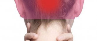
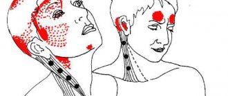
Often these symptoms occur in the feet, in the neck, especially on the side, on the hands, and in the head area. Normally, such sensations gradually disappear without treatment. But if they are repeated again and again and become pronounced and long-lasting, this clearly indicates the development of the disease. In such cases, you need to consult a doctor for diagnosis and treatment.
1.General information
The term "paresthesia" in the broad sense of the word means "false tactile sensations." These are the same skin deceptions that we usually describe as goosebumps, tingling, electrical discharges, coldness, heat, causeless tickling in the absence of visible irritants, numbness, etc. It would seem that what could be simpler? However, the phenomenon of paresthesia is studied within the framework of more than serious disciplines: neurology, psychiatry, vertebrology, neurophysiology, psychology...
When it comes to the human body, and especially its neurophysiological organization, technical analogies do not always work. However, in this case, such an analogy is quite appropriate and accurate. The central nervous system exercises control and control through complex electrochemical reactions in the conductive nerves; from this point of view, paresthesia is a breakdown of insulation (as an electrical engineer would say, “to ground”), as a result of which the brain receives a false signal about skin irritation. In essence, such a violation of sensitivity is not a disease, but a symptom that is not causeless. The mechanisms of its occurrence are very diverse, but it is always based on either disturbances in nerve conduction or a malfunction in the “control panel” (when the brain itself begins to produce false sensations). In the International Classification of Diseases ICD-10, however, a separate nosological code is allocated for the designation of cutaneous paresthesia under the heading “skin sensitivity disorders.”
There are no statistical data on the incidence of paresthesia, and it is difficult to imagine collecting this kind of information, given the huge number of possible options and causes. Let us only note that most sources traditionally (and not unreasonably) classify paresthesia primarily as a neurological disorder, and interpret it precisely in this aspect.
A must read! Help with treatment and hospitalization!
Diagnosis of the disease
To determine the disease, the doctor conducts a visual examination, studies the patient’s history and complaints. For example, the condition of the skin, its color and temperature are checked. Laboratory diagnostic procedures are also carried out:
- blood analysis;
- toxicological analysis;
- MRI of the spine, head;
- Ultrasound of cervical vessels;
- X-ray of legs;
- diagnosis of the thyroid gland.
Treatment of paresthesia of the upper and lower extremities
To treat limb paresthesia, it is important to identify the underlying disease. This condition is only a conical symptom and does not require separate therapy. Moreover, treatment of paresthesia of the upper extremities (as well as the lower ones) separately from the underlying disease will not bring a positive result.
Thus, if paresthesia of the lower extremities is associated with the development of an intervertebral hernia in the lumbar spine, then treatment must begin with a procedure of traction traction of the spine. During this effect, the intervertebral spaces will be increased and the compression exerted on the radicular nerves will be completely eliminated. The pain will go away and the paresthesia syndrome will disappear. Then the doctor develops an individual course of treatment for osteochondrosis.
It may include osteopathy and massage, reflexology, kinesiotherapy, therapeutic exercises and much more. Paresthesia of the upper extremities with brachial plexitis is treated in a similar way. First, the doctor finds the cause of the damage to the structure of the brachial nerve plexus. Then he applies techniques to restore physiological nerve conduction. And only after this begins treatment of the identified disease.
If you require treatment for paresthesia of the lower or upper extremities, then make an appointment for a free appointment with a neurologist at our manual therapy clinic in Moscow. After the examination and diagnosis, the doctor will tell you about all the possibilities and prospects for using manual therapy methods in your individual case.
Treatment
Pre-hospital assistance
If your legs become numb due to overload or vascular disease, it is recommended to limit the amount of time you stay on your feet, take regular breaks, wear comfortable shoes, and avoid hypothermia. Patients with radicular syndrome are advised to reduce the load on the back, use painkillers and local warming agents.
In case of traumatic injuries, immobilization is required, and sometimes a bandage and anesthesia are required. Signs of circulatory problems in the spinal cord or brain are an indication to immediately call an ambulance.
Conservative therapy
For polyneuropathies, treatment of the underlying pathology is required. Patients with neuropathies, plexitis, and radicular syndrome are prescribed anti-inflammatory, vasodilator, painkillers, and vitamins. Persistent pain syndrome that is not relieved by analgesics is an indication for therapeutic blockade. Patients are referred for physiotherapeutic procedures, massage, exercise therapy, manual therapy, and acupuncture.
The treatment program for vascular pathologies includes antispasmodics, antithrombotic therapy, and vitamin therapy. Acute occlusion is eliminated using anticoagulants and thrombolytics. For severe pain, blockades are performed. Ozone therapy is performed as part of physiotherapeutic treatment. ILBI, hyperbaric oxygen therapy. Hydrotherapy provides a good effect.
Surgery
For pathologies accompanied by numbness of the leg, interventions such as:
- Traumatic injuries
: suturing or plastic surgery of the nerve trunk, decompression of the spinal cord and fixation of fragments in spinal cord injuries. - Neuropathies
: decompression, isolation of the nerve from the adhesions, excision of a benign neoplasm. - Lesions of the central nervous system:
removal of hematomas and herniated discs, vascular surgery to restore blood supply to nerve structures. - Vascular diseases
: removal of blood clots from arteries and venous trunks, bypass surgery, stenting and arterial replacement, various options for phlebectomy.
What complications can arise without timely treatment?
In the absence of timely treatment or violation of the course, life-threatening conditions arise for the patient:
- meningitis, encephalitis, encelophopathy
- stroke, heart attack, circulatory disorders, thrombovasculitis
Symptoms that require close attention and immediate consultation with a neurologist include:
- localized pain that lasts for a long time
- hacking cough that is not controlled by medications
- paresthesia
- non-systemic dizziness
- sudden loss of consciousness
- convulsive syndrome
- disturbances of coordination, speech, gait, swallowing, behavior, sleep, thermoregulation
- progression of any of the symptoms
Unexplored pathogenetic mechanisms
According to scientists, the mechanism of occurrence of post-Covid neurological symptoms is presumably associated with inflammatory changes in the walls of small vessels and the formation of microthrombi. Tissue hypoxia and organ ischemia also have a negative impact.
Due to increased neutrotropism, the virus enters the central nervous system transneurally. The most affected brain structures are those responsible for:
- for emotional and behavioral reactions, motor learning
- regulation of neuroendocrine brain activity
- coordination of movements of muscle groups, maintaining balance
- vision
- connections between the cerebral hemisphere, cerebral cortex and spinal cord
Complications include disturbances in sensitivity, thermoregulation, speech and gait, partial loss of hearing and vision, insomnia and other symptoms that require a visit to a neurologist.
Providing medical care
At the first symptoms of dysesthesia, you should definitely see a doctor. The onset of the disease may be an indicator of the presence of serious problems in the body. Doctors will determine the cause and prescribe effective therapy. And to make your well-being easier, you can use the following tips:
- move a lot , taking walks before bed, since it is walking that enhances chemical reactions, freeing the brain from endorphins, promoting overall restful and sound sleep;
- If you experience discomfort in the lower extremities, you can get up and walk around the room ;
- don't overeat before bed;
- take multivitamins - according to research, the cause of this disease lies precisely in the lack of folic acid or iron;
- taking Aspirin - doctors could not explain exactly how it works, but in some patients, after taking it, their general condition improved significantly;
- avoid stressful situations;
- alcohol should not be taken as a sedative or pain reliever.
Fans of traditional methods of treatment claim that foods enriched with magnesium help improve well-being for various types of dysesthesia: soy, milk, wheat bran, almonds, nuts, pumpkin seeds.