More detailed information about other diseases starting with the letter “R”: Radiation damage to the nervous system; Multiple sclerosis; Radiculitis; Spina bifida; Disseminated encephalomyelitis; Retrobulbar neuritis; Retinal migraine
What is retrobulbar neuritis?
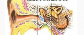
Retrobulbar neuritis is an inflammatory disease that affects the optic nerve, located between the orbit and the visual area. If there is an anomaly, the patient experiences a decrease in the level of visual acuity, the occurrence of prolapses, minimization of boundaries, and painful sensations during motor reflexes with the eyeballs. As research methods, doctors use complex neurological and ophthalmological techniques. Therapeutic therapy is performed using antibacterial/antiviral medications, glucocorticosteroids, diuretics and neuroprotective drugs. In some cases, detoxification procedures and physical therapy are prescribed.
Symptoms
Introtrobulbar optic neuritis
An acute disorder of visual function is characteristic, and their nature/severity of inflammation of the optic nerve is determined by the degree of damage to the trunk and membranes. Neuritis of the optic nerve is manifested primarily by a decrease in visual acuity, the indicators of which depend on changes in the papillomacular bundle. With partial damage, visual acuity may not be impaired, but spots appear in the field of vision - central/paracentral, round or arch-shaped scotomas, but can also be significantly reduced (at the initial stage, myopia up to 2 diopters).
Symptoms of optic neuritis are manifested by a narrowing of the boundaries of the visual field, a decrease in dark adaptation and color perception, a high frequency of flicker fusion is noted, as well as a decrease in electrical sensitivity and a low level of n lability. Opticus. With total damage to the diameter of the nerve, visual acuity can drop sharply up to complete blindness. Symptoms of the pathology appear predominantly in one eye.
At the initial stage of neuritis, the pathognomonic picture is represented by changes in the optic nerve head: blurred boundaries, moderate vasodilation, hyperemia, exudative swelling, the presence of streak-shaped hemorrhages in the tissue of the disc/peridiscal region. When the vascular funnel is filled with exudate and the adjacent layers of the vitreous are immobilized, unclear visualization of the fundus is noted.
In the acute period, symptoms of inflammation of the optic nerve last 3-5 weeks, and then gradually the swelling/hyperemia of the disc disappears, hemorrhages resolve and the boundaries of the disc acquire clear outlines again. In severe cases, ophthalmoscopy reveals a pale disc with pronounced vasoconstriction and clear boundaries and atrophy of n. opticus.
Retrobulbar optic neuritis
Symptoms of damage to the optic nerve with retrobulbar neuritis n. opticus. determined by the type of inflammatory changes.
With axial inflammation, the pathological process primarily involves a bundle of axons passing through the optic nerve. Characterized by a pronounced disorder of central vision with the formation of central scotomas throughout the entire visual field and a significant decrease in functional tests.
With the peripheral type of retrobulbar neuritis, the inflammatory process in the nerve sheaths spreads deep into the nerve trunk. A significant amount of exudate accumulates under the optic nerve sheath, causing the appearance of “shell-like” pain in the eye, which increases during movement of the eyeball. Central vision is not affected, but there is a concentric narrowing of the visual fields.
In the most severe form - transversal type of retrobulbar neuritis , the inflammatory process completely covers all tissues of the optic nerve. Functional tests with extremely low scores. In the acute course, visual acuity decreases sharply, in some cases to the point of blindness, while in chronic retrobulbar neuritis the decline in vision occurs gradually.
However, regardless of the type of retrobulbar neuritis, there are no changes in the optic nerve head and only 1-2 months after the manifestation of the disease can decolorization of the disc, as well as signs of partial/total atrophy of the n. opticus.
Useful facts
Typically, retrobulbar neuritis acts as a secondary consequence after any abnormality occurring in the brain. In very rare situations, the disease is diagnosed as a primary true inflammatory process. In the neurological field, doctors are still studying the issue of this disorder in detail. Neuritis that is localized in the intraorbital region is called intrabulbar (it is examined by ophthalmologists). In both cases, therapy requires a joint solution of the problem between the neurologist and the ophthalmologist. The risk group includes people aged 20 to 40 years. According to average statistics, this type of disease occurs twice as often in females as in males.
Factors causing retrobulbar neuritis
Various problems provoke the development of abnormal vision impairment. Inflammation itself begins as a result of damage to the body by infectious diseases, chronic diseases caused by immunodeficiency. Etiological reasons include:
- Chronic stages of viral infections - the main causative agent is cytomegalovirus, which provokes the development of mononucleosis. The stable location of harmful agents causes deformations in immune properties. Due to the production of antibodies for one's own tissues, the process of autoimmune inflammation begins.
- Chronic infectious points - most often the anomaly is caused by unfavorable foci that are located nearby (for example, sinusitis, otitis media and others), much less often - by distant places (cystitis). The infection enters the nervous structure of the visual apparatus through blood transportation.
- Inflammatory phenomena in the orbit - provocateurs are inflammatory processes. Spread occurs in two ways: hematogenous and perineural, which causes damage to the retrobulbar perimeter of the nerve. Quite often, total optic neuritis is diagnosed.
Secondary progression of optic nerve disorder begins as a result of an anomaly that is localized in the cerebral region. The main etiofactors include the following diseases:
- Multiple sclerosis - selective damage to the myelin sheath of nerve fibers moves to the optical areas, which causes an inflammatory reaction. Neuritis of a retrobulbar nature is considered the initial stage in more than 65% of cases of disease.
- Brain tumors - when voluminous tumors form in the area of the chiasm glioma, objects in another place begin to actively pressure the optic chiasm, disrupting the process of blood circulation in the body and metabolism in tissues. As a result, the patient develops symptoms of neuritis.
- Neuroinfections - Lyme disease, neuroAIDS, tuberculous meningitis can provoke abnormalities in the retrobulbar zone. Inflammatory phenomena are associated with the entry of infectious microorganisms into the body. In some cases, the disease may appear due to intoxication. Exogenous lesions are caused by the influence of toxic chemicals, methyl alcohol, iodine substances. Toxins appear as a result of dysmetabolic processes, renal failure or during pregnancy.
List of sources
- Borisevich E. S. Clinical manifestations, diagnosis and outcomes of optic neuritis / Borisevich E. S., Shamal D. Yu., Klyuiko Yu. D., Kachan T. V. // Young scientist. - 2021. - No. 13 (147). — pp. 146-149.
- Zhaboedov G.D. Lesions of the optic nerve / G.D. Zhaboyedov, R.L. Skripnik // GEOTAR-Media. - 2006. - 286 p.
- Gustov A.V. Practical neuro-ophthalmology / Gustoe A.V., Sigriansky K.I., Stolyarova Zh.P. // Nizhny Novgorod State Medical Academy, 2014. - pp. 260-263.
- Kruzhkova G.V. Pathology of the fundus and optic nerve/G.V. Kruzhkova, M.S. Agronovich //MEDpress-inform, 2014. - pp. 32-35.
- Kanski, D. Clinical ophthalmology: a systematic approach. - M.: Logosphere., 2006. - 744 p.
Mechanism of occurrence
When examining the visual department, one can understand that the nerve contains a large number of fibers (more than a million), which begin their journey from the retina and end in the subcortical ganglia of the eye. Various forms of pathogens can cause serious disorders, for example: ischemia, inflammation and tissue destruction. As a result, the patient experiences detrimental changes that affect the functions of transmitting information from the retinal receptors that receive the external image.
The clinical picture appears as a decrease in the level of vision, impaired color perception, and loss of boundaries. If you ignore the doctor’s recommendations or completely refuse treatment, the patient begins to experience irreversible abnormalities that fill the cavities with connective tissue growths. The last stage of the disease leads to complete loss of visual abilities.
Forecast
In mild cases, optic nerve neuritis is benign and in 75% of cases, visual acuity is restored after 4-6 weeks. However, at the same time, other parameters of the visual analyzer function (contrast sensitivity/color vision) are often not fully restored. Progressive chronic disease leads to persistent disabling disorders - optic nerve atrophy and irreversible vision loss, the development rates of which vary between 22-25%.
Types of neuritis
There are different types of symptoms, which depend solely on the location of the lesion (central or peripheral lobe). In neurology, retrobulbar neuritis is divided into three classes:
- Axial - a common form of inflammatory reaction, localized only in the axial fascicle, characterized by a sharp decrease in central visual acuity and the appearance of scotomas.
- Peripheral - originates in peripheral structures, accompanied by the formation of intrathecal fluid, which provokes pain. A narrowing of the optical fields is characteristic.
- Transversal - a detrimental anomaly progresses in the periphery or axial region and then reaches the diameter of the nerve center. The patient is completely blind.
Another area of division of retrobulbar neuritis depends on the variability of the course:
- Acute form - manifests itself in the rapid loss of visual acuity. Most often it is diagnosed in the primary version of the lesion, which is typical for young people.
- Chronic form - symptoms have a gradual scale of increase, which are caused by a cerebral disorder. Patients experience some periods of remission or exacerbation. There is a possibility of the acute form becoming chronic.
How does it manifest itself?
The main manifestations of the anomaly are various disturbances of optical perception. According to average statistics, in 60% of situations patients have vision problems. When diagnosing an acute form of neuritis, visual acuity decreases within a short time (from a couple of hours to two days), and in the chronic form it will take 7-14 days. Patients, talking about their sensations, report a “veil before their eyes” and “blurring of all objects”; some experience “light flashes”. The slow progression of the disease causes deterioration of visual adaptation at night.
Deformations of the optical boundaries provoke the appearance of dark dots before the eyes, narrowing the visible trajectory. They can be combined with a decrease in vision. Retrobulbar neuritis occurs with impaired color perception. Dychromatopsia is practically not noticed by patients, but is only detected during certain tests. Doctors say that more than 50% of patients complain of a feeling of pain in the area of the eye orbits, which increases with activity, in particular when turning their gaze upward.
Examination methods
The main goal of the diagnostic procedure is to make a correct conclusion with a clear identification of the etiology and the mandatory exclusion of the presence of multiple sclerosis in the patient. The study will require clarification of all symptoms, the rate of their development and previously experienced diseases, as well as full ophthalmological and neurological testing. Diagnostics:
- Visometric analysis helps determine vision loss; emerging problems can be corrected by wearing lenses.
- Color vision test - designed to accurately identify anomalies in the absence of changes in the optic disc.
- Perimetric scanning - scan the central, paracentral and peripheral sections.
- Ophthalmoscopy - looks for swelling, bleeding and the possibility of atrophy.
Tests and diagnostics
The diagnosis of optic neuritis is made based on a thorough history (pre-existing diseases, symptoms, rate of progression) and ophthalmological/neurological examination of the patient.
Ophthalmological examinations include:
- Visometry. Characteristic is a decrease in visual acuity to 0.1-0.2 diopters, less often to light perception, which cannot be corrected by lenses.
- Determination of color perception using color tables. Characterized by a decrease in color saturation/brightness.
- Perimetry. Characteristic is the presence of scotomas (central, peripheral, paracentral) in the visual fields.
- Ophthalmoscopy. Allows you to determine hyperemia/swelling of the optic disc, the presence of petechial hemorrhages, disc pallor (with the development of atrophy).
- Tonometry. In order to identify increased IOP, characteristic of a number of diseases that can contribute to the development of neuritis.
Neurological studies to assess the neurological status of the patient, allowing to identify cerebral/focal cerebral:
- MRI of the brain/orbit of the eye. To visualize brain pathology (areas of demyelination, inflammatory foci, tumor) and identify compression/edema/damage to the orbital part of the n. Opticus. X-ray of the paranasal sinuses/chest organs.
- Ultrasound of the optic nerve.
- Study of cerebrospinal fluid (chemical/cellular composition of the cerebrospinal fluid).
To identify the etiology of optic neuritis, blood cultures for sterility, ELISA/PCR studies, consultations with an immunologist, neurologist, infectious disease specialist, endocrinologist and other specialists may be prescribed.
Differential diagnosis of optic neuritis is carried out with intrabulbar lesions, ischemic optic neuropathy, and neuromyelitis optica. With the gradual development of atrophy n. Opticus it is necessary to exclude the hereditary mitochondrial disease Leber's disease (syn. Leber's syndrome).
How to get rid of it?
Therapeutic measures for retrobulbar neuritis are based on an etiopathogenetic principle, which is selected taking into account the etiology of the disease. A secondary lesion requires elimination of the primary source, that is, removal of the tumor, therapy aimed at restoring immune abilities in multiple sclerosis, and getting rid of neuroinfections. The main treatment complexes include:
- Pharmacological antibiotics with a wide range of applications.
- Antiviral components.
- Introduction to a course of glucocorticosteroid therapy.
- To reduce swelling, diuretics and antihistamines are prescribed.
- A course of vitamin complexes (vitamins B and C, nicotinamide and others) is prescribed.
In case of toxic damage, detoxification will be required; for this, intravenous administration of saline solutions and dextran through a dropper is used. If the patient has no restrictions, it is recommended to carry out physiotherapeutic procedures.