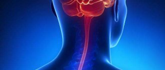
Purulent meningitis is an inflammation of the soft membrane of the brain, which is caused by pyogenic microorganisms (meningococci, pneumococci, streptococci, etc.) embedded in it.
The disease can be diagnosed in all age categories of people, but most often in children under the age of five. The development of purulent meningitis is facilitated by a weakened state of the immune system, so the peak of the disease occurs in the winter-spring period.
With purulent meningitis, patients have increased body temperature, intense headache, nausea, vomiting, disorders of the cranial nerves, early appearance of meningeal syndrome, hyperesthesia, disturbance of consciousness, and psychomotor agitation.
Thanks to the development of modern medicine, today the incidence of purulent meningitis has decreased significantly, the number of severe complications and deaths has decreased.
Services for the diagnosis and treatment of meningitis are offered by the neurology clinic of the Yusupov Hospital, a leading multidisciplinary medical center in Moscow. Thanks to the modern equipment of the clinic, the impressive medical experience of our specialists and the use of the latest technologies, the Yusupov Hospital achieves high efficiency in the treatment of meningitis, including purulent meningitis.
The diagnosis of purulent meningitis in the Yusupov Hospital is based on a typical clinical picture and data from a study of cerebrospinal fluid.
For purulent meningitis, patients at the Yusupov Hospital are required to undergo antibacterial therapy. In addition, the use of decongestants, glucocorticosteroids, tranquilizers, anticonvulsants and other symptomatic treatment is prescribed.
Causes of purulent meningitis
The main pathogen leading to infection with purulent meningitis is meningococcal infection. However, in recent years, scientists have proven that not only it leads to the disease, but also Haemophilus influenzae and pneumococci. In half of all cases, the causative agent of purulent meningitis will be Haemophilus influenzae, while meningococcus accounts for no more than 20%, and pneumococci - only 12-13%.
Purulent meningitis also occurs in newborns. The reason for this will be streptococcal infection or salmonellosis. Additionally, infected E. coli may also be a major pathogen.
Regarding the routes of penetration of the pathogen into the human body, purulent meningitis is classified into:
- primary: development is facilitated by the hematogenous spread of the pathogenic rod from the nasal cavity, where it enters when a person breathes; it is easy to become infected through contact with sick people (airborne or contact); direct infection of the meninges occurs in the case of an open type of traumatic brain injury, skull fracture or any other open injury to the paranasal sinuses (or mastoid process); failure to comply with hygiene rules during neurosurgical operations is another possible cause of infection;
- secondary: occurs on the basis of an already existing primary focus, from which the infection spreads to the brain area; The nature of the spread of infection can be either contact (with a brain abscess or osteomyelitis of the cranial bones) or hematogenous, spread from any source of infection is typical, but most often with sinusitis or otitis media.
If the causative agent of purulent meningitis enters the body through the blood-brain barrier, then the immune system is weakened. The latter can also be caused by acute respiratory viral infections or frequent stressful situations, as well as physical fatigue and changes in climatic zones.
Symptoms
Meningitis is characterized by pronounced symptoms. The first symptoms of the disease in adults are as follows:
- Specific headache – constant, unbearable, aching. Any head tilt enhances this feeling.
- Severe tension in the muscles of the back of the head. The sick person is unable to lie in a relaxed position; he involuntarily throws his head back, since its natural position causes severe pain.
- Chills and fever. This is the dominant symptom and one of the first to appear.
- Painful sensitivity to light and sound. Bright light and even not very loud sounds have an irritating effect on the patient.
- Lethargy and drowsiness. The patient is not always able to quickly and accurately formulate a thought or answer the question posed.
- Vomit. Moreover, it is repeated and often not accompanied by a feeling of nausea. Vomiting with meningitis is distinguished by the fact that it is not associated with eating, and therefore does not bring relief and often leads to dehydration.
These are the main and most characteristic signs of the disease. However, certain forms of meningitis may also cause:
- Rash. These rashes are always irregular in shape and often form extensive hemorrhages.
- Cramps. They are observed less frequently in adults than in children.
- Mental disorders. They can manifest themselves both at the initial stage of the disease and in a later period.
Important! The first symptoms of meningitis appear unexpectedly, and the disease develops rapidly.
If the disease is not diagnosed in time and treatment is not started immediately, the consequences of meningitis can be tragic for an adult patient.
All of the above-described manifestations of the disease in question are not considered specific, as they may indicate other possible pathologies.
Classification of purulent meningitis
Regarding the severity of symptomatic manifestations, the following forms of purulent meningitis are distinguished:
- light;
- medium-heavy;
- severe (typical for people with severely reduced immunity or those who have previously had their spleen removed).
Regarding the characteristics of the course of the disease, they speak of purulent meningitis:
- fulminant (characterized by a very rapid development of symptoms in the form of increased cerebral edema, which contributes to confusion and the onset of vital functions);
- abortive (has erased symptoms, where the first place is given to intoxication);
- acute (occurs more often than others; it is characterized by traditional cerebral and meningeal symptoms);
- recurrent (typical in the case of an advanced form, with untimely treatment or in the case when a chronic type of purulent infection is observed in the body).
Symptoms of purulent meningitis
- The onset of purulent meningitis is usually acute and is characterized by a sharp increase in body temperature to 39-40°C. Along with the temperature, characteristic chills, severe headache of increasing character, nausea and vomiting appear. The patient's condition may be characterized by psychomotor agitation, confusion, and manifestations of delirium.
- About 40-45% of patients have a convulsive syndrome. The so-called shell symptoms: stiff neck, Kernig's sign, Guillain's sign, usually appear in the first hours of the disease, becoming more and more intense on the 3-4th day.
- Additional symptoms of purulent meningitis will be hyperesthesia, decreased abdominal reflexes, which is observed against the background of already increased activity of deep reflexes. It is possible that a diffuse rash of a hemorrhagic nature may occur.
- The focal symptoms that accompany purulent meningitis are understood, first of all, as a dysfunction of the cranial nerves. Most often, the oculomotor nerves are affected, which leads to double vision, further development of strabismus, the upper eyelid may droop or anisocoria (a noticeable difference in the size of the pupils) may appear. Neuritis of the facial nerve is much less common; damage to the trigeminal nerve or dysfunction of the optic nerves may occur, which is expressed in partial loss of the visual field and a decrease in its acuity. There is also a disruption in the functioning of the vestibulocochlear nerve, which in medicine is called progressive hearing loss.
- Severe focal symptoms indicate the continued spread of inflammatory processes occurring in the brain, as well as the development of vascular abnormalities similar to manifestations of ischemic stroke, which is explained by the presence of vasculitis, thrombosis of cerebral vessels or reflex spasm.
- If the inflammatory process completely extends to the substance of the brain, then we are talking about meningoencephalitis. In this case, purulent meningitis develops with the addition of special focal symptoms inherent in encephalitis (paresis or paralysis, impaired sensitivity, slurred speech, the occurrence of pathological reflexes, muscle irritability).
- Symptoms such as hallucinations, vestibular ataxia, hyperkinesis, sleep disturbance, memory and behavior disorders cannot be excluded.
- If the purulent process further spreads to the ventricles of the brain, a spastic attack may occur, unfolding like hormetonia with flexion contractures of the arms or extension of the legs.
Results and discussion
In the group of patients we studied, young and middle-aged persons predominated: from 16 to 20 years old there were 55, from 21 to 30 years old - 77, from 31 to 50 years old - 78, over 50 years old - 65; There were 194 men, 87 women. Note that viral infections with damage to the meninges are more common at the age of 15-20 years, and in the age group over 30 years, bacterial infections with purulent meningitis syndrome are more common (meaning a 2-fold increase in comparison of these groups); in persons over 50 years of age this ratio reaches 5:1.
The group of bacterial neuroinfections should be classified as typical multiseasonal diseases. Thus, in January - March, 27.1% of patients were admitted, in April - June - 21.1%, in July - September - 27.1%, in September - December - 24.7%, while in non-endemic viral infections with Serous meningitis syndrome showed a certain seasonality with a decline in January - March - 7.2% of patients and a peak in July - September - 50.0% [30].
Patients with specific diseases such as tuberculosis and syphilis were excluded from the total number of patients. Therefore, the following groups of diseases are discussed below: meningococcal infection, other bacterial infections with purulent meningitis syndrome, pathology of the ENT organs with purulent meningitis, bacterial infections with purulent meningitis and other septic foci.
Meningococcal infection
This infection constitutes category A 39 in ICD-10.
In total, there were 38 patients aged from 16 to 90 years in the meningococcal infection group. Patients were admitted to the hospital on days 1-3 (26), days 4-7 (10), and days 8-10 (2). The condition upon admission was assessed as moderate in 15 cases, severe in 17 and extremely severe in 6. The onset of the disease was acute in all patients. 2 patients a week ago, and 6 patients 2-4 days before had a runny nose, cough, pain or sore throat; 2 also had an increase in body temperature to 37.2-37.6 °C.
Generalized forms of meningococcal infection include cases with a clinical picture of purulent meningitis and meningococcemia. These two syndromes can develop independently of each other, or they can occur simultaneously.
Meningococcal infection with meningitis
In ICD-10, this form of the disease is defined by heading A 39.0. There were 27 patients in this group. Signs of the development of the disease were an increase in body temperature to 38 °C - in 9 patients, to 39-40 °C - in 15 (thermometry was not carried out in 3 patients), chills, muscle pain, aches - in 8, weakness - in 16, headache - in 27, nausea, vomiting - in 25, depression of consciousness - in 8, psychomotor agitation - in 2.
Meningeal signs were detected in all 27 patients. The cellular composition of the CSF was neutrophilic: leukocytes completely covered all fields of view - in 17 patients, pleocytosis above 1000 was in 7 patients, 636-852 cells per 3 mm3 - in 3. High protein level (2.64-6.6 g/l ) was noted in 14 patients. The glucose content in the cerebrospinal fluid was less than 1.0 mmol/l (the norm is 2.0 and above) in 12 patients.
Characteristic changes in peripheral blood: leukocytosis 14-32 thousand, a significant shift of the formula to the left were in 21 patients, leukocytosis 9-14 thousand - in 6, less than 9 thousand - in 2. Acceleration of ESR to 38-61 mm/h was noted only in 10 patients.
Meningococcal infection with coccemia and meningitis
According to ICD-10, section A 39.2. Meningococcemia is a pathognomonic sign of this disease; other manifestations were observed in 11 patients. These cases typically began with chills, severe headache and vomiting in 10 patients. These phenomena were accompanied by severe weakness in 8 patients. An increase in body temperature occurred in the first hours to 39.6-41 °C in 8 patients, to 38 °C in 2; 1 patient did not have relevant data.
A small-spotted hemorrhagic rash appeared 9-12 hours after the onset of the disease in 3 patients, by the end of the first day in 6, and not clearly established in 2 (late admission on the 7th day of illness). The rash was noted on the arms (hands, elbows) in 9 patients, on the legs (hips, legs) in 8 patients, on the face in 4 patients, and on the abdomen in 2 patients. The rash was “star-shaped”, but not profuse and did not increase subsequently - in 6 patients, including 2 that began to fade on the 3rd day. However, in 5 patients, hemorrhages continued to increase; along with the rash, pale skin, marbling (in 1 patient), acrocyanosis (in 3 patients), and a drop in blood pressure to 70/50-60/40 mm Hg were noted. (in 5), shortness of breath up to 36-56 per minute (in 2), hypothermia (in 1), oligo- and anuria (in 2), confusion (in 1), stunning (in 1), convulsive syndrome (in 1 ). In general, the deterioration of the patients’ condition until the development of the terminal phase of the disease occurred with clear consciousness.
Meningeal symptoms were clearly or severely expressed in 10 of 11 patients, however, in 5 patients, lumbar puncture was postponed due to resuscitation measures. The cellular composition of the CSF was not much different from that in purulent meningitis without coccemia: neutrophilic pleocytosis of 540 cells in 3 mm3 was in 1 patient, 3696 cells in 3 mm3 in 1, cells covered all fields of vision in 3. In 1 patient out of 2- On the 1st day of illness, the cerebrospinal fluid was colorless, transparent with lymphocytic cytosis 12/3.
In the peripheral blood, leukocytosis up to 20 thousand was in 1 patient, 10-13 thousand in 4, 4.5-7.5 thousand in 3. Only in this group, 3 patients had leukopenia 2.6-2, 7 thousand, at the same time in 2 - with a band shift of up to 25%. In patients with high leukocytosis, the content of juvenile and band forms reached 17-31%.
If in the group of patients with meningococcal infection with meningitis, stabilization of the condition was achieved on the 1st-3rd days of treatment, then 8 of the patients with meningococcemia died. Death occurred by the end of the 1st day in 3 patients, on the 2nd-3rd day - in 3, on the 6-7th day - in 2. The deceased were aged 23, 24, 29, 43, 55, 68, 69 and 90 years, respectively.
Other bacterial infections with purulent meningitis syndrome
These cases, according to ICD-10, are determined by the headings A 40.0 - A 41.9 or by the code of a particular nosological unit, for example, for listeriosis - A 32.9.
The majority of our patients - 128 people - belonged to this group of infections. They were traced during the development of the disease, which makes it possible to compare the data obtained in these cases with previously published observations [30] regarding viral infections with serous meningitis.
In the initial examination of CSF, pleocytosis in the chamber was below 500/3 in only 1 (0.8%), from 500 to 2000 cells in 3 mm3 with approximately the same frequency as in patients with viral infections with serous meningitis syndrome (33.6 and 41.5%, respectively), but high and very high pleocytosis was in 84 (65.6%), while in viral infections with serous meningitis, cytosis exceeded 2000/3 only in 10 (6.3%) patients (see. drawing).
Figure 1. Distribution of patients with bacterial and viral infections with meningitis according to the size of cytosis. The ordinate axis is the number of patients (in%), the abscissa axis is the number of cells per 3 mm3. Black columns are patients with bacterial infections with purulent meningitis, white columns are patients with viral infections with serous meningitis syndrome. Curves are polynomial trend lines. Low and low (from 100/3 to 500/3), as well as very high (when cells cover all fields of view) cytosis can help distinguish between the viral or bacterial nature of the disease. Still, more than half of the patients (58.8 and 54.4%, respectively) are in the zone of intersection of the indicator values and the initial cytosis from 500/3 to 2000/3 in itself has no diagnostic value. Data on 10 patients with viral diseases with cytosis 2080-4722/3 are indicative: in 9 of them lymphocytes predominated (52-81%) and only 1 had 60% neutrophils. The protein content is normal (in 6) or moderately increased (0.66-0.99 g/l). Of the 26 patients with bacterial infections with initial pleocytosis from 2100/3 to 6520/3, 22 had a predominance of neutrophils (72-98%), the number of lymphocytes from 53 to 92% was in 4 patients (their protein content was 0.495-0.99 g/l). In 58 patients, neutrophils completely covered all fields of view (cytosis above 10,000/3).
The composition of CSF at different stages of the disease is presented in Table. 1.
In the first days of bacterial diseases, cytosis was an order of magnitude higher than in viral infections with serous meningitis (5700 versus 800 in 3 mm3), the average protein content was 2.1 and 0.52 g/l, respectively. Often, in bacterial diseases with purulent meningitis, the protein level in 1-7 does not increase (Table 2).
At week 2, almost normal protein content (0.264-0.495 g/l) was already in 82% of patients. The protein content in the CSF of at least 0.99 g/l at any time requires one to be very careful when diagnosing a viral disease.
Lymphocytic cytosis at the onset of the disease was noted only in 14 (10.9%) patients in this group, while in viral infections with meningitis, a “neutrophilic phase” of pleocytosis was detected in 42 (28%) patients [30], but later than the 8th day this phenomenon had never occurred to them before.
With a favorable course in the 2nd week of the disease, parallel to the normalization of protein levels, the number of cells also sharply decreases (on average to 653/3; for viral infections - up to 540/3), the neutrophilic nature of pleocytosis in the majority is replaced by lymphocytosis: from 53 to 74% of lymphocytes in 23, from 75 to 90% in 47 and above 90% in 19 patients. In a control study against the background of antibacterial therapy, in 11 of them, already on the 7-10th day, the cytosis was 13/3-84/3, and in 12 on the 11-13th day of illness from 11/3 to 78/3, which practically not observed in the natural course of viral infections with serous meningitis syndrome (in isolated observations on days 8-14, cytosis decreased to 85-94 cells per 3 mm3). Of the 128 patients, sanation of the cerebrospinal fluid was observed in 6 on the 7th day, in 73 on the 8-14th day, in 21 on the 15-21st day. The course became protracted in 12 patients (see Table 2); 9 patients died.
The nature of symptoms in this group of patients differed little from the previously described characteristics of patients with bacterial diseases with purulent meningitis syndrome [30].
Damage to the ENT organs with purulent meningitis
According to ICD-10, they are classified under headings J 01.8 and H 66.8.
In this group there were 42 patients (30 of them men) aged 15 to 67 years. Patients were hospitalized evenly throughout the year, of which 33 were hospitalized on days 1-5 from the moment of illness. The condition of the patients upon admission was assessed as severe in 18 cases, moderate in 24. The main signs of the disease were an increase in body temperature to 38 °C in 6 patients, to 39-40 °C in 36 patients, often with chills and body aches - in 13, intense headache - in 39, nausea, vomiting - in 27, general weakness - in 22, gross meningeal signs - in 26, depression of consciousness, stupor - in 6, confusion, psychomotor agitation - in 7, convulsive seizures - in 2, signs of focal damage to the central nervous system - in 5. Symptoms such as sore throat, cough, runny nose were present in 40%, pain and discharge from the ear canals - in 21%.
57% of patients were sent to the hospital with diagnoses of “acute respiratory viral infection (ARVI)”, “ARVI, influenza, severe course”, “ARVI with symptoms of meningism”. 17 (40%) patients were admitted with suspected “meningitis” or “serous meningitis”, including 5 with obvious clinical signs of otitis.
When examining CSF, neutrophils covered all fields of view in 19 (45.3%) patients; in other cases, pleocytosis in the chamber from 1000 to 3000 was in 7 (16.7%), from 500 to 1000 in 8 (19.4% ), 155-448 cells - in 5 (11.9%) and from 63 to 94 cells per 3 mm3 - in 3 (7.1%), and in 7 patients lymphocytes predominated (from 57 to 82%). Normal protein levels were observed in 11 patients, a slight increase to 0.495-0.66 g/l in 7, an increase to 0.99-1.8 g/l in 9. Higher protein levels (2.64-3.3 -6.6 g/l and higher) was in 15 patients. Glucose content in the cerebrospinal fluid less than 2.0 mmol/l was observed only in 9 patients. In the blood, leukocytosis up to 9 thousand was observed in 10 patients, an increase in the number of leukocytes from 9 to 12 thousand in 3, from 12 to 20 thousand in 14, and more than 20 thousand in 13.
A feature of this group of patients was the presence of purulent diseases of the ear - in 7 and of the adnexal cavities - in 35, including purulent sinusitis - in 26, maxillary sinusitis - in 2, frontal sinusitis - in 2, maxillary ethmoiditis - in 2, frontal sinusitis - in 2 -ethmoiditis - in 1, sphenoiditis - in 1. X-ray examination revealed a decrease in the transparency of one or intense uniform darkening of both maxillary sinuses (sometimes with a horizontal fluid level) or frontal sinuses and cells of the ethmoid labyrinth. During puncture of the maxillary sinus, pus was obtained in 27 patients. 34 patients in this group were transferred to an ENT hospital on days 1-3 for further treatment, 8 were discharged for outpatient follow-up treatment on days 5 to 24, 1 patient died.
Additional diagnostic measures
During the diagnosis, a blood test and analysis of the separated elements of the skin rash are also carried out. If a specialist suspects the presence of secondary purulent meningitis, then an additional examination is prescribed, the purpose of which is to identify the primary infectious focus. For this, the patient is sent for consultation to a pulmonologist, otolaryngologist or therapist. Effective diagnostic measures here would be otoscopy, x-ray of the paranasal sinuses, and x-ray of the lungs.
Differential diagnosis is carried out with viral meningitis, subarachnoid hemorrhage, meningism, which is also observed in typhus, leptospirosis and severe cases of influenza.
Treatment
Otogenic meningitis requires urgent hospitalization and surgical intervention, which depends on the nature of the inflammatory process. These may be the following types of ear operations, listed from least surgical impact and volume to greater: paracentesis (artificial perforation of the eardrum with a paracentesis needle), anthrotomy (treponation and surgical opening of the mastoid process), extended mastoidotomy, general cavity surgery with wide exposure of the dura mater. After the operation, massive antibacterial, dehydration, detoxification therapy is prescribed in combination with immune correction, vitamin therapy, antihistamines and other medications.
Complications of purulent meningitis
The most serious complication of purulent meningitis is cerebral edema, which compresses the brain stem, disrupting the functioning of vital centers located in it. Acute swelling occurs on the 3rd day from the onset of the disease. In the case of a lightning-fast course, the acute form appears already in the first hours.
According to the clinic, complications of purulent meningitis are most often expressed in motor restlessness, confusion, disruption of normal breathing and functioning of the cardiovascular system (in the form of tachycardia, bradycardia, arterial hypotension or arterial hypertension).
Other complications of purulent meningitis may include:
- septic shock;
- subdural empyema;
- adrenal insufficiency;
- purulent arthritis;
- pneumonia;
- pyelonephritis;
- infective endocarditis;
- cystitis;
- septic panophthalmitis.
Treatment of purulent meningitis
For purulent meningitis, patients are treated only in a hospital. Patients should immediately undergo a lumbar puncture with further bacterioscopic examination of the cerebrospinal fluid. After the etiology of meningitis has been established, the patient is prescribed a course of antibiotics. The latter often involves the use of ampicillin with drugs from a number of cephalosporins, including cefotaxime, ceftriaxone and ceftazidime. If the causative agent of purulent meningitis is not identified, initial therapy consists of intramuscular administration of aminoglycosides or their combination with ampicillin. In severe forms of purulent meningitis, intravenous antibiotics may be prescribed.
To reduce brain swelling, dehydration therapy with mannitol and furosemide can be prescribed. If we talk about the pathogenetic type of treatment, it involves the use of dexamethasone or prednisolone, in a word, glucocorticosteroids. The prescribed doses will depend on the severity of the disease. In addition, doctors prescribe symptomatic therapy. If the patient also has a sleep disorder, then tranquilizers are prescribed. In order to relieve psychomotor agitation and eliminate seizures, lytic mixtures, as well as valproic acid or diazepam, are prescribed. Infusion therapy will be prescribed in the presence of infectious-toxic shock.
Diagnostics
Diagnosis is based on an examination by a pediatrician and a pediatric neurologist , collecting a history of the disease and the results of laboratory tests.
General and biochemical analysis of venous blood allows us to determine an increased number of leukocytes, as well as protein markers of inflammation.- A lumbar puncture, performed in a hospital setting, allows a certain amount of cerebrospinal fluid to be collected through a puncture in the lumbar region for further examination.
- Analysis of the cerebrospinal fluid reveals its turbidity and sedimentation, increased content of leukocytes and inflammatory proteins, and also allows you to detect the pathogen.
- Serological techniques (ELISA, PCR, RIF) in laboratory conditions help to establish an increase in the titer of specific antibodies in the blood, as well as to identify bacterial antigens in the cerebrospinal fluid.
- MRI of the brain is performed when there is suspicion of involvement of the brain substance in inflammation.
- Determining the sensitivity of the identified pathogen to a spectrum of antibiotics facilitates the prescription of the correct drug.
Prognosis of purulent meningitis
According to statistics, about 15% of all cases of purulent meningitis are fatal. If the diagnosis was made in a timely manner and treatment was started urgently, then the prognosis of the disease will be favorable.
It should be said that after a person has had purulent meningitis, he may develop asthenia with characteristic liquor-dynamic disturbances and inherent hearing loss of a sensorineural nature. Also, in some cases, subtle focal symptoms may be observed.
Such severe complications after suffering purulent meningitis, such as complete deafness, hydrocephalus, amaurosis, dementia or epilepsy, are very rare today.
Prevention of purulent meningitis
A proven surefire way to prevent purulent meningitis is vaccination. Typically, the goal will be to combat infection by major pathogens such as Haemophilus influenzae, pneumococci, or meningococci. Such vaccination is not mandatory, but highly recommended.
Vaccination is usually carried out among young children (usually under 5 years of age), as well as among people who have an immunodeficiency state due to the development of HIV infection in the body.
Interestingly, vaccination is also prescribed to people after removal of the spleen, thymus, or after immunosuppressive therapy for cancer patients. If we talk about vaccination against meningococcal infection, it is indicated for children older than 18-20 months, and is mandatory if the disease has been diagnosed in at least one family member. In those regions that are considered most susceptible to the spread of meningococcal purulent meningitis, vaccination should be carried out regularly, especially for people with immunodeficiency, as well as those who have traumatic brain injuries. Vaccinations should be carried out among children who often suffer from otitis media, pneumonia, or simply have low immunity.