Tumors of the pineal gland parenchyma are tumor formations of the brain that arise in the parenchyma of the pineal gland, which have the form of a dense node of gray-red color and are located in the posterior parts of the third ventricle (the so-called pineal region). Pinealomas account for about 30% of the total number of tumors of the pineal gland parenchyma.
Causes of the disease
Tumors of the pineal gland are rare and affect people mainly in adolescence (13-20 years).
Classification of pineal gland tumors:
- Pineocytoma is a slow-growing tumor that consists of mature pinealocytes. It is observed in approximately 45% of patients with tumors of the pineal gland. Develops at the age of 25-30 years equally often in men and women.
- Pineoblastoma is a high-grade tumor. Accounts for about 45% of all pineal gland tumors. Has a tendency to metastasis. It is detected mainly in adolescence (childhood), although it is very rare in adults.
- A tumor of the pineal gland parenchyma is characterized by the least predictable course. They are more often observed in adults and account for only 10% of all tumors of the pineal gland.
Symptoms and course of the disease
The type and location of the pineal affects symptoms. The slow growth of pineocytoma causes a gradual increase in symptoms with a generally satisfactory condition, while with pineoblastoma there is rapid development and a sharp deterioration in the patient’s condition.
Due to compression of the posterior part of the third ventricle and the cerebral aqueduct, symptoms associated with increased intracranial pressure, hydrocephalus and midbrain syndrome (oculomotor disorders) occur:
- headache;
- attacks of nausea and vomiting;
- fatigue, weakness, drowsiness;
- visual disturbances: deterioration of the pupil's reaction to light, double vision;
- memory impairment;
- hearing disorders;
- violations of oral and written speech;
- cerebellar disorders: unstable statics and difficulty walking;
- weight loss or gain.
The presence of neurological symptoms (including spinal) indicates the development of metastases in the spinal cord.
Treatment of the disease
The choice of therapy is determined by the histology of the diagnosis. The main treatment method for parenchymal tumors is surgery, during which complete or maximum possible removal of the tumor is performed.
For pineocytoma, radical removal of the tumor is resorted to.
For pineoblastoma, excision of the tumor is performed to the maximum extent possible in combination with irradiation of both the brain and spinal cord.
For a tumor of the pineal parenchyma of intermediate differentiation, therapeutic therapy is carried out by analogy with that used for pineoblastoma.
For parenchymal tumors in children under 3 years of age, chemotherapy is used, and for children over 3 years of age, radiation therapy is used. In case of hydrocephalus, ventriculoperitoneal shunting or endoscopic ventriculostomy of the third ventricle is necessary.
With pineocytomas, radical excision of the tumor ensures a cure for the patient.
Partial tumor removal helps achieve 85% 5-year survival rate.
Comprehensive and adequate treatment for pineoblastoma helps to achieve 5-year survival in 58% of cases and 3-year survival in 78% of cases.
The prognosis for intermediate-grade pineal parenchyma tumors is similar to that for pineoblastoma.
1. Pineal body, or otherwise pineal gland
The pineal body, or otherwise the pineal gland
, an organ located in the central part of the brain between the hemispheres, and producing hormones - melatonin, serotonin, adrenoglomerulotropin.
In addition, the regulation of human circadian rhythms depends on the functioning of the pineal gland, i.e. the relationship between wakefulness and sleep, correction of growth hormone production, control over puberty and behavior. In addition, the pineal gland is believed to inhibit the growth of tumors. It should be noted that the functions of this organ are still not fully understood.
Tumors and cysts occur in the pineal area
. Tumors, according to various sources, are registered in approximately 1-2% of cases of all brain tumors. Cysts are a more common pathology.
A must read! Help with treatment and hospitalization!
Prices
| Disease | Approximate price, $ |
| Prices for thyroid cancer screening | 3 850 — 5 740 |
| Prices for examination and treatment for testicular cancer | 3 730 — 39 940 |
| Prices for examinations for stomach cancer | 5 730 |
| Prices for diagnosing esophageal cancer | 14 380 — 18 120 |
| Prices for diagnosis and treatment of ovarian cancer | 5 270 — 5 570 |
| Prices for diagnosing gastrointestinal cancer | 4 700 — 6 200 |
| Prices for breast cancer diagnostics | 650 — 5 820 |
| Prices for diagnosis and treatment of myeloblastic leukemia | 9 600 — 173 000 |
| Prices for treatment of Vater's nipple cancer | 81 600 — 84 620 |
| Prices for treatment of colorectal cancer | 66 990 — 75 790 |
| Prices for treatment of pancreatic cancer | 53 890 — 72 590 |
| Prices for treatment of esophageal cancer | 61 010 — 81 010 |
| Prices for liver cancer treatment | 55 960 — 114 060 |
| Prices for treatment of gallbladder cancer | 7 920 — 26 820 |
| Prices for treatment of stomach cancer | 58 820 |
| Prices for diagnosis and treatment of myelodysplastic syndrome | 9 250 — 29 450 |
| Prices for leukemia treatment | 271 400 — 324 000 |
| Prices for thymoma treatment | 34 530 |
| Prices for lung cancer treatment | 35 600 — 39 700 |
| Prices for melanoma treatment | 32 620 — 57 620 |
| Prices for treatment of basal cell carcinoma | 7 700 — 8 800 |
| Prices for the treatment of malignant skin tumors | 4 420 — 5 420 |
| Prices for treatment of eye melanoma | 8 000 |
| Prices for craniotomy | 43 490 — 44 090 |
| Prices for thyroid cancer treatment | 64 020 — 72 770 |
| Prices for treatment of bone and soft tissue cancer | 61 340 — 72 590 |
| Prices for treatment of laryngeal cancer | 6 170 — 77 000 |
| Testicular cancer treatment prices | 15 410 |
| Bladder cancer treatment prices | 21 280 — 59 930 |
| Prices for cervical cancer treatment | 12 650 — 26 610 |
| Prices for treatment of uterine cancer | 27 550 — 29 110 |
| Prices for treatment of ovarian cancer | 32 140 — 34 340 |
| Prices for treatment of colon cancer | 45 330 |
| Prices for lymphoma treatment | 11 650 — 135 860 |
| Prices for kidney cancer treatment | 28 720 — 32 720 |
| Prices for breast reconstruction after cancer treatment | 41 130 — 59 740 |
| Prices for breast cancer treatment | 26 860 — 28 900 |
| Prices for prostate cancer treatment | 23 490 — 66 010 |
3. Pineal cyst and its symptoms
>The cyst is a round-shaped formation filled with liquid contents produced by the pineal gland and is completely benign. It should be noted that the cyst never develops into a malignant tumor. In very rare cases, this neoplasm can have a negative impact on the functions of adjacent brain structures, and therefore is generally asymptomatic. Cysts are usually diagnosed accidentally during a medical examination.
In cases where the cystic formation reaches a large size and begins to put pressure on the surrounding brain structures, the following symptoms may occur:
- causeless headaches;
- nausea, vomiting;
- double vision, unfocused picture;
- impaired motor coordination, unsteady gait;
- hydrocephalus (if the circulation of cerebrospinal fluid is impaired due to compression of the duct);
- mental disorders.
About our clinic Chistye Prudy metro station Medintercom page!
Diagnostics
A treatment regimen for pineocytoma is developed by a neurologist. To confirm a preliminary diagnosis, a consultation with an ophthalmologist will be required. The latter will evaluate the functioning of the visual organs and rule out eye pathologies.
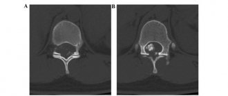
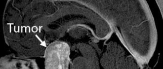
In case of brain damage, it is necessary to visualize the problem area, for which CT and MRI are used. Using these methods, it is possible to determine the location of the tumor and assess the condition of neighboring structures. Ventriculography is prescribed for a similar purpose.
Why can our articles be trusted?
We make health information clear, accessible and relevant.
- All articles are checked by practicing doctors.
- We take scientific literature and the latest research as a basis.
- We publish detailed articles that answer all questions.
As part of the examination of the patient, cerebrospinal fluid is collected. When the pineal gland is damaged, an increased concentration of protein and cellular elements is noted in the cerebrospinal fluid.
The analysis also helps to exclude inflammatory diseases of the brain. The presence of tumor cells in the cerebrospinal fluid indicates the development of a malignant neoplasm.
In addition, the complex of diagnostic measures includes a biopsy of the problem area. In extreme cases, the stereotactic method is used for these purposes. A biopsy allows you to determine the type of tumor with high accuracy.
If the patient has had epileptic seizures, the diagnosis is complemented by electroencephalography.
What are the symptoms of bone tumors?
Most patients with bone tumors complain of pain. The pain, as a rule, is long-lasting, causing discomfort with low intensity, the so-called “dull” pain. The pain persists even when the patient is resting, and it intensifies at night. Trauma is not the cause, but pain increases after injury. In weakened bones, a pathological fracture occurs, which increases pain. Some tumors cause fever and night sweats. Sometimes neoplasms are painless. Some tumors are discovered by chance during x-rays after ankle injuries.
What should be done in case of this kind of painful situation?
If a person thinks they may have a bone tumor, they should see a doctor right away.
What measures are taken at the diagnosis stage?
The doctor takes a detailed history of the patient in order to know the patient's medical history. The medical history includes all the details - from the drugs used to all previous diseases. The size and mobility of the tumor, its relationship to the joints and whether it has invaded are examined, and other systems are examined if necessary. First, the patient is given an x-ray. Different bone tumors produce different X-ray images. Some show excess calcification, some show bone resorption. Sometimes we see a combination of the two.
Is x-ray enough to identify a tumor, or is some other method necessary? Some tumors can be seen on X-rays, but we use detailed imaging techniques such as tomography, MRI, scintigraphy, PET scans and pulmonary tomography to determine the type of tumor. We use CT scans to see details of the bone, and MRIs to see how the tumor is growing in the bone or to see whether the tumor has spread to other distant sites. Bone scintigraphy provides information about the biological activation of the tumor and whether there are metastases.
What are the most common bone tumors?
The most common primary bone tumors are:
- Multiple myeloma: This is the most common type of primary bone tumor. This is a malignant bone marrow tumor. Every year it is detected in 20 people per million. It is most common between the ages of 50 and 70 and can affect any bone.
- Osteosarcoma: This is the second most common type of primary tumor of bone origin. Often found in teenagers and located in the knee area, it is fatal in 2-3 cases per million each year. Less commonly, this tumor is localized in the hip and shoulder area.
- Ewing's Sarcoma: Mostly occurs between the ages of 5-20 years. It is characterized by extensive neoplasm in soft tissues and destroys bone. Most often localized in the area of the upper and lower extremities, pelvic bone and chest.
- Chondrosarcoma: Most often occurs between the ages of 40-70 years. It provokes neoplasms in the hip, pelvic bone and shoulder.
Types of tumors of the epiphyseal region
Several types of tumors can affect the pineal gland area:
- pinealomas, which in turn are divided into pineoblastomas and pineocytomas,
- germinomas,
- non-germinoma tumors.
Pinealomas
- tumors developing from parenchymal tissues of the epiphysis. Pinealomas primarily affect males during adolescence. Tumors are nodular in shape and have a dense consistency. There are two forms of pinealoma: pineoblastoma (malignant) and pineocytomas (benign). Pineoblastomas grow aggressively and spread to adjacent tissues along the cerebrospinal fluid pathways. But pineocytomas may not cause symptoms for a long time, and only when they grow to large sizes do they compress vital brain structures. Both forms can be encapsulated, that is, located in a capsule, in which case they are much easier to remove without affecting the surrounding tissue.
Germinomas
- The most common malignant tumors of the pineal gland. This type of tumor begins to develop from about ten years of age and may indicate disorders of embryonic development. Due to their location, germinomas sometimes lead to visual impairment.
Non-germinoma tumors
are divided into: embryonal carcinoma, choriocarcinoma, teratoma.
The type of tumor can be determined only after the results of histological examination.
How is bone cancer treated?
Treatment of bone tumors is always the result of teamwork. The main members of this team are an orthopedic oncologist, an oncologist, a radiologist, a radiation oncologist, and a pathologist. The goal of treatment is to overcome the cancer and protect the affected limbs. Are there improvements in the treatment of bone tumors as medicine advances? Are there changes in surgical techniques? Previously, in order to remove a tumor from the body, there was mainly a method of amputation of limbs. But a surgical approach is now available that can both remove the tumor and protect the limbs. Surgery can be performed by removing the center of the tumor or removing it along with a small area of healthy skin. The goal of treatment is to ensure the functioning of the limb after cancer therapy. This is facilitated, in particular, by the development of reconstructive surgery. Also often used are prosthetics, bone grafts, which are pieces of bone taken from other parts of the skeleton, used to heal the affected area, and biological reconstruction techniques.
Incidence (per 100,000 people)
| Men | Women | |||||||||||||
| Age, years | 0-1 | 1-3 | 3-14 | 14-25 | 25-40 | 40-60 | 60 + | 0-1 | 1-3 | 3-14 | 14-25 | 25-40 | 40-60 | 60 + |
| Number of sick people | 0.001 | 0.001 | 0.001 | 0.01 | 0.01 | 0.03 | 0.02 | 0.001 | 0.001 | 0.001 | 0.01 | 0.01 | 0.03 | 0.02 |
Symptoms
It is difficult to differentiate pineocytoma from other brain tumors due to the similarity of symptoms. The following phenomena indicate damage to the pineal gland:
- frequent headaches of varying degrees of intensity;
- feeling tired;
- sudden weight loss or gain ;
- deterioration of handwriting;
- attacks of nausea and vomiting.
The growth of a tumor in the pineal gland provokes hypertension and increases intracranial pressure.
The development of the tumor over time causes visual disturbances, which is expressed in the form of the following phenomena:
- inability to raise the eyes (gaze paresis or Parinaud syndrome);
- strabismus;
- diplopia;
- sensation of the upper eyelid.
If the tumor process affects the cerebellum, the patient experiences:
- lack of measure in movement;
- impaired coordination of movements;
- intermittent speech;
- inability to maintain balance while standing;
- nystagmus.
Some patients suffer from epileptic seizures, convulsions, and muscle hypertonicity. A sharp increase in sleep duration is possible. If a tumor in the pineal gland develops in children, then the latter experience premature puberty.
Prevention
Due to the fact that the true causes of the development of pineocytoma have not been established, there are no specific methods for preventing tumors in the pineal gland.
To reduce the likelihood of tumors appearing in the pineal gland, it is recommended to avoid consuming carcinogenic foods. You should also avoid exposure to radiation and toxins.
Pregnant women should give up smoking and alcoholic beverages. In the case of traumatic brain injuries, patients must undergo a comprehensive examination of the brain and, in order to prevent complications, repeat these procedures every six months.
Pineocytoma is a benign neoplasm that grows from the tissues of the pineal gland. The tumor is characterized by slow growth and provokes the development of neurological disorders.
Large formations cause compression of brain structures, which manifests itself in the form of dysfunction of the visual organs and respiratory system. Treatment for pineocytes is carried out through surgical removal.
Causes
The true reasons for the formation of a tumor of the pineal gland have not been established. Researchers suggest that pineocytoma occurs under the influence of a group of factors.
Among the possible causes of the appearance of a neoplasm, genetic predisposition is distinguished. However, a direct relationship between heredity and the likelihood of pineocytoma has not been identified.
The group of predisposing factors also includes the adverse effects of the external environment: toxic damage to the body, consumption of preservatives, etc. In children, pineocytomas occur due to abnormal development of the fetus, which is facilitated by maternal smoking, intrauterine infections, and others. These factors stimulate the growth of atypical cells, which trigger the tumor process.
If abnormal tissues are present in the structures of the pineal gland, then the appearance of pineocytoma due to traumatic brain injury cannot be ruled out.
Under the influence of these factors, the processes responsible for uncontrolled cell division are launched. The tumor forms between the hemispheres of the brain in the area where the outflow of cerebrospinal fluid from the third to the fourth ventricle occurs. Due to the fact that the pineal gland is small, changes in the tissues of the pineal gland lead to dysfunction of the latter and neighboring structures.
At the initial stage of development of pineocytoma, the flow of cerebrospinal fluid is disrupted, which eventually accumulates in the ventricles of the brain.
As the tumor grows, it affects the visual centers of the brain, which manifests itself in the form of visual impairment. In the future, the development of cerebellar syndrome is possible due to compression of the cerebellar department.
Possible complications
As the volume of the tumor increases, fluid accumulates between the hemispheres of the brain. This leads to blocking the outflow of cerebrospinal fluid, which provokes hypertensive crises. This condition is characterized by frequent vomiting, headache and confusion.
Subsequently, intracranial pressure increases, due to which the patient loses the ability to perceive new information and sleeps longer. Large pineocytomas compress the brain, resulting in the death of local tissue.
Prolonged development of the tumor process leads to irreversible consequences: persistent neurological disorders occur.
Compression also causes dislocation syndrome, in which brain tissue is displaced. Such processes are accompanied by multiple symptoms, which are determined depending on the severity of the case.
Brain damage negatively affects the performance of internal organs. Compression provokes pulmonary edema, pneumonia, tachycardia, arterial hypotension, and apnea. If the tumor process causes dysfunction of the medulla oblongata, respiratory and heart failure occurs. Such complications lead to the death of the patient.
Possible complications of pineocytoma include the risk of tumor malignancy. Malignant transformation of the tumor is characterized by the spread of cancer cells throughout the internal organs and increased growth of pineoblastoma.
What is pineocytoma
The pineal gland produces melatonin and serotonin. The first hormone regulates the change in biorhythms of sleep and wakefulness. Serotonin is involved in the functioning of the endocrine, central nervous, digestive and excretory systems. Therefore, when the pineal gland is damaged, symptoms appear that indicate dysfunction of various parts of the body.
Pineocytoma (also known as pinealoma) is a benign tumor of the pineal gland that develops from the glandular tissue of the pineal gland. More often the neoplasm is localized in the area of the third ventricle.
The tumor is characterized by slow development. Externally, pineocytoma resembles a node with smooth edges. A tumor of the pineal gland is difficult to diagnose due to the small size of the pineal gland (on average 12 mm in diameter).
The risk group for developing pineocytoma includes people aged 25-40 years. However, the possibility of a neoplasm occurring in older people and children cannot be ruled out.
A neoplasm of this type does not pose a danger to the body, since tumor cells do not grow into neighboring structures. Clinical signs of damage to the pineal gland become pronounced when the pineocytoma begins to compress nearby tissue.
Sometimes tumor cells degenerate, and pineoblastoma forms in its place. This neoplasm is characterized by rapid development.
Pineoblastoma structures include dead tissue. In the absence of treatment, a malignant tumor gives metastases, which spread along with the cerebrospinal fluid into the internal organs.