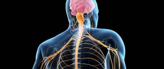
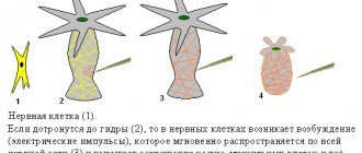
The article will discuss one of the structural elements of the nervous system - oligodendrocytes, which are participants in the transmission of nerve impulses.
Their structure, as well as their functions, are examined in detail. The pathology associated with damage to these structures is described.
Oligodendrocytes are one of the structural elements representing neuroglia. They are part of the components of the central nervous system and are present in both the brain and spinal cord.
The heir of the great histologist and brain phagocytes
The term “microglia” was introduced by a student of the famous histologist Santiago Ramon y Cajal, Pio del Rio Gortega, back in the 1920s, when he divided the glial cells of the brain into macro- and microglia.
Subsequently, they began to be called Gortega cells, and so the scientist forever wrote his name in history. Pio del Rio Gortega (1882-1945).
Credit: Wikipedia
Pio del Rio-Gortega first introduced the concept of microglia as a specific cellular element of the central nervous system on June 1, 1920 in the article La microglía y su transformación en células en bastoncito y cuerpos granuloadiposos.
- Functions[ | code]
- Synaptic connections
- Morphological components of the reflex arc.
- Microscopic structure of a neuron. Classification of neurons.
- Structure and functions of neuroglia (astrocytes, oligodendrocytes, microglia, ependyma). Relationships between neurons and neuroglia.
- Neuron structure
- Cell membrane
- Damaged wrapper
- Pathologies
- What is neuroglia
Gortega's first work
He later developed these ideas in the chapter Microglia
, written for the book that later became iconic -
Cytology and Cellular Pathology of the Nervous System
(Cytology and Cellular Pathology of the Nervous System). It was published under the editorship of Wilder Penfield in 1932. In this chapter, Rio-Gortega talked about the fact that microglia migrate to the central nervous system in the early stages of embryogenesis and in the developing brain there is a temporary form of it - amoeboid. It, actively phagocytizing (absorbing foreign inclusions), performs a protective function in the early postpartum period, when the blood-brain barrier is not yet fully developed and substances from the blood easily enter the brain. Moreover, such cells have a greater ability to migrate and multiply.
Origin and development of microglia. Credit: public domain
It should be noted that Gortega had predecessors and he was not the discoverer of Gortega cells. It is still unclear who was first. It seems that in 1878, Karl Frohmann identified cellular changes in certain areas of the brain and spinal cord of a 22-year-old patient who died of multiple sclerosis. We know for sure that at the end of the 19th and beginning of the 20th centuries these cells were seen and sketched by Franz Nissl, Alois Alzheimer and Ludwig Merzbacher. And Gortega’s teacher himself, Santiago Ramon y Cajal, also made his mark in this field.
Sketches by Alois Alzheimer It is now known that microglia are fundamentally different from the bone marrow-derived monocytes/macrophages that are often found in peripheral tissues. The difference is that its cells originate from primitive macrophages, which originate from the wall of the yolk sac, during embryogenesis (8th week of embryonic development) and enter the brain rudiment through the circulatory system.
These precursors surround the neuroepithelium of the developing brain around the 9th week of embryonic development and on the 64th day enter the neuroepithelium, beginning to populate the tissue of the central nervous system. Indeed, microgliocytes at this stage of development have an amoeboid rather than a branching shape.
It is interesting that the scientist back in the 20s of the 20th century in the chapter “ Microglia
“wrote that migrating along the choroid plexus that entwines the neural tube and the pathways of the white medulla, microglial cells penetrate into all parts of the central nervous system. In the formed brain, they are similar in appearance to astrocytes (macroglia cells), and in this form they can be distinguished by a body and many processes that are not intertwined with each other (this state of microglia is known today as “resting” microglia). That is, the idea of them has not changed much after decades.
Microglia completely populate the central nervous system only by day 28 of postnatal development. The development and survival of microglia depends on several factors, including the transcription factor PU.1 as well as CSF1R.
In the already formed brain, microglial cells are distributed evenly in all its parts and, with rare exceptions, show little variability. But as soon as a pathological process occurs, these cells, when activated, acquire an amoeboid shape, which is characteristic of them in the early stages of embryogenesis.
Del Rio-Gortega introduced his postulates based on research published in a series of papers between 1919 and 1927, in which he used an improved technique of silver impregnation (a special stain invented by Camillo Golgi, for which he shared with Ramón y) to label microglial cells. Kahalem received the Nobel Prize). The imaging technique was tedious and quite time-consuming, but it was worth it - high-quality and clear images of microglial cells were obtained.
In addition to the term microglia, del Rio-Gortega also coined the term microgliocyte. In neuroscience textbooks published after World War II, these cells were already called microglial cells. The name, although slightly changed to microglial cells, remains relevant to this day.
An excerpt characterizing Oligodendrocytes
The horses were brought in. “Bonjour, messieurs, [Here: farewell, gentlemen.],” said Dolokhov. Petya wanted to say bonsoir [good evening] and could not finish the words. The officers were whispering something to each other. Dolokhov took a long time to mount the horse, which was not standing; then he walked out of the gate. Petya rode beside him, wanting and not daring to look back to see whether the French were running or not running after them. Having reached the road, Dolokhov drove not back into the field, but along the village. At one point he stopped, listening. - Do you hear? - he said. Petya recognized the sounds of Russian voices and saw the dark figures of Russian prisoners near the fires. Going down to the bridge, Petya and Dolokhov passed the sentry, who, without saying a word, walked gloomily along the bridge, and drove out into the ravine where the Cossacks were waiting. - Well, goodbye now. Tell Denisov that at dawn, at the first shot,” said Dolokhov and wanted to go, but Petya grabbed him with his hand. - No! - he cried, - you are such a hero. Oh, how good! How great! How I love you. “Okay, okay,” said Dolokhov, but Petya did not let him go, and in the darkness Dolokhov saw that Petya was bending down towards him. He wanted to kiss. Dolokhov kissed him, laughed and, turning his horse, disappeared into the darkness. X Returning to the guardhouse, Petya found Denisov in the entryway. Denisov, in excitement, anxiety and annoyance at himself for letting Petya go, was waiting for him. - God bless! - he shouted. - Well, thank God! - he repeated, listening to Petya’s enthusiastic story. - Why don’t I sleep because of you! - Denisov said. - Well, thank God, go to bed now. We'll rise again until the end. “Yes... No,” said Petya. – I don’t want to sleep yet. Yes, I know myself, if I fall asleep, it’s over. And then I got used to not sleeping before the battle. Petya sat for some time in the hut, joyfully recalling the details of his trip and vividly imagining what would happen tomorrow. Then, noticing that Denisov had fallen asleep, he got up and went into the yard.
Functions[ | code]
Oligodendrocytes are closely associated with nerve cells, and like other glial cells, oligodendrocytes provide scaffolding as well as trophic support to neurons by producing glial-derived neurotrophic factor (GDNF), brain-derived neurotrophic factor (BDNF), and insulin-like growth factor-1 (IGF-1). . Additionally, the mammalian nervous system relies heavily on myelin sheaths to reduce ion leakage and reduce cell membrane capacitance. Myelin also increases the speed of nerve impulses when action potentials jump along nodes of Ranvier between Schwann cells (in the peripheral nervous system) or oligodendrocytes (in the central nervous system). In addition, the impulse velocity of a myelinated axon increases proportionally to its diameter, whereas the impulse velocity of unmyelinated fibers increases only as the square root of the axon diameter. The thickness of the insulation should be proportional to the diameter of the fiber. The optimal ratio for the maximum speed of impulse conduction is 0.6, divided by the diameter of the entire fiber (including the myelin sheath).
Satellite oligodendrocytes are functionally different from other oligodendrocytes. They are not attached to neurons and therefore do not serve an insulating role. They regulate the composition of extracellular fluid. Satellite oligodendrocytes are considered part of the gray matter, whereas myelinating oligodendrocytes are part of the white matter.
Important Neuroplasticity of the brain: how to develop the most important organ
Myelination is an important factor influencing intelligence. Neuroscientist Vincent J. Schmithorst has suggested that there is a relationship between the volume of white matter in the brain and intelligence. People with more white matter have higher IQ scores. A study conducted by Janice M. Juraska on rats found that rats raised in an enriched environment had a greater degree of myelination in their corpus callosum.
Classification [edit]
Oligodendrocytes are a type of glial cell. They arise during development from oligodendrocyte progenitor cells (OPCS), [6] which can be identified by their expression of a number of antigens, including GD3 gangliosides, [7] NG2 chondroitin sulfate proteoglycans and platelet-derived growth factor subunit alpha ( PDGF-alphaR). [8] Mature oligodendrocytes are broadly classified as myelinating or non-myelinating satellite oligodendrocytes. The progenitors and both mature types are usually identified by their expression of the transcription factor OLIG2.[9]
Synaptic connections
The formation of neural networks is based on electrical excitation, which consists of two processes:
- the launch of electrical excitation from the energy of external influences occurs due to the special sensitivity of the membranes located on the dendrites;
- triggering cellular activity based on the received signal and influencing other structural units of the nervous system.
Neurons are connected to each other through special structures - synapses. They consist of presynaptic and postsynaptic membranes, between which there is a synaptic cleft filled with fluid.
By the nature of their action, synapses can be excitatory or inhibitory. Signal transmission can be chemical or electrical.
In the first case, neurotransmitters are synthesized on the presynaptic membrane, which arrive at the receptors of the postsynaptic membrane of another cell from special vesicles - vesicles. After their influence, ions of a certain type can massively enter a neighboring neuron. This occurs through potassium and sodium channels. In the normal state, they are closed, there are negatively charged ions inside the cell, and positively charged ones outside. Consequently, a voltage difference is formed across the shell. This is the resting potential. After positively charged ions enter, an action potential occurs - a nerve impulse.
Cell balance is restored with the help of specialized proteins - potassium-sodium pumps.
Properties of chemical synapses:
- excitation is carried out only in one direction;
- the presence of a delay of 0.5 to 2 ms during signal transmission, associated with the duration of the processes of mediator release, its transmission, interaction with the receptor and the formation of an action potential;
- fatigue may occur due to depletion of the transmitter supply or the appearance of persistent depolarization of the membrane;
- high sensitivity to poisons, drugs and other biologically active substances.
Electrical transmission is characterized by a narrow synaptic gap and reduced resistance between membranes. In this case, the potential created on the presynaptic membrane causes excitation to spread to the postsynaptic membrane.
Properties of electrical synapses:
- the speed of information transfer is higher than in chemical synapses;
- Both one-way and two-way signal transmission (in the opposite direction) is possible.
There are also mixed synapses, in which excitation can be transmitted both with the help of neurotransmitters and with the help of electrical impulses.
Memory includes the storage and reproduction of received information. As a result of training, so-called memory traces remain, and their sets form engrams - “records”. The neural mechanism is as follows: certain impulses pass through the chain many times, structural and biochemical changes are formed in the synapses. This process is called consolidation. Repeated use of the same contacts creates specific proteins - these are memory traces.
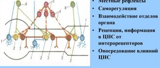
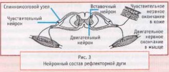
Morphological components of the reflex arc.
reflex arc
- several interconnected neurons, ensures the organ’s response to irritation. consists of three links: afferent (receptor), associative (insert) and effector. the intercalary may be absent.
Autonomous (vegetative) arc: the afferent link is represented by a pseudounipolar neuron. its body lies in the spinal ganglion, and the dendrites form endings with tissues, organs, vessels, and glands. the axon enters the spinal cord as part of the dorsal roots, goes into the lateral horns of the gray matter, where it forms synapses with the intercalary link. interneurons are multipolar, their dendrites are located in the lateral horns of the gray matter, and the axon through the anterior roots rushes to the autonomic ganglion, where it ends in a synapse with the effector link. the neurons of this link are also multipolar, and the axon is directed to the cells of the working organs (smooth muscle, glands, heart)
somatic arc: everything is the same with the receptor link, only the dendrites are in contact with the skin and skeletal muscles. The axons go to the dorsal horns of the gray body, where the interneurons are located. some collaterals pass into the anterior horns, where they contact motor neurons. the intercalary link differs only in location (posterior horns). the neurons of the effector link lie in the spinal ganglia, from where, as part of the mixed nerve, they go to the skeletal muscles, forming motor plaques on them.
Microscopic structure of a neuron. Classification of neurons.
neuron body
- nucleus and nearby cytoplasm + any grEPR, AG, etc. There are a lot of receptors on the plasmalemma. the nucleus is large, with 2-3 nucleoli; Barr’s body lives near the nucleus in women. The plasmalemma is capable of conducting impulses and has na/k pumps (they say that it is not sodium that is pumped in, but calcium). The grEPR forms clumps, which is why the whole thing is called a tigroid (Nissl body). they disintegrate with prolonged irritation or damage to the neuron. everything else is uninteresting. many mitochondria, developed AG, active endosomes... cytoskeleton - MCT (neurotubes), microfilaments, PF (neurofilaments). neurophile and neurotubules are connected by bridges, forming neurofibrils.
dendrite
. conducts impulses from neuron to neuron through axo-dendritic synapses located in the area of protrusions of the cytoplasm - dendritic spines. There are many dendrites, they are short and highly branched. neurotubules and neurofilaments carry out dendritic transport.
axon
. the process is long and extends from the axon hillock on the body of the neuron (where the impulse is generated). most of it is covered with glia. The cytoplasm of the axon is even named in a special way - axoplasm. it, of course, contains MKT and PF, as well as gLEPR, AG tanks, mitochondria and all that. axons can have perpendicular branches - collaterals. at the end, the axon often breaks up into small branches (telodendria), everyone knows how it ends.
the movement of substances along the axon is carried out due to dynein and kinesin, there is retrograde (from the axon to the body), and there is anterograde (from the body to the axon)
Important Diseases of the pituitary gland and hypothalamic-pituitary system
classification
- morphological
unipolar (amacrine neurons of the retina and interglomerular neurons of the olfactory bulb)
- bipolar (have an axon and a dendrite. These also include pseudounipolar)
- multipolar (axon and several dendrites)
functional
- sensitive (afferent)
- motor (efferent)
- associative (insert)
biochemical
- cholinergic (mediator - acetylcholine)
- adrenergic (transmitter norepinephrine)
- serotonergic (everything is clear)
- dopaminergic
- GABAergic (gamma-aminobutyric acid)
- purinergic (ATP and derivatives)
- peptidergic (mediators - substance P, enkephalins, endorphins and a bunch of other neuropeptides)
Neuron structure
Each structure in the human body consists of specific tissues inherent to the organ or system. In nervous tissue - neuron (neurocyte, nerve, neuron, nerve fiber). What are brain neurons? This is a structural and functional unit of nervous tissue that is part of the brain. In addition to the anatomical definition of a neuron, there is also a functional one - it is a cell excited by electrical impulses, capable of processing, storing and transmitting information to other neurons using chemical and electrical signals.
The structure of a nerve cell is not as complex as that of specific cells of other tissues; it also determines its function. A neurocyte consists of a body (another name is soma), and processes - axon and dendrite. Each element of a neuron performs its own function. The soma is surrounded by a layer of fatty tissue, allowing only fat-soluble substances to pass through. Inside the body there is a nucleus and other organelles: ribosomes, endoplasmic reticulum and others.
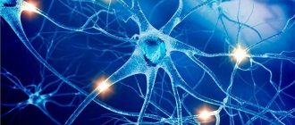
In addition to the neurons themselves, the following cells predominate in the brain, namely: glial cells. They are often called brain glue for their function: glia serve as a support function for neurons, providing an environment for them. Glial tissue provides nerve tissue with the ability to regenerate, nourish, and assist in the creation of nerve impulses.
The number of neurons in the brain has always interested researchers in the field of neurophysiology. Thus, the number of nerve cells varied from 14 billion to 100. Recent studies by Brazilian specialists revealed that the number of neurons averages 86 billion cells.
Processes
The tools in the hands of a neuron are the processes, thanks to which the neuron is able to perform its function as a transmitter and storer of information. It is the processes that form a wide nerve network, which allows the human psyche to reveal itself in all its glory. There is a myth that a person’s mental abilities depend on the number of neurons or on the weight of the brain, but this is not so: those people whose fields and subfields of the brain are highly developed (several times more) become geniuses. Due to this, fields responsible for certain functions will be able to perform these functions more creatively and quickly.
Axon
An axon is a long extension of a neuron that transmits nerve impulses from the nerve soma to other similar cells or organs innervated by a specific part of the nerve column. Nature has endowed vertebrates with a bonus - myelin fiber, the structure of which contains Schwann cells, between which there are small empty areas - nodes of Ranvier. Along them, like a ladder, nerve impulses jump from one area to another. This structure makes it possible to speed up the transmission of information several times (up to about 100 meters per second). The speed of movement of an electrical impulse along a fiber that does not have myelin is on average 2-3 meters per second.
Dendrites
Another type of nerve cell extension is dendrites. Unlike the long and solid axon, the dendrite is a short and branched structure. This process is not involved in transmitting information, but only in receiving it. Thus, excitation reaches the neuron body using short dendritic branches. The complexity of information that a dendrite is capable of receiving is determined by its synapses (specific nerve receptors), namely its surface diameter. Dendrites, thanks to the huge number of their spines, are capable of establishing hundreds of thousands of contacts with other cells.
Metabolism in a neuron
A distinctive feature of nerve cells is their metabolism. Metabolism in the neurocyte is distinguished by its high speed and predominance of aerobic (oxygen-based) processes. This feature of the cell is explained by the fact that the work of the brain is extremely energy-intensive, and its need for oxygen is great. Although the brain weighs only 2% of the body's weight, its oxygen consumption is approximately 46 ml/min, which is 25% of the body's total consumption.
The main source of energy for brain tissue, in addition to oxygen, is glucose, where it undergoes complex biochemical transformations. Ultimately, large amounts of energy are released from the sugar compounds. Thus, the question of how to improve neural connections in the brain can be answered: eat foods containing glucose compounds.
Structure
Externally, they have an oval shape, from which processes extend.
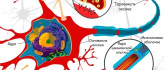
The number of processes can be small, and it can be difficult to determine the exact number due to the fact that they wrap one of the parts of a nearby axon. This creates a close anatomical arrangement. Under microscopy, the oligodendrocyte creates a kind of insulation for axons. The body of the oligodendrocyte is much smaller in size than the body of astrocytes.
According to their internal structure, they have a chalky-sized core, which, when grown, acquires a dark color, resembling the shape of a circle or oval. It also contains small nucleoli. The cytoplasm is represented by a weakly oxyphilic element, which, with nonspecific staining methods, is not visualized as a separate structure, but by merging, it becomes invisible with other cellular components of the nervous system.
Each of the organelles has a structural origin similar to neurons. Electron microscopy can detect a large number of mitochondrial cells, endoplasmic reticulum, polysomes, ribosomes, elements of the Golgi apparatus, as well as microfilaments.
Cell membrane
Thanks to the membrane, the cell has its potential. When it is transmitted along the chain, excitable tissue is innervated. Contact between the membranes of neighboring neurons occurs at synapses. Maintaining a constant internal environment is an important component of the life of any cell. And the membrane subtly regulates the concentration of molecules and charged ions in the cytoplasm. At the same time, they are transported in the required quantities for metabolic reactions to occur at an optimal level.
Important: Main symptoms and clinical picture of Parkinson's disease
The main component of the human or other mammal's brain is the neuron (also known as neuron). It is these cells that form the nervous tissue. The presence of neurons helps to adapt to environmental conditions, feel, and think. With their help, a signal is transmitted to the desired area of the body. Neurotransmitters are used for this purpose. Knowing the structure of a neuron and its features, one can understand the essence of many diseases and processes in brain tissue.
Damaged wrapper
Figure 5. Sensory impairment of the polyneuritic type. The name "sock-glove" is due to the fact that the anatomical areas corresponding to nerve damage are similar to the areas covered by these items of clothing.
It seems to me that the following rule is quite suitable for the human body: if there is an organ, then there must be a disease associated with it. In principle, this rule can be extended to molecular processes: if there is a process, there are also diseases associated with disruption of this process. In the case of myelin, these are demyelinating diseases. There are quite a few of them, but I will talk in more detail about two - Guillain-Barré syndrome and multiple sclerosis. In these disorders, damage to myelin leads to disruption of adequate signal transmission along the nerves, which causes the symptoms of the disease.
Guillain-Barré syndrome (GBS) is a disease of the peripheral nervous system in which the myelin sheath formed by Schwann cells is destroyed. GBS is a classic autoimmune disease. As a rule, it is preceded by an infection (often caused by a microbe). The presence of various pathogens in the human body triggers autoimmune damage to the myelin of nerve fibers by T- and B-lymphocytes. Clinically, this is manifested by muscle weakness, sensory impairment of the “socks-gloves” type (polyneuritic type) (Fig. 5). In the future, muscle weakness can increase until complete paralysis of the limbs and damage to the trunk muscles. Damages to the sensitive nervous system can also be varied: from a decrease in the ability to distinguish one’s own movements (impaired deep sensitivity) to severe pain. In severe forms of GBS, the main danger is the loss of the ability to breathe independently, requiring connection to a mechanical ventilation device (ALV). To treat GBS, plasmapheresis (purification of plasma from harmful antibodies) and intravenous infusions of human immunoglobulin preparations are currently used to normalize the immune response. In most cases, treatment leads to lasting recovery.
Figure 6. Lesions of the central nervous system in multiple sclerosis appear as white plaques on MRI.
Figure 7. Depending on the location of damage to the nervous system in multiple sclerosis, there may be different symptoms: from tremor and ataxia with damage to the cerebellum to emotional disorders with localization of lesions in the frontal lobes.
Links[edit]
- ^ a b
Carlson, Neil (2010).
Physiology of Behavior
. Boston, MA: Allyn and Bacon. pp. 38–39. ISBN 978-0-205-66627-0. - Baumann, Nicole; Pham-Dinh, Daniel (2001-04-01). "Biology of Oligodendrocytes and Myelin in the Mammalian Central Nervous System". Physiological Reviews
.
81
(2):871–927. DOI: 10.1152/Physrev.2001.81.2.871. ISSN 0031-9333. PMID 11274346. - Richardson, W. D.; Kessaris, N; Pringle, N. (January 2006). "Oligodendrocyte Wars". Nature Reviews.
Neurology .
7
(1): 11–8. DOI: 10.1038/nrn1826. PMC 6328010. PMID 16371946. - Thomas, J.L.; Spassky, N; Perez Villegas, EM; Olivier, K; Cobos, I; Goujet-Zalc, C; Martinez, S; Zalc, B (February 15, 2000). "Spatiotemporal development of oligodendrocytes in the embryonic brain". Journal of Neurological Research
.
59
(4): 471–6. DOI:10.1002/(SICI)1097-4547(20000215)59:4<471::AID-JNR1>3.0.CO; 2-3. PMID 10679785. - Perez-Cerda, Fernando; Sanchez-Gomez, Maria Victoria; Matute, Carlos (2015). "Pio del Rio Hortega and the discovery of oligodendrocytes". Frontiers of Neuroanatomy
.
9
: 92. DOI: 10.3389/fnana.2015.00092. ISSN 1662-5129. PMC 4493393. PMID 26217196. - Cameron-Curry, Patricia; Le Doirin, Nicole M. (December 1995). "Oligodendrocyte precursors originate from both the dorsal and ventral portions of the spinal cord". Neuron
.
15
(6):1299–1310. DOI: 10.1016/0896-6273 (95) 90009-8. PMID 8845154. - Curtis et al., 1988; Levin and Goldman, 1988; Hardy and Reynolds, 1991
- Pringle, NP; Mudhar, H.S.; Collarini, E. J.; Richardson, W. D. (June 1992). "PDGF receptors in the rat CNS: during late neurogenesis, PDGF receptor alpha expression appears to be restricted to glial cells of the oligodendrocyte lineage" (PDF). Development
.
115
(2):535–51. PMID 1425339. - Yokoo N, Nobusawa S, Takebayashi N, Ikenaka K, Isoda K, Kamiya M, Sasaki A, Hirato J, Nakazato Y (2004). "Anti-human Olig2 antibody as a useful immunohistochemical marker of normal oligodendrocytes and gliomas". Am J Pathol
.
164
(5):1717–25. DOI: 10.1016/S0002-9440(10)63730-3. PMC 1615653. PMID 15111318. CS1 maint: multiple names: list of authors (link) - Vallstedt etc., 2004
- ^ a b
Menn, B;
Garcia-Verdugo, J.M.; Yaschina, C; Gonzalez-Perez, O; Rowitch, D; Alvarez-Builla, A (July 26, 2006). "Origin of oligodendrocytes in the subventricular zone of the adult brain". Journal of Neuroscience
.
26
(30): 7907–18. DOI: 10.1523/JNEUROSCI.1299-06.2006. PMC 6674207. PMID 16870736. - Hardy and Reynolds, 1991; Levison and Goldman, 1993
- Barateiro, Andrey; Fernandez, Adelaide (September 2014). "Oligodendrocyte timeline progression: in vitro models of proliferation, differentiation and myelination". Biochimica et Biophysica Acta (BBA) - Molecular cell research
.
1843
(9):1917–1929. DOI: 10.1016/j.bbamcr.2014.04.018. PMID 24768715. - Barres et al., 1992
- Ren et al., 1992
- Gonzalez-Perez, O, B; Romero-Rodriguez, R. Soriano-Navarro, M; Garcia-Verdugo, J.M.; Alvarez-Builla, A (2009). "Epidermal growth factor induces subventricular zone type B cell progeny to migrate and differentiate into oligodendrocytes". Stem cells
.
27
(8):2032–43. DOI: 10.1002/stem.119. PMC 3346259. PMID 19544429. - Gonzalez-Perez, O, B; Alvarez-Builla, A (24 June 2011). "Oligodendrogenesis in the subventricular zone and the role of epidermal growth factor". Brain Research Reviews
.
67
(1–2): 147–56. DOI: 10.1016/j.brainresrev.2011.01.001. PMC 3109119. PMID 21236296. - Sokol, Stacy. "Physiology and pathophysiology of multiple sclerosis". Multiple Sclerosis: A Textbook of Physiology
. Archived from the original on 2012-07-16. Retrieved April 29, 2012. - ^ a b c
Fields, Douglas (February 18, 2008).
“White matter matters.” Scientific American
.
298
(March 2008): 54–61. Bibcode: 2008SciAm.298c..54D. DOI: 10.1038/Scientificamerican0308-54. PMID 18357821. - Yuraska JM; Coccyx JR (1988). "Sex and environment influence the size and ultrastructure of the rat's corpus callosum." Brain Research
.
450
(1–2):1–8. DOI: 10.1016/0006-8993 (88) 91538-7. PMID 3401704. S2CID 2720782. - Bradl (2010). "Oligodendrocytes: biology and pathology". Acta Neuropathol
.
119
(1):37–53. DOI: 10.1007/s00401-009-0601-5. PMC 2799635. PMID 19847447. ... oligodendrocytes can provide trophic support to neurons through the production of glial cell line-derived neurotrophic factor (GDNF), brain-derived neurotrophic factor (BDNF), or insulin-like growth factor-1 (IGF-1). - Jump up
↑ Philips T, Rothstein JD (2017).
"Oligodendroglia: metabolic scaffolds of neurons". J Clin Invest
.
127
(9):3271–3280. DOI: 10.1172/JCI90610. PMC 5669561. PMID 28862639. - Jump up
↑ Baumann and Pham-Dinh, 2001 - Szuchet S, Nielsen JA, Lovas G, Domowicz MS, de Velasco JM, Maric D, Hudson LD (2011). "Genetic signature of perineuronal oligodendrocytes reveals their unique phenotype". Eur J Neurosci
.
34
(12): 1906–22. DOI: 10.1111/j.1460-9568.2011.07922.x. PMC 4286392. PMID 22132705. CS1 maint: multiple names: list of authors (link) - Kinney, H. C.; Back, S. A. (September 1998). "Development of human oligodendroglia: relation to periventricular leukomalacia". Seminars in Child Neurology
.
5
(3): 180–9. DOI: 10.1016/s1071-9091(98)80033-8. PMID 9777676. - ^ a b
Káradóttir et al., 2007 - Tkachev D., Mimmack M.L., Ryan M.M. and others (September 2003). "Oligodendrocyte dysfunction in schizophrenia and bipolar disorder." Lancet
.
362
(9386):798–805. DOI: 10.1016/S0140-6736(03)14289-4. PMID 13678875. S2CID 7511585. - "Chemotherapy-induced CNS injury as a disease of progenitor cells." Archived December 27, 2011, at the Wayback Machine by Dr. Mark D. Noble, University of Rochester.
- Khan, R; Yang, YM; Dietrich, J; Lubke, A; Mayer-Proschel, M; Noble, M. (2008). "Systemic treatment with 5-fluorouracil causes delayed myelin destruction syndrome in the central nervous system". Journal of Biology
.
7
(4): 12. DOI: 10.1186/jbiol69. PMC 2397490. PMID 18430259.
Bibliography
- Raine, C. S. (1991). Oligodendrocytes and myelin of the central nervous system. In Textbook of Neuropathology, Second Edition, R.L. Davis and D.M. Robertson, ed. (Baltimore, MD: Williams and Wilkins), pp. 115–141.
- Káradóttir, R.; D. Attwell (14 April 2007). "Neurotransmitter receptors in the life and death of oligodendrocytes". Neurology
.
145
(4):1426–1438. DOI: 10.1016/j.neuroscience.2006.08.070. PMC 2173944. PMID 17049173. - Gonzalez-Perez, O, B; Romero-Rodriguez, R. Soriano-Navarro, M; Garcia-Verdugo, J.M.; Alvarez-Builla, A (2009). "Epidermal growth factor induces subventricular zone type B cell progeny to migrate and differentiate into oligodendrocytes". Stem cells
.
27
(8):2032–43. DOI: 10.1002/stem.119. PMC 3346259. PMID 19544429. - Wallstedt, A; Klos JM; Erickson F (January 6, 2005). "Multiple dorsoventral origins of oligodendrocyte formation in the spinal and hind cord". Neuron
.
1. 45
(1): 55–67. DOI: 10.1016/j.neuron.2004.12.026. HDL: 10616/40454. PMID 15629702. S2CID 7971750. - Thomas, JL; Spassky N; Perez Villegas E.M.; Olivier C; Cobos I; Goujet-Zalc C; Martinez S; Salz, B. (February 15, 2000). "Spatiotemporal development of oligodendrocytes in the embryonic brain". Journal of Neurological Research
.
59
(4):471–476. DOI:10.1002/(SICI)1097-4547(20000215)59:4<471::AID-JNR1>3.0.CO; 2-3. PMID 10679785. - Richardson, W. D.; Kessaris, N; Pringle, N. (2006). "Oligodendrocyte Wars". Nature Reviews Neuroscience
.
1. 7
(1): 11–18. DOI: 10.1038/nrn1826. PMC 6328010. PMID 16371946.
Pathologies
Damage to oligodendrocytes is observed in demyelinating diseases such as multiple sclerosis and various leukodystrophies. Oligodendrocyte dysfunction may also be associated with the pathophysiology of schizophrenia and bipolar disorder.
Injuries to the body, such as spinal cord injuries, can also cause demyelination. Cerebral palsy (sometimes developing from periventricular leukomalacia, which is local or widespread aseptic necrosis of the white matter of the cerebral hemispheres and occurs most often in premature infants) is mainly congenital or caused by damage to the newly formed brain (trauma to brain structures during fetal development and childbirth). In cerebral palsy, spinal cord injury, stroke and possibly multiple sclerosis, oligodendrocytes are thought to be damaged by excessive release of the neurotransmitter glutamate. Damage has also been shown to be mediated by NMDA glutamate receptors.
Oligodendrocytes are also susceptible to infection by human polyomavirus (JC virus), which causes progressive multifocal leukoencephalopathy (PML), a condition that specifically affects the white matter, usually in immunocompromised patients.
Tumors of oligodendroglia are called oligodendrogliomas. The chemotherapeutic agent fluorouracil (5-FU) causes oligodendrocyte damage in mice, leading to both acute acute central nervous system damage and delayed central nervous system degeneration that gradually worsens over time.
Development[edit]
Most oligodendrocytes develop during embryogenesis and early postnatal life from limited periventricular germinal regions. [10] Oligodendrocyte formation in the adult brain involves glia-restricted progenitor cells known as oligodendrocyte progenitor cells (OPCs). [11] SVZ cells migrate from the germinal zones [11] to populate the developing white and gray matter, where they differentiate and mature into myelin-forming oligodendrocytes. [12] However, it is unclear whether all oligodendrocyte progenitors undergo this sequence of events.
Between mid-gestation and full-term newborns, three successive stages of the classical human oligodendrocyte lineage are found in the white matter of the human brain: OPCs, immature oligodendrocytes (nonmyelinating), and mature oligodendrocytes (myelinating). [13] It has been suggested that some of them undergo apoptosis, [14] and others fail to differentiate into mature oligodendrocytes but persist as adult OPCs. [15] Notably, the subventricular zone-derived oligodendrocyte population can be significantly increased by administration of epidermal growth factor (EGF). [16] [17]