Brachiocephalic vessels are the main vessels of the brain, which are responsible for its saturation with blood. These include the carotid, vertebral and subclavian arteries, as well as their connection, which forms the brachiocephalic trunk.
Ultrasound of brachiocephalic arteries from RUB 3,500.
All prices Make an appointment
The condition of the brachiocephalic arteries of the neck and head can be determined using ultrasound. This diagnostic method is the most accurate and accessible means for identifying atherosclerosis and other vascular diseases.
In our International Hemostasis Clinic, a similar procedure is performed using the VOLUSON E8 device (USA). We guarantee high accuracy and affordable cost of the procedure; you can undergo diagnostics any day, from 7:30 to 20:00.
Ultrasound of cerebral vessels
Dopplerography of cerebral vessels is prescribed for suspected neurological diseases. The following symptoms may indicate this:
- Panic attacks.
- Difficulty speaking.
- Difficulties in perceiving information.
- Impaired coordination of movements.
- Decreased visual, auditory, kinesthetic sensations.
Based on the results of the ultrasound, the doctor can determine what changes in the structure of the blood vessels could cause these symptoms and prescribe treatment, if necessary.
The procedure is simple and safe and has no age restrictions. It can be carried out at home using a portable scanner.
What does a head ultrasound show?
Ultrasound can reveal abnormalities such as impaired conductivity of the cerebrospinal fluid spaces of the brain, pathological processes of infectious and inflammatory etiology (brain abscess, meningitis, encephalitis), benign and malignant tumors, cysts, hematomas, hydrocephalus, hemorrhages in brain tissue, cerebrovascular accident.
Please note that this diagnostic technique is not the main one in determining the diagnosis. If any abnormalities are detected, the doctor refers the patient for additional studies. Only after this the doctor makes a final diagnosis and prescribes appropriate treatment.
The essence of Doppler ultrasound
Based on the name of the procedure, it is performed using ultrasonic waves. Their work is based on the Doppler effect. The procedure differs from a standard ultrasound: a vessel with blood passing through it is displayed on the monitor screen. Looking at the resulting image, the specialist assesses the state of blood flow and vascular patency, which affects the quality of blood supply to the brain.
Dopplerography of cerebral vessels makes it possible to study the work of the veins, which “supply” blood to the brain, and the arteries, which are responsible for the outflow of waste products from it. The vessels being examined are located in the cervical and cranial regions. The type of ultrasound will depend on their location:
- Dopplerography of extracranial vessels located on the surface of the neck (jugular veins, carotid, vertebral and subclavian arteries). Any disturbances in their structure affect the state of the brain, which is why these vessels are called “maternal” vessels.
- Dopplerography of transcranial, or “daughter” vessels. The doctor installs an ultrasound sensor in the thinnest parts of the skull and evaluates the functioning of the veins and arteries that supply the brain.
How is the examination carried out?
The patient lies down on the couch, and the doctor applies a thin layer of conductive gel to the surface of the body in the areas of projections of the organs that need to be examined. Then the specialist brings the scanner to the skin and moves it over the body with slight pressure. At this time, the screen of the diagnostic station displays a picture according to which the doctor makes a conclusion.
Modern ultrasound units have many additional functions, such as “Clear picture” or “3D image”, as well as a huge database of diagnostic parameters, which is already immediately entered into the computer.
During the ultrasound, the patient does not experience any painful or unpleasant sensations. At the end of the diagnosis, the gel is easily removed from the skin with a towel without staining clothes.
In what cases is ultrasound ultrasound prescribed?
The main symptoms signaling a cerebrovascular accident are:
- Impaired coordination of movements.
- Deterioration of hearing and memory.
- Frequent attacks of headache.
- Impairment or partial loss of taste.
- Insomnia, difficulty falling asleep.
- Noise, whistling and ringing in the ears.
- Deterioration of visual function, manifested in loss of visual fields.
- Difficulty concentrating.
- Decreased sensitivity and motor activity of the arms and legs.
A doctor may prescribe a procedure in the following cases:
- The patient has diabetes mellitus.
- For arrhythmia, as this increases the risk of cardiac clot rupture and artery blockage.
- The patient has suffered a heart attack or stroke.
- Osteochondrosis of the cervical spine.
- Before planned heart surgery.
- If the patient is a smoker with many years of experience.
- An area with strong pulsation is visible on the neck (in this case, an ultrasound of the neck is prescribed).
Based on the above symptoms, the doctor can determine the location and name of the vessel in which the patency is impaired. Several pronounced symptoms indicate poor blood flow in a large vessel. In this case, extracranial Doppler ultrasound is prescribed. Violation of only one function will be the reason for conducting transcranial Doppler ultrasound.
Prices for ultrasound of the head, neck and thyroid gland
Our prices for ultrasound of the vessels of the head and neck, as well as for examination of the thyroid gland, are traditionally among the most affordable in Khabarovsk. At the same time, when finding out how much a thyroid ultrasound costs in other clinics, you must definitely check the model and year of manufacture of the scanner on which the diagnosis is carried out. We examine patients only with the latest devices that provide the highest quality images.
| Code | Name of procedure | price |
| 7.21 | Doppler ultrasound of brachiocephalic vessels (neck vessels) | 2200 |
| 7.22 | Doppler ultrasound of intracranial vessels of the head | 2200 |
| 7.29 | Ultrasound of the thyroid gland | 1000 |
| 7.30 | Ultrasound of lymph nodes (one group) | 800 |
| 7.34 | Ultrasound of superficial formations (neoplasms, soft tissues, salivary glands) | 800 |
| 7.37 | Ultrasound of the thymus | 800 |
| 7.38 | Neurosonogram | 900 |
| 7.39 | Ultrasound of the cervical spine (children) | 900 |
Our clinic often runs promotions for ultrasound examinations of the head and neck, so to take advantage of the discount, regularly check our website for updates.
Rules for preparing for research
On the day of the procedure, you must refrain from:
- Taking antispasmodics such as “Riabal”, “No-shpa”, “Drotaverine”, “Papaverine”, “Baralgin”, “Cinnarizine”.
- Smoking.
- Drinking black tea and drinks containing caffeine.
- Staying in unventilated, stuffy rooms with large crowds of people, which can have a detrimental effect on vascular tone.
If the patient is undergoing treatment with cardiovascular medications, then the advisability of discontinuing them before the procedure must be agreed with a neurologist. In any case, the sonologist must be informed about the treatment being performed before the ultrasound.
Stages of extracranial Dopplerography:
1. In the office, remove all jewelry from your neck and head.
2. Remove clothing from those parts of the body through which the sensor will be passed: neck, collarbones, shoulder blades.
3. Lie on the couch with your head facing the monitor. The doctor will apply acoustic gel to open areas of the body and begin to move the device over them, placing it in areas where medium and large arteries and veins are located. To obtain more accurate data on the state of vascular tone, the patient will need to hold his breath at the doctor’s request, take special medications and change body position.
Doppler ultrasound of vessels located in the cranial area has a different specificity. Acoustic gel is applied to the temples, the back of the head and the area above the eyes. The device is installed there because in these places the bones of the skull are the thinnest and are able to transmit ultrasonic waves.
How to properly prepare for a head ultrasound
To ensure that information about the state of the brain and blood vessels is not distorted and reliable, the patient will need to follow certain recommendations. The day before the scheduled ultrasound examination, try to give up coffee, strong black tea and other tonic drinks, this can affect your blood pressure.
You should not drink alcohol 2 days before the test.
Smoking is prohibited 5 hours before the procedure.
The study should be carried out when the patient is relaxed - there is no need to exercise or be nervous.
If you have any diseases or regularly take medications, be sure to inform your doctor about this. If it turns out that these medications can change the condition of blood vessels or blood flow, they can be discontinued a day before the ultrasound examination, provided that these drugs are not vital.
By what principle is the received data decrypted?
Each vessel has its own parameters, on the basis of which calculations are made and the degree of their discrepancy with the norm is assessed.
Transcranial and extracranial Dopplerography of arteries compares digital data in each vessel segment:
- Artery wall thickness.
- Vessel diameter.
- Phases of blood flow, its features.
- The degree of symmetry of blood flow through identical arteries located on different sides.
- Diastolic blood velocity.
- Maximum systolic blood flow velocity.
- Pulsation and resistive indices, systole and diastole ratio.
- The degree of pathological narrowing of blood vessels (stenosis), the condition of the artery beyond its limits.
Dopplerography of veins evaluates the following parameters:
- Condition of the venous wall of the vessel.
- The nature of blood movement through the studied vessels.
- Vein diameter
Data obtained at rest and after functional tests are compared with normal values. Any deviations are marked with a special abbreviation of letters and numbers. Based on them, the neurologist prescribes further studies or forms a course of treatment.
Advantages and disadvantages of head ultrasound
Ultrasound is safe and diagnostics can be performed as many times as necessary. The examination can be recommended not only for adults, but also for children, as well as nursing mothers and pregnant women. Ultrasound is one of the most accessible diagnostic methods, since special equipment is available in most medical institutions.
There are also disadvantages - insufficient information content and the need for additional research if deviations are detected.
Where can I get Doppler ultrasound of cerebral vessels?
The study is carried out in municipal clinics and public hospitals, specialized clinics, and general medical centers. Depending on the location of the ultrasound, the cost of each type of Doppler ultrasound will be in the range of 500-6000 rubles. The procedure has many advantages, including painlessness and the absence of side effects. Within 20 minutes, a sonologist will be able to determine the presence of anomalies in venous or arterial inflow and assess the degree of development of danger to blood vessels.
However, the procedure will not help determine the cause of compression of the thin arteries, since the capabilities of the device do not allow them to be seen from the outside.
Ultrasound of the hip joints
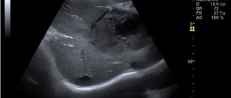
It should be performed on all children in the first month of life, regardless of the presence or absence of indications. Ultrasound allows you to identify pathology of the hip joints (delayed development of the joint, dislocation, subluxation, dysplasia - underdevelopment of one or both joints) and begin treatment even before the appearance of clinical signs. Starting treatment at a later date significantly worsens the child’s quality of life and leads to the need for surgical intervention.
Unlike X-ray examination, which shows only bone tissue and joint spaces, ultrasound of joints allows you to examine ligaments, tendons, joint capsule, cartilage, and synovium. It is extremely important for early diagnosis, because it records the initial changes in the joints.
Preparing for the study. No special preparation is required for ultrasound of the hip joints. The procedure takes about 5 minutes and is completely painless for the child.
If you are worried about dizziness, tinnitus, ringing in the head, headaches, heaviness, weather dependence, etc. - this may be a sign of impaired, insufficient blood circulation to the brain.
In such a situation, it is necessary to study the blood flow in the vessels of the brain. The most accessible and informative method for assessing the state of cerebral circulation is ultrasound examination of blood vessels, which is aimed at identifying obstacles to blood flow to the brain as the cause of vascular diseases of the brain.
Previously, ultrasound equipment made it possible to study vessels only “blindly” - without visualizing the vessel itself, only by the nature of the blood flow in it (using the Doppler effect, the speed and direction of blood flow was determined. This method is called “USDG” - “Ultrasound Dopplerography of Vessels”) .
Currently, the Doppler ultrasound method is not used independently; it is part of a more modern ultrasound examination of blood vessels, which is called “Vascular Duplex Scanning” (vascular DS). Some doctors continue to call this method in the old way: “USDG”. Therefore, there is confusion in the name of this method of ultrasound examination of blood vessels.
The term “ Duplex scanning of vessels ” means “double scanning of vessels” and allows not only to determine the nature of blood movement through the vessels using the Doppler effect (this is ultrasound scanning), but also to examine the vessels in detail: determine their size, the structure of the vascular wall (which becomes denser and thickens with atherosclerosis, thickens with inflammation of the vessel, etc.); determine the course of blood vessels (which can be smooth or tortuous, up to “loop-shaped”), identify intravascular formations - atherosclerotic plaques, blood clots that create an obstacle to blood flow.
Modern ultrasound machines add to these two components of the study the ability to color vessels depending on the direction of blood flow . Therefore, the term “ Triplex vascular scanning ” (that is, triple scanning) appeared.
The terms “Duplex scanning of vessels”, “DS of vessels”, “Triplex scanning of vessels”, “USDG of vessels” mean one thing - ultrasound examination of blood vessels, which is carried out in triplex mode on modern ultrasound machines.
When performing an ultrasound examination of the vessels carrying blood from the heart to the brain, it is customary to distinguish two levels: the vessels of the neck and the vessels of the brain.
For more reliable information about the state of cerebral circulation, it is necessary to examine both levels of blood flow to the brain, since the “neck and head” are one whole: blood flow in the arteries of the neck is an intermediate stage in the movement of blood from the heart to the brain, and blood flow in the cerebral vessels - this is the final result. Blood flow disturbances can occur at any of these levels.
The first stage of ultrasound diagnostics is the examination of the arteries and veins of the neck :
- common carotid arteries and their branches (internal carotid arteries - carry blood to the brain, external carotid arteries - supply blood to the face)
- vertebral arteries, which at the level of the neck pass through the transverse processes of the cervical vertebrae and carry blood to the posterior parts of the brain.
Obstacles to the flow of blood to the brain at the level of the neck can be tortuosity of blood vessels - congenital (up to the formation of a “loop” by the vessel), or acquired (for example, an uneven tortuous course of the vertebral arteries, formed against the background of spinal osteochondrosis); or there may be narrowing of the arteries - congenital (narrow diameter of the artery), or acquired (narrowing of the lumen of the arteries due to inflammation of the vascular wall, atherosclerotic plaques, blood clots, etc., up to complete blockage of the lumen of the artery). The veins of the neck - jugular and vertebral - are also examined to identify disturbances in the outflow of venous blood from the brain to the heart.
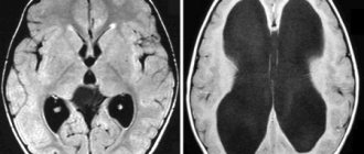
The second stage of ultrasound diagnostics is a study of the arteries and veins of the brain , which is performed through the bones of the skull (transcranial). The arteries of the brain are a continuation of the arteries of the neck on the surface of the brain inside the skull, and form two pools of blood circulation in the brain: “Carotid pool” - from the terminal branches of the internal carotid artery (ultrasound examination is carried out through the temporal bone); and the “Vertebro-basilar basin” - consisting of the terminal segments of the vertebral arteries, which, after entering the cranial cavity, merge with each other into the main (or basilar) artery of the brain (ultrasound examination is carried out through the foramen magnum).
Ultrasound examination of cerebral vessels reveals the resulting indicators of cerebral blood flow: either the blood supply to the brain is not impaired, or there are disturbances in arterial blood flow in one or another circulatory system of the brain (arterial spasm, insufficient blood flow to the brain, etc.) or there are signs of obstructed outflow venous blood from the brain (which leads to increased intracranial pressure). This study is called “Transcranial ultrasound examination of cerebral vessels”, its synonyms: “TCDG” “Transcranial Dopplerography”.
Therefore, it is impossible to study, for example, only transcranial blood flow through the vessels of the brain, without knowing the characteristics of blood movement through the arteries of the neck, where there may be various obstacles to the blood flow to the brain.
And vice versa - the study of only the vessels of the neck will be incomplete in its information content without taking into account the resulting indicators of blood flow in the cerebral vessels.
Carrying out an ultrasound examination of the neck arteries separately is only possible to track the growth dynamics of atherosclerotic plaques identified earlier in them (for timely referral of the patient to vascular surgeons).
There are cases when doctors prescribe a blood flow study in only one of the levels of blood flow to the brain: either in the vessels of the neck or in the vessels of the brain. It is not right.
This may be due to terminological confusion in the name of the method of ultrasound examination of blood vessels (out of habit, they continue to call it “USDG of the neck”).
Or it may be due to mistrust in the results of transcranial examination of cerebral blood flow, which developed in the era of “blind” Doppler examination of cerebral vessels. But those days are already gone. Modern ultrasound equipment allows you to clearly locate cerebral vessels and the movement of blood through them.
The method of ultrasound examination of blood vessels is fast, simple, informative and safe for the patient’s health.
To clarify the state of cerebral blood flow, it is necessary to conduct an ultrasound examination of the vessels of the neck and brain for such complaints as:
- headache
- dizziness
- tinnitus, ringing in the head
- decreased vision and hearing
- memory loss
- sleep disorders
- emotional disorders, psycho-emotional overload, etc.
To prevent the development of stroke, thanks to the identification of atherosclerotic plaques in the arteries, ultrasound examination of the vessels of the neck and brain is indicated for all patients:
- over 40 years of age
- with obliterating atherosclerosis of the arteries of the lower extremities
- for coronary heart disease, heart rhythm disturbances
- for arterial hypertension
- diabetes mellitus
- kidney diseases
- patients who have had a stroke or myocardial infarction.
No other studies other than Duplex Vascular Scanning provide information about the functional state of the vessels of the head and neck and the movement of blood through them. Including this study is not replaced by MRI or CT scan of the brain (since they characterize the structure and condition of the brain tissue itself, and do not characterize cerebral blood flow). Ultrasound examinations of cerebral vessels, MRI or CT scans of the brain complement each other in their information content.
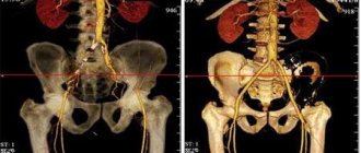
In our department, ultrasound diagnostics of blood vessels is carried out by very experienced and responsible specialists using modern ultrasound equipment. In addition to studies of the vessels of the neck and brain, studies are carried out of the arteries and veins of the upper and lower extremities, arteries of the kidneys, and vessels of the abdominal cavity.
| Name of service by nomenclature | Price |
| Functional diagnostics | |
| Code: A04.12.001.002 Duplex scanning of the renal arteries | 1050.00 |
| Code: A04.12.003 Duplex scanning of the aorta | 1050.00 |
| Code: A04.12.003.002 Duplex scanning of the abdominal aorta, iliac and common femoral arteries | 1600.00 |
| Code: A04.12.005.002 Duplex scanning of the arteries of the upper extremities | 1500.00 |
| Code: A04.12.005.003 Duplex scanning of the brachiocephalic arteries with color Doppler mapping of blood flow | 1700.00 |
| Code: A04.12.005.004 Duplex scanning of the veins of the upper extremities | 1500.00 |
| Code: A04.12.006.001 Duplex scanning of the arteries of the lower extremities | 1500.00 |
| Code: A04.12.006.002 Duplex scanning of the veins of the lower extremities | 1500.00 |
| Code: A04.12.018 Duplex scanning of transcranial arteries and veins | 1100.00 |