The brain is a complex organ consisting of gray and white matter, blood vessels, nerve fibers, membranes, and venous sinuses. To study anatomical structures, one diagnostic method is not enough. A computed tomography scan can be performed if hard tissue pathology is suspected. Verification of soft tissue changes is best done using magnetic resonance imaging (MRI). Oncological search is carried out comprehensively using both procedures.
Residents of large megalopolises in Russia (Moscow, St. Petersburg) have access to the services of public and private clinics, the addresses of which can be found on the global network. When choosing an establishment in St. Petersburg, you need to focus on reviews from previous clients of the centers. The addresses are distributed on the global network and in the media. In the city, more than a hundred clinics are equipped with radiation diagnostic methods.
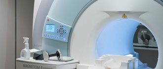
Magnetic resonance imaging scanners are open and closed, low- and high-field. Gradation is carried out based on the structure of the electromagnet location zone.
Equipment for performing computed tomography can be step-by-step, spiral (multispiral). The separation is based on the principles of the scanning procedure.
A radiation diagnostics doctor will be able to determine the type of examination and choose the right type. It is also necessary to take into account the harm to health and the diagnostic benefits of the method of studying the brain. Read the article for details.
CT scan of the brain - what will it show?
X-ray radiation, which underlies the functioning of computed tomography, detects dense formations with a diameter of more than 1 mm. After intravenous administration of an iodine-containing drug, radiology doctors are able to use tomograms to identify vascular anomalies, aneurysms (wall dissection), hemorrhages in the brain parenchyma, and strokes (hemorrhagic, ischemic).
Contrast CT scan of the brachiocephalic arteries shows changes in thin sections. During one scan, from 200 to 500 tomograms are obtained, performed at 1 mm (or other interval). Through consistent study, the specialist determines the pathology. Analysis takes a long time.
Innovative software for CT machines allows for spatial reconstruction of the brain. The spatial model shows the smallest anatomical details. 3D reconstruction is used by neurosurgeons and oncologists before planning surgery.
CT scan of the brain - why do it?
Clear medical indications have been developed, on the basis of which you can determine when to do a computer examination of the brain:
- Speech disorders;
- Prolonged headaches, dizziness;
- Suspicion of micro- and macro-strokes;
- Identification of the causes of increased intracranial pressure;
- Muscle cramps;
- Tumors;
- Cerebral edema, displacement of structures due to hydrocephalus.
CT angiography (vascular contrast) is performed to study intracerebral blood supply, identify hemorrhagic foci, if MRI is contraindicated. The main purpose is to detect small cancerous tumors.
CT brain perfusion provides information about tissue oxygen saturation and gas diffusion through nerve sheaths.
Advantages
Computed tomography has some advantages over other methods of studying the brain. The main ones include:
- Painless. Some discomfort may occur only after the administration of a contrast agent. This must be reported to your doctor.
- Accuracy. A layer-by-layer study of the brain allows us to detect initial changes in its structure. Contrast enhancement increases the information content of the method.
- Speed of research. The procedure takes on average one hour. Within an hour after diagnosis, the results are delivered to your hands.
- Possible for people suffering from claustrophobia.
CT or MRI of the brain - what to do
Magnetic resonance scanning is used to study structures containing water. The brain consists mostly of soft tissue, which makes magnetic resonance examination a popular method for verifying nosological forms. The study determines vascular malformations (pathological joints between arteries and veins), congenital anomalies without contrast. The procedure is harmless and can be performed on children who are able to remain still for 30-40 minutes (duration of the examination). Combined MRI and MSCT of the brain are done when tumors are suspected, metastases are determined, and perifocal structures around the cancer focus are studied.
When is it better to do an MRI of the vessels of the brain and neck:
- Diagnosis of inflammatory processes – abscess, encephalitis, meningitis;
- Mental disorders;
- Stuttering, convulsions;
- Sudden loss of consciousness.
- When is spiral tomography preferable:
- Determination of the state of the ventricles;
- Verification of fluid accumulations;
- Diagnosis of cystic cavities;
- Identification of areas of accumulation of cerebrospinal fluid (CSF).
Difficulties arise with prescribing a diagnostic procedure for suspected stroke. In the ischemic variant, it is better to use MRI, which records changes in the physicochemical properties of gray and white matter. If clinical symptoms indicate a hemorrhagic form, it is more rational to prescribe CT.
The essence of the method
Computed tomography of the brain is a research method based on the use of x-rays. Using a series of images, a three-dimensional image of the brain is created. Various pathological conditions are diagnosed in this way. The advantage of CT is the ability to study the organ layer by layer. Tomography is used in various fields of medicine. The method has a small number of contraindications, which allows it to be used for various conditions. To determine damage to the blood vessels of the brain, a computed tomography scan is performed using a contrast agent. This type of study allows you to most accurately determine the affected area.
Computed tomography of the vessels of the brain and neck with contrast - how it is performed
Contrast examination is used to visualize the vessels that tightly envelop the brain. Examination of the cerebral arterial network and neck (carotid, vertebral, brachiocephalic artery) allows us to identify the causes of pathological symptoms (dizziness, headaches, short-term loss of consciousness).
Native scanning does not show the soft tissue component. After an intravenous injection of an iodine-based enhancing drug, specialists are able to study the arterial and venous network of the brain. The appearance of pathological circulation indicates neoplasms that have their own capillaries. Atypical vessels appear in the early stages, when the lesion is several millimeters in size. Innovative MSCT of the brain has the ability to verify lesions with a diameter of 0.5 mm. When determining where to do a CT scan, you need to take into account the specifics of the devices.
Using the bolus contrast method, radiology doctors are able to monitor the structure of the vessels of the neck and brain as the contrast spreads.
The CT perfusion method involves studying small capillary networks after the contrast agent has spread through them. The study is used for partial visualization of soft tissue components when it is impossible to use a magnetic resonance procedure. The contrast is excreted by the kidneys after 1.5 days. After CT angiography, women should not breastfeed.
MSCT of the head: what is it, indications, preparation - MEDSI
Table of contents
- What is MSCT for brain research?
- MSCT of the head allows
- How is MSCT performed?
- Differences between MSCT and MRI
- Contraindications and risks
- Advantages of carrying out the procedure at MEDSI
MSCT (or CT)
– multislice computed tomography. A radiation diagnostic method that allows you to achieve highly detailed images of an organ.
What is MSCT for brain research?
MSCT of the brain is a multi-slice scan of brain structures. Depending on the settings of the tomograph, it can record both soft tissues of different densities (vessels, glands, brain tissue and meninges) and bone structures of the skull. The procedure lasts up to 20 minutes, depending on the purpose and scope of the study.
MSCT of the brain is prescribed:
- If a stroke is suspected - in order to clarify the focus and extent of the lesion (ischemia, hemorrhage, hematoma)
- For the diagnosis of neoplasms (cysts, tumors, infiltrates), their location and size
- For obsessive headaches, dizziness, double vision and other meningeal symptoms of unknown origin - in order to clarify the diagnosis
- If you have symptoms of increased intracranial pressure
- When performing a biopsy of neoplasms of brain structures - for the purpose of better visualization, for greater accuracy of the procedure
- After a head injury to assess the degree of damage to the brain and membranes, the presence of bleeding, foreign bodies
- To assess the condition of blood vessels and the level of blood supply to the brain (for atherosclerosis, ischemia, aneurysm)
To study the condition of the vessels, a radiopaque substance is used, which makes it possible to trace the vessel along its entire length: to assess its integrity, filling, and patency. With the blood flow, it passes through the vessels, highlighting them in the picture. CT uses iodine-based drugs, which are then eliminated from the body by the kidneys.
Contraindications for the use of contrast:
- Kidney failure
- Continuous use of Metformin (for diabetics)
- Intolerance to iodine-containing drugs
- History of hypersensitivity reactions (anaphylactic shock, angioedema), asthma
MSCT devices administer a contrast agent as a bolus - automatically, in a strictly dosed mode. This method allows you to reduce the dose of the drug.
MSCT of the head allows:
- examine the bone structures of the skull for microdamages, structural changes, metastases
- examine the temporal bones in the presence of pathologies of the auditory or vestibular system
- conduct an oncological search: determine the location of the tumor in the organ (if its presence is suspected) and diagnose small focal neoplasms up to 1 mm in size
- monitor antitumor therapy
- establish the causes of impairment or lack of consciousness
- diagnose stroke, cerebral edema, intracranial bleeding, hematoma or hemorrhage, vascular disorders
- assess the condition of the hypothalamic-pituitary system
How is MSCT performed?
- The device is adjusted to the desired structure depending on its density
- The patient changes into loose clothing without metal parts (usually a light disposable gown)
- All jewelry, watches, and dentures are removed during the procedure.
- The subject lies comfortably on a flat tomograph table
Further movements will be performed only by the device:
- The table progressively moves into the device as the desired area of the body is scanned, and the sensors rotate around it, illuminating the selected structures
- The patient does not experience any sensations during the procedure
- When a contrast agent is injected, a slight metallic taste in the mouth and a feeling of spreading warmth may occur, but this quickly passes
Differences between MSCT and MRI
- Imaging method: MSCT is x-rays, MRI is magnetic waves that are reflected from tissues
- Oncological diagnostics: MSCT is preferable
- Indications: MSCT is the method of choice for visualizing bone structures, MRI – soft organs and tissues
- Contrast agent: in MSCT – iodine preparation, in MRI – gadolinium-based (less allergenic than iodine-based)
- Frequency: MSCT – according to indications, MRI – without restrictions
- The presence of metal and electronic implants in the body, tattoos with metal-based paint: MSCT - no contraindications, MRI - contraindicated
- Children's age, pregnancy: MSCT is contraindicated, MRI is allowed from the 12th week of pregnancy (children under 7 years old are performed under anesthesia)
- Duration of the procedure: MSCT – 5–15 minutes, MRI – up to 30 minutes
Contraindications and risks
The main limitations to CT scanning relate to the use of contrast media.
But there are some risks that the patient should be aware of:
- Frequent CT scans at a young age increases the risk of development, worsening or recurrence of cancer
- Research may lead to malfunctions of electronic implants
Advantages of carrying out the procedure at MEDSI
- Modern MSCT devices allow you to carry out the procedure as quickly as possible and minimize the radiation dose
- High accuracy and detail of images allow diagnosing pathology at an early stage, which improves the prognosis of the disease
- Tests are carried out by qualified and experienced radiologists
- More than 25 types of research
Make an appointment by calling 24/7 8 (495) 7-800-500.
MR angiography of the neck - types, indications
Anomalies, aneurysms, and malformations determine the clinical symptoms of dizziness and unexplained headaches. To verify these nosologies, MRI is performed, and if contraindications are performed, contrast-enhanced MSCT is performed.
The value of both studies is determined individually after analyzing the patient's medical history. A special type is cisternography of the brain, which allows one to estimate the size of the intracerebral ventricles.
For example, with an unsteady gait and double visual objects, vestibulopathy is suspected. Nosology is best determined using MRI of the brain and neck. The presence of metal objects is a contraindication. An alternative is contrast tomography of the brain and neck.
When do you need MRI and MSCT?
During an oncological search, doctors need comprehensive information about the consistency of tissues, the location of anatomical structures, visualization of additional shadows, and the condition of blood vessels (presence of plaques, blood clots, malformations).
The norm in the study of white and gray matter is rare. Specialists often find minor pathological abnormalities that do not require treatment. MRI of the brain and neck is indispensable for verification:
- Aneurysmal dilatations;
- Multiple sclerosis;
- Alzheimer's and Huntington's diseases.
The diagnosis of a tumor is more rational than its formation after a person undergoes an MRI-MSCT complex. To prescribe methods for examining the brain and neck, clear indications are required.
If an MRI shows cancer, there is no reason to despair. The final conclusion is formed only after a systemic examination and the use of all necessary diagnostic methods. High-field MRI and CT are the basis of oncological diagnostics. Radiation imaging is informative and highly specific.
When choosing a clinic in St. Petersburg where you can do MSCT, you should pay attention to the availability of important software modes:
- Selective fluid exclusion (Flair);
- Visualization of the total diffusion of molecules;
- 3D modeling;
- Assessment of perfusion parameters;
- Mapping of nerve centers;
- Determination of physicochemical composition (MRI).
The full complex will allow you to obtain maximum information about the structure of brain structures.
MSCT angiography of the arteries of the lower extremities
Patients with atherosclerosis and diabetes mellitus often experience severe damage to the arteries of the lower extremities. Intermittent claudication, trophic changes in the skin, ulcers, secondary infections, impaired sensitivity and motor function, up to gangrene, with the need for limb amputation - this is an incomplete list of the suffering of these patients. The only possible radical treatment for such patients is open surgery or endovascular surgery to restore blood supply.
MSCT angiography allows a detailed assessment of the condition of the vascular bed and provides invaluable assistance in planning surgical treatment. The study is carried out in case of damage to the vessels of the lower extremities to identify aneurysms; hemodynamically significant narrowing (stenosis) and occlusion of the iliac, femoral arteries, and arteries of the leg. After surgery, MSCT angiography is necessary to monitor the patency of vascular stents or shunts; condition of vascular prostheses, as well as, if necessary, planning re-interventions.
Radiation imaging of the neck – when is it performed?
An MSCT of the neck can be done if there are symptoms of intervertebral hernias or compression of the vertebral artery by unstable vertebrae. Neurologists can determine whether a nerve fiber is pinched in the cervical spine. Radiation imaging is used to assess the degree and nature of cerebral circulatory disturbances. The methods identify nosology at an early stage of development, when there are no symptoms.
MRI of the brain and neck is performed to identify or exclude blood clots, atherosclerotic deposits, and blockage of the lumen of the vessel.
Procedures are not used for mass examinations. Scientists have only theoretically substantiated the need for MSCT and MR imaging for early verification of atherosclerotic lesions of the heart arteries. The widespread spread of harmful radiation and the high cost of procedures are limited.
Computer scan of the neck and brachiocephalic trunk
The maximum number of cases of insufficiency of cerebral blood supply occurs in the brachiocephalic complex, which includes the carotid, vertebral, subclavian arteries, and brachiocephalic trunk.
Atherosclerotic deposits in the wall and a blood clot clog the lumen of the vessel. Non-contrast MSCT of the brain reveals abnormalities, helps assess the degree of microcirculation impairment, and visualize enlarged lymph nodes.
Late detection of BC damage is highly likely to lead to disability or death.
The use of multispiral tomography of the brain with contrast detects aneurysms, pathological anastomosis between arteries and veins (malformations). The study helps determine the volume and prevalence of death of brain parenchyma (atrophy), and areas of lack of oxygen supply (ischemia). Information is necessary for specialists when planning surgical interventions to install vascular stents, perform embolization, and remove aneurysmal dilatations.
What MSCT of the brachiocephalic arteries shows:
- Ischemic areas;
- Areas of narrowing of the arteries that do not manifest clinical symptoms;
- Discirculatory encephalopathies;
- Anomalies in the course and structure of blood vessels;
- history of stroke;
- Neoplasms of the neck;
- Thromboembolism.
The information content of spiral scanning is beyond doubt, but there are contraindications:
- Kidney failure (with contrast examination);
- Excessive secretion of thyroid hormones;
- Allergy to iodine;
- Carrying a child;
- Decompensated diabetes.
Vascular stenosis is the leading pathology leading to ischemic strokes. To prevent nosology, surgeons introduced endoscopic revascularization operations. Their use requires early diagnosis using MRI and MSCT of the brain.
Long-term persistence of ischemia due to lack of blood supply contributes to the expansion of the capillary areas located in front of the block. Tissues receive more oxygen, which leads to the development of hyperperfusion syndrome. If such areas are detected on computer angiography, an additional examination is prescribed - CT or MR perfusion, which evaluates the parameters of blood passage through the capillaries. The choice of method is determined by diagnostic tasks.
The option of choice is MSCT perfusion - a method with a quick analysis of the passage of blood through the capillaries without loss of tomogram quality.
Indications for CT scan of the head and neck
Computed tomography of the head carries a certain radiation load on the body, so scanning is recommended no more than 3 times a year for emergency indications.
The interval between studies should not be less than 4 weeks. For preventive purposes, experts do not recommend undergoing examination, as the X-ray load on the body increases. The main indications for CT of the head and neck are:
- dizziness and tinnitus, the causes of which have not been established;
- anomalies in the development of bone structures of the head, blood vessels;
- suspicion of the development of a hernia in the cervical spine;
- neck and head injuries;
- suspected tumors of the head and neck;
- headache;
- identifying dilation of blood vessels in the neck and head;
- signs of cerebrovascular insufficiency.
When contacting specialists at the Yusupov Hospital with these signs, patients receive advice on the scope of diagnostic measures to identify the causes of the pathology, after which they are sent for diagnosis.
Make an appointment
MSCT angiography of the aorta – what it shows
The procedure determines the narrowing and expansion of the aorta located below the neck. The vessel is large, so serious blockage is accompanied by hypoxia of the organ complex - heart, brain.
MSCT angiography shows even minor areas of narrowing of the aorta and arteries. The study is characterized by intravenous administration of a contrast agent excreted by the kidneys. Enhancement with iodine-containing products causes restrictions - it cannot be done for people with kidney disease.
Doctors have developed clear criteria that limit the capabilities of multislice tomography:
- Blood creatinine level is more than 130 µmol per liter;
- Excess thyroid hormones;
- Hypersensitivity to iodine;
- Planning for radioiodine scintigraphy.
If any of the described criteria are present, contrast MSCT of the brain is not performed. The procedure can cause disability or death of a person if prescribed unreasonably.
CT scan of the neck and larynx with contrast is not an alternative to MRI and is prescribed for people with contraindications to the procedure.
Contraindications for computed tomography
CT head
, as practice shows, cannot always be prescribed to patients for diagnosing brain diseases.
Contraindications for CT are as follows:
- pregnancy and lactation;
- diabetes mellitus (complicated form);
- liver and kidney diseases (severe form);
- bronchial asthma;
- excess weight – over 150 kg.
For a head CT scan using contrast, a significant contraindication is an allergic reaction to the constituent elements of an intravenously administered auxiliary substance.
MRI of the pituitary gland - what it shows
The pituitary gland is a soft tissue gland located in the area of the sella turcica. A tumor in this zone, an adenoma, leads to damage to the entire endocrine sphere. A computer scan does not show a neoplasm, but with its help it is possible to determine changes in the bone structure (expansion of the bed, calcification).
The pituitary gland is studied using MRI. The procedure shows intrasellar, laterosellar, intrasellar formations. Verification of lesions is achieved with dynamic contrast. The study is based on assessing the accumulation of contrast in the area of the corpus callosum, cerebral ventricles, and sellar zone.
Radiologists identify microadenomas by analyzing the accumulation curve at the initial stage. A pathological focus can be suspected by assessing contrast enhancement of the neck area immediately after injection.
For large formations of the fossa turcica, the features of the contrast accumulation curve during the peak are studied. For example, craniopharyngioma can be traced only 10 minutes after intravenous injection of the drug. MSCT of the brain does not have similar capabilities, but if the patient has metallic inclusions, there is no alternative to computed tomography.
How is the procedure done?
A CT scanner is a square or round X-ray machine with a gantry ring in the center. During the scanning process, the CT scanner table on which the patient lies moves through the tunnel, and a ring located in the walls of this tunnel, rotating around the patient’s body (in particular, his head), emits X-rays. These rays pass through the skull bones and brain tissue at different angles and are then captured by the detector. Information from the detector is sent to a computer, on the screen of which an image of cross sections of the examined part of the body is created. The CT procedure lasts approximately 5-10 minutes.
CT head with contrast
In some cases, a more accurate diagnosis can be determined using head computed tomography (CT), which is performed with contrast. This is a substance that the patient can receive in the form of a drinking solution or by injection. Thanks to the contrast, tumors and their outlines are most accurately visualized in the images. This research method is most often used if it is necessary to exclude or confirm the presence of cancer in a patient. After a contrast-enhanced CT scan, it is recommended to drink plenty of fluids, this will help to quickly remove the auxiliary drug from the human body.