Monitoring a child, especially in the first year of life, is a troublesome task.
A pediatrician, neurologist, ophthalmologist, orthopedist - this is just the minimum list of doctors who should regularly examine your child.
But even the best and most highly qualified specialist cannot do without ultrasound examinations, which provide invaluable assistance in monitoring the baby. And sometimes ultrasound, in particular neurosonography (NSG), is the only method that allows timely detection of pathology, for example, malformations of the brain. Ultrasonic waves are high frequency mechanical vibrations. The patient does not receive any radiation during an ultrasound examination. The only effect that ultrasound has on tissue is thermal, and it is completely insignificant. In practice, there was not a single complication or deterioration in the child’s condition associated with NSG. The baby’s well-being, both during and after the study, does not change. And children who undergo neurosonography during sleep do not even wake up.
What is neurosonography of the newborn brain?
Neurosonography is a method of ultrasound sectoral scanning of the head
the child’s brain through the fontanelle (including in newborns).
Synonyms for the word “ neurosonography ” are “ neurosonography of newborns , neurosonography of the brain , ultrasound neurosonography , transcranial neurosonography , ultrasound of the brain , ultrasound examination of the brain, NSG .”
Decoding the result
data of the brain in newborns
are considered in conjunction with what other research methods show. The norm greatly depends on the stage of pregnancy at which the baby was born. Therefore, the results of ultrasound of the brain of newborns require careful decoding and verification with tables of normative indicators. However, ultrasound of the brain has standard indicators common to all children:
- The structure of the ventricles of the brain is homogeneous.
- Stem structures without displacement.
- The convolutions and furrows are clearly visible.
- Symmetry of brain structures.
- The subcortical nuclei and thalamus have medium echogenicity.
Intracranial hematomas
Hematomas can appear as a result of asphyxia during childbirth or intrauterine hypoxia, or due to mechanical impact on the head during childbirth. Hemorrhage is diagnosed by a specialist during examination; ultrasound of the brain confirms the diagnosis in newborns.
Intracranial pressure
A number of diseases, including hydrocephalus, trauma, hemorrhage, tumors and various infections, can lead to increased intracranial pressure. The diagnosis is made based on a set of data; ultrasound of the newborn’s head is performed as an additional examination.
Hydrocephalus
The disease leads to increased intracranial pressure. The child suffers from headaches and gets tired quickly. Without treatment, hydrocephalus leads to delays in physical and mental development. Neurosonography is performed to diagnose the disease as an additional examination.
Ischemia
Ischemia in newborns develops as a result of lack of oxygen during pregnancy or childbirth. In mild cases, the disease leads to excessive excitability or depression in the child. More severe forms are characterized by intracranial hypertension and severe seizures. Severe ischemia leads to cerebral edema and coma. For the health of the baby, it is important to diagnose the disease early and begin treatment. To identify the disease, a study of blood vessels and ultrasound of the newborn’s brain
.
Cysts
Cysts are fluid-filled spherical cavities that arise at the site of death of nervous tissue. Cysts can appear in different areas of the brain as a single tumor or in a group. Ultrasound of the brain is considered the most reliable method for diagnosing such diseases for children under one year of age.
How is neurosonography done?
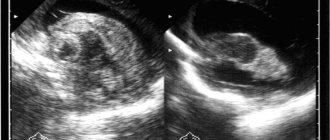
Neurosonography is performed only on a child (1 month, 2 months, 3, 4, 5 months, 6, 7, 8, 9, 10, 11 months) whose fontanels or sutures are not closed. The fontanelle is an area on the head that is not covered by bone tissue. Fontanas transmit ultrasound very well. The study is often carried out through the anterior large fontanelle, which is located between the parietal bones and the frontal bone. The foramen magnum is used less frequently; in full-term patients (through a thin bone), the anterolateral fontanel and posterolateral fontanel can be used. By the way, neurosonography can be performed on children even during sleep. Neurosonography is performed at an ultrasound frequency of 5–75 mHz in the coronal and sagittal planes. The study visualizes the ventricular system, periventricular structures, formations of the posterior cranial fossa, middle cranial fossa, anterior cranial fossa, and cerebral vessels (arteries and veins). The bones that form the skull, gyri, sulci of the brain, choroid plexuses of the ventricles and cerebellum have a high echogenic density. The ventricles of the brain containing cerebrospinal fluid, the cavity of the transparent septum, Verge's cavity, and cisterns are anechoic. The brain parenchyma has low echodensity, with the exception of the basal ganglia, which have increased echogenicity.
Evaluation of results
Ultrasound examination allows you to study:
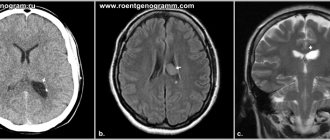
- Condition and structure of brain tissue. Darkening in the image allows one to suspect the presence of tumors, cystic formations, and hemorrhages. Increased tissue density can also be a symptom of inflammatory processes.
- Large vessels and choroid plexuses. Enlargement of some areas of the vessels may indicate the presence of an aneurysm.
- Ventricles of the brain. Changing their size makes it possible to diagnose pathologies such as rickets or hydrocephalus.
- Symmetry of brain structures. Slight asymmetry is considered a variant of the norm.
Identification of any abnormalities is a reason for the doctor to prescribe other studies, such as electroencephalography or rheoencephalography.
Timely diagnosis of neurological pathologies allows treatment to begin in the early stages of the disease, on which its effectiveness directly depends.
Neurosonography is an accurate type of diagnosis
Neurosonography is very important in the diagnosis of pathology in children in the first month of life in identifying various hemorrhages. Both isolated subependymal hemorrhages and hemorrhages in the ventricles of the brain with their spread to the brain parenchyma are well defined. Neurosonography has high sensitivity. Neurosonography is an accurate type of diagnosis. Neurosonography allows you to diagnose focal and multifocal necrosis, foci of periventricular leukomalacia, foci of subcortical leukomalacia.
In the acute phase, ischemic foci in the gray matter of the brain on the echogram are represented by areas of increased echo density. Sarklinik believes that the diagnosis of damage to the thalamus opticus and subcortical nuclei is better determined in the presence of hemorrhagic necrosis. Neurosonography is of great importance in the diagnosis of infectious and inflammatory processes. Neurosonography makes it possible to monitor the development of inflammatory and infectious processes and to promptly identify complications such as hydrocephalus, subdural hygroma, ventricular abscess, and cerebral abscess.
Neurosonography perfectly identifies congenital malformations of the central nervous system, changes in the size, shape, location of the ventricular system, and other anatomical structures of the brain. Using neurosonography, it is possible to diagnose various forms of pathology such as congenital hydrocephalus, Dandy-Walker malformations, holoprosencephaly, aneurysm of the vein of Galen, agenesis of the corpus callosum, Arnold-Chiari syndrome. Neurosonography plays a certain role in the diagnosis of brain tumors. Sonographic signs of tumors are nonspecific. They can manifest themselves in the form of cystic formations against the background of hydrocephalus, or areas of increased echo density. Often the tumors are asymmetrical and are accompanied by displacement of the midline structures of the brain.
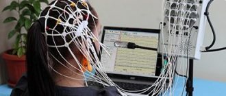
Carrying out the procedure
Neurosonography in a child is performed using special ultrasound equipment. The doctor places the sensor in the area of open fontanelles, which makes it possible for ultrasound waves to freely penetrate into the structures of the brain. Thanks to this, an image corresponding to the reflected waves appears on the monitor, on the basis of which a conclusion is drawn about the state of the brain.
Neurosonography does not require any preparation and can be performed even on a sleeping baby. The procedure does not bring any discomfort and lasts 10-15 minutes.
Neurosonography can determine the pathology of the brain of newborns and infants
What does neurosonography show in children? Hypoxic damage to the central nervous system, traumatic damage to the central nervous system, trauma, periventricular edema, increased peripheral vascular resistance, vascular spasm, periventricular leukomalacia, periventricular hemorrhage, cyst (diameter 1, 2, 3 or more cm), pseudocyst, vascular abnormalities, hydrocephalus, hypertensive -hydrocephalic syndrome, hypertensive syndrome, infectious brain lesions (infections), congenital malformations of the brain, central nervous system, dilatation of the ventricles of the brain, subependymal cysts, cysts of the choroid plexus of the brain, expansion of the subarachnoid space, expansion of the external cerebrospinal fluid spaces, hemorrhages in the substance of the brain , hemorrhage into the ventricles (intraventricular hemorrhage, IVH grades 1, 2, 3, 4), cerebral ischemia.
Indications for neurosonography
Since the procedure allows us to identify abnormalities in brain development, it is included in the list of mandatory screening studies in the first days after birth (in the maternity hospital), then at 1, 3 and 6 months (according to indications).
Risk factors for disorders of the central nervous system that require mandatory examination of the state of the brain:
- premature birth (birth before term);
- low birth weight;
- violations of the structure of the facial skeleton or irregular shape of the child’s head;
- suspicion of intrauterine infections;
- hereditary abnormalities in brain development;
- fetal hypoxia during pregnancy;
- Rhesus conflict;
- asphyxia at birth;
- birth injuries;
- neurological diseases.
Neurosonography is also prescribed for frequent regurgitation, convulsions, strabismus, and unexplained crying.
Ultrasound of the brain with Dopplerography
Thanks to the features of the ultrasound equipment of our clinic, along with neurosonography, it is possible to additionally perform ultrasound Dopplerography - a study of the vessels of the child’s brain. The operating principle of this diagnostic method is associated with the Doppler effect, according to which ultrasonic waves, reflected from a moving object, change their frequency depending on the speed of movement of this object. In this way, the waves probe the movement of blood cells, producing a color display of blood flow activity on the screen.
This research method is quite informative and allows you to identify disorders of the cerebral arteries, including hypoplasia (underdevelopment) of blood vessels, stenosis, tortuosity, and other pathologies.
Neurosonography Center
Neurosonography centers in Russia necessarily determine the size of the ventricles of the brain (the lateral ventricles may be enlarged), the area of the ventricles, the contours of the ventricles, cerebral vessels, vascular plexuses, and convexital spaces. With pathology, the sizes are reduced or increased. Send us letters with your stories about neurosonography and your beloved children. We will publish them on the medical website sarclinic.ru. You will be able to read patient reviews and discuss all problems.
The sooner the necessary treatment for a child is started, the greater the chance of his recovery.
Photo: (©) Prometeus | Dreamstime.com \ Dreamstock.ru The child depicted in the photo is a model, does not suffer from the diseases described and/or all similarities are excluded.
Related posts:
The child does not sit, what time do children sit, when does the child begin to sit, at what months
Puberty boys, girls, teenagers, adolescence, puberty
Which clinic best treats nocturnal enuresis in children in Russia
Children who involuntarily shout out words, why does the child scream
Birth trauma of newborns, treatment of birth injuries, rehabilitation, recovery from injuries
Comments ()
Who undergoes neurosonography?
Neurosonography is mandatory for all premature babies. Children who required intensive care or resuscitation after birth, with birth trauma or after traumatic obstetric care, large newborns, and babies with intrauterine growth retardation. In addition, the study is prescribed for children with defects and anomalies of other organs and systems. But, in general, every child needs to have neurosonography done at least once. To exclude changes that may manifest themselves only after a year, when the fontanel has already closed.