Neurologist
SEMENOVA
Olga
5 years experience
neurologist, head of the office for diagnostics and treatment of cognitive disorders
Make an appointment
A pathology characterized by an increase in the volume of fluid in the patient’s brain is called hydrocephalus. This condition increases the pressure inside the skull, causing a person to develop numerous brain disorders. The disease affects all age groups, from prenatal children to the elderly.
Basic information about pathology
A special cerebrospinal fluid, or cerebrospinal fluid, is produced in the choroid plexuses of the brain, called the ventricles, and fills the subarachnoid space. Excess cerebrospinal fluid is released into the bloodstream through the arachnoid membrane. When problems with absorption, circulation occur, or excessive secretion of cerebrospinal fluid occurs, hydrocephalus develops. It can be congenital or acquired as a result of certain diseases. In addition, the following forms of the disease are distinguished:
- internal hydrocephalus - overflow of one or more ventricles inside the brain with cerebrospinal fluid;
- external hydrocephalus - fluid injection between the brain and the enveloping subarachnoid membrane.
In its acute form, the disease develops over several days. Subacute course involves the development of decompensation within a month from the onset of the first symptoms, chronic - longer than six months. In addition, the classification of hydrocephalus includes stable and progressive forms of pathology, characterized, respectively, by stability or a gradual increase in pressure inside the skull.
Ependymomas
Most often localized within the ventricular system of the brain. Symptoms of ependymoma directly depend on the location of the tumor and its size, the presence and severity of hydrocephalus.
Diagnosis : the gold standard is MRI without and with contrast enhancement. Imaging of the brain and all levels of the spinal cord is necessary due to the high incidence of metastasis. It is advisable to perform FLAIR in addition to standard modes (T1, T2, T1 with contrast).
Signs of the disease in adults
As a rule, hydrocephalus of the brain in an adult or a teenager is directly related to increased intracranial pressure, which causes the patient to feel:
- severe headache that does not improve with analgesics;
- feeling of internal pressure on the eyeballs;
- nausea, sometimes leading to vomiting;
- impaired coordination, due to which the gait becomes unstable, hand movements become sharp and sweeping, handwriting changes, etc.;
- tinnitus, impaired visual acuity, loss of certain areas of space from the field of vision.
Sometimes changes affect the patient’s emotional sphere, causing rapid changes in mood for no apparent reason, neurasthenic manifestations and unmotivated aggression.
Magnetic resonance imaging (MRI) in St. Petersburg
The following brain tumors can be located inside the ventricles: ependymoma, subependymoma, choroid plexus papilloma, central neurocytoma, meningioma, giant cell tumor.
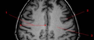
Most of these tumors are localized in the lateral ventricles. The optimal method for diagnosing intraventricular tumors is MRI of the brain.
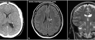
Ependymoma accounts for 2-9% of intracranial tumors and 6% of gliomas. Of the ependymal tumors in adults, subependymoma occurs. It makes up about 8% of ependymal tumors and is located near the ventricular wall. The peak incidence of ependymoma itself occurs at the ages of 5 years and 34 years. The tumor originates from ependymocytes. These are usually benign forms. In 60-70% of cases, the tumor is localized infratentorially in the region of the fourth ventricle and in 5-8% in the cerebellar hemispheres. Much less commonly, supratentorial localization of ependymoma is detected: in the hemispheres near the ventricles (on coronal MRI sections it looks like a “cauliflower”), in the region of the third ventricle. Benign ependymomas have an expansive growth pattern. The internal structure is heterogeneous due to cysts (43-83% of cases) and calcification (50% of cases). Contrast enhancement is observed in a third of cases. Anaplastic ependymoma (grade III) metastasizes along the cerebrospinal fluid pathways.
Meningioma is the most common non-glial brain tumor and belongs to the meningeal tumors. In the area of the choroid plexuses of the ventricles they are extremely rare and may be a manifestation of neurofibromatosis type 2. Intraventricular meningiomas occur in 20% of children, while in adults they are 10 times less common. Meningiomas have typical features on MRI , usually homogeneous, with a clear contour, and are well contrasted.
Benign formations of the lateral ventricles include ependymal cysts. They represent the lacing of the lining of the ventricle. On MRI , they have a cerebrospinal fluid signal, are normal, have a clear contour and are not contrasted.
Ventricular choroid plexus tumors overall account for approximately 3% of pediatric brain tumors. In the first year of life their frequency reaches 20%. They are usually localized in the lateral ventricles (80%) and rarely in the IV and III ventricles, as well as the cerebellopontine angle. In adults, on the contrary, they are usually found in the fourth ventricle. Bilateral tumors have been described.
Papillomas are benign tumors. Their frequency is 0.4-0.6% of all brain tumors in all age groups and 1.5-6% of brain tumors in children. In the first 2 months. 40% of papillomas are observed in life. Clinically, the tumor manifests itself in the form of developmental delay, epilepsy and manifestations of mass effect (hydrocephalus, loss of visual fields, etc.). MRI within the ventricle reveals a low signal intensity mass on T2-weighted images and iso- or hypointense on T1-weighted images. On the side of the tumor, the lateral ventricle is sharply dilated, and a high-intensity signal is observed around it, which is associated with transependymal fluid resorption. The formation protrudes from the ventricle, infiltrating its edges, and passes into the paraventricular white matter. Edema around the tumor is pronounced. The internal structure of the tumor may be heterogeneous due to calcification (about a third of cases). Contrast enhancement is good.
Carcinomas, which make up about 20% of choroid plexus tumors, disseminate throughout the ventricular system and subarachnoid spaces, invading brain tissue and bone. On T2-weighted tomograms they are brighter than papillomas and are clearly heterogeneous. MRI reveals a hypointense or isointense mass inside the ventricle on T1-weighted tomograms. On T2-weighted tomograms the tumor has low signal intensity. On the part of the tumor, pronounced hydrocephalus and a high signal around the ventricles themselves are determined due to transependymal fluid resorption. The mass protrudes from the ventricle, infiltrating its edges with transition to the periventricular white matter. Swelling around the tumor is pronounced. The internal structure is often heterogeneous due to calcification (about a quarter of cases). Contrast enhancement is good. Carcinomas are brighter on T2-weighted tomograms and are markedly heterogeneous.
Central neurocytoma accounts for 0.25-0.5% of brain tumors and belongs to neuroglial tumors. Has gradation II. The prognosis is favorable.
A giant cell tumor is also typically located in the 3rd ventricle. Giant cell astrocytoma is one of the characteristic manifestations of tuberous sclerosis, a disease from the group of phakomatoses. In addition, this is a typical site for a colloid cyst of the 3rd ventricle, which is a developmental anomaly and requires differential diagnosis with intraventricular tumors. Unlike a colloid cyst, it has a heterogeneous structure on MRI and CT due to calcifications.
In the 4th ventricle, papillomas of the choroid plexus are rarely found in adults. Epidermoid and dermoid cysts may also be located there. Metastases and hemangioblastomas are initially localized in the brain stem or cerebellar hemispheres, but can spread to the 4th ventricle. Tumors of the 4th ventricle are optimally examined using MRI , since it, unlike, does not produce artifacts. All tumors are well contrasted. In children, tumors of the 4th ventricle are much more common than in adults. These are astrocytomas (usually pilocytic), medulloblastomas and ependymomas. Pediatric stem tumors can also grow towards the 4th ventricle.
Medulloblastoma is a primitive neuroectodermal tumor. It accounts for 15-20% of all brain tumors in children and over a third of tumors of the posterior cranial fossa in this age group. In approximately 75% of cases, medulloblastoma is found in children in the first 10 years of life. The sex ratio is M:F as 2-4:1. The tumor is malignant and grows rapidly in the midline along the cerebellar vermis, obstructing the fourth ventricle and causing occlusive hydrocephalus. Going down, the tumor can pass through the foramen magnum into the cistern magna. Medulloblastoma metastasizes along the cerebrospinal fluid pathways and in 5% of cases hematogenously to the lymph nodes, bones and liver. Clinical manifestations consist of nausea, vomiting, diplopia, and ataxia. MRI usually reveals a homogeneous midline mass in the inferior cerebellum that is hypointense on T1-weighted scans . It is usually hyperintense on T2-weighted MRI The internal structure of the tumor is usually homogeneous. Calcifications, areas of necrosis and cysts are observed in 10-15% of cases. The contrast is good and uniform. Hydrocephalus is present in almost all patients. Leptomeningeal metastases are common.
Cerebellar astrocytoma is usually benign (grade I) of the pilocytic subtype. Peak frequency occurs at ages 5-9 years. The diffuse fibrillary subtype is much less common, usually in adolescents. It is characterized by infiltrative growth and an unfavorable prognosis. The primary source of astrocytoma is the cerebellar vermis. The tumor is located in the midline, but can also grow in the cerebellar hemisphere. Only 10% of tumors are purely solid, the rest have a cystic necrotic center or consist almost entirely of a cyst with tumor tissue inside its wall. Clinical manifestations include weakness, ataxia and tremor.
On MRI , the tumor is hypointense on T1-weighted MRIs and hyperintense on T2-weighted MRIs . It can be very difficult to distinguish a cystic component from a solid one, since the cyst contains a lot of protein. Contrast helps, in which the solid part is enhanced. The node can be round or flat. The walls of the cyst are not contrasted. Unlike a cyst, the walls of the necrotic cavity are contrasted. Occlusive hydrocephalus is often observed.
Ependymoma accounts for 9-16% of CNS tumors in children, with 60-70% of them localized infratentorially. In children, ependymoma most often occurs before the age of 5 years. A rare embryonic variant is ependymoblastoma, a very malignant tumor. The degree of differentiation of ependymomas can be different; a benign subtype is more often observed. Being initially located inside the IV ventricle, it tends to grow into the trunk, move to the spinal cord, or, less commonly, to grow through the foramina of Luschka into the cerebellopontine angle. Clinical manifestations consist of nausea, vomiting, ataxia, diplopia, and loss of cranial nerve function.
On MRI , ependymoma is almost indistinguishable from medulloblastoma or astrocytoma. It is usually inhomogeneous due to microhemorrhages, calcification, small cysts and vessels. CT helps confirm the presence of calcifications (over 40% of cases), which are noticeably less common in medulloblastoma. The cystic component occurs in 20% of cases. In some cases, the “melted wax” symptom helps: the tumor seems to flow along the edges of the fourth ventricle, enveloping the trunk and without causing a noticeable mass effect. In 90% of cases, the tumor is well contrasted.
During MRI in St. Petersburg, we carry out differential diagnosis of tumors of the posterior cranial fossa in children with vascular malformation, abscess, epidermoid and dermoid. These tumors and other formations are better visible in high fields, somewhat worse in low-field open MRIs. Examination of children by MRI in St. Petersburg makes it possible in specialized children's institutions, which is preferable.
Leave feedback.
MRI in St. Petersburg USA
Signs in children
Thanks to the mobility of the cranial bones, children, especially newborns and toddlers in the first years of life, do not experience an excessive increase in pressure inside the skull. Manifestations of hydrocephalus of the brain in a child are:
- enlarged head;
- bulging fontanelle without pulsation (in children in the first months of life);
- bulging veins on the head;
- limited mobility of the eyeballs when moving upward;
- expansion of cranial sutures.
With the congenital development of the disease or the appearance of pathology in newborns, the child almost always lags behind his peers in his development: later he begins to hold his head up, sit up, and reacts poorly to attempts to communicate.
In children with severe pathology, characteristic external signs of hydrocephalus develop: a large spherical head with thin skin, deep-set eyes, protruding ears. Such children are characterized by inactivity, which is why they often suffer from obesity. Almost always, the disease leads to intellectual and emotional deficiency: children grow up lethargic, lack initiative, and do not experience attachment to relatives.
Are you experiencing symptoms of hydrocephalus?
Only a doctor can accurately diagnose the disease. Don't delay your consultation - call
What causes the disease?
There are many factors that influence the mechanism of cerebrospinal fluid circulation, and they are different for intrauterine development and for acquired forms of pathology.
The causes of congenital hydrocephalus include:
- deviations in the formation of the brain’s liquor system;
- structural defects of the subarachnoid space;
- anomalies of the craniovertebral region;
- influence of intrauterine infections;
- birth injuries.
In cases of acquired hydrocephalus, the causes of impaired circulation of cerebrospinal fluid differ sharply. This:
- infectious inflammation of the tissue and membranes of the brain;
- traumatic brain injury;
- pathological processes in the vessels of the brain (strokes, hemorrhages, hematomas);
- intracerebral tumors, ventricular cysts.
Sometimes there is an atrophic form of the disease, which develops as a result of age-related or traumatic atrophy of brain tissue due to cerebrovascular accidents.
How is it diagnosed?
As a rule, diagnosing hydrocephalus is not difficult: the doctor determines the presence of pathology during the first examination. However, to identify the causes of the development of the disease, the nature and extent of the damage, instrumental studies may be needed:
- X-ray of the skull;
- tomography (computer or magnetic resonance imaging);
- echography;
- lumbar puncture and subsequent examination of the cerebrospinal fluid.
In addition, ophthalmic examinations may be necessary to evaluate visual impairment.
How are they treated?
Depending on the etiology and severity of the disease, conservative or surgical treatment of hydrocephalus may be chosen. Conservative therapy is most effective in cases of development of disorders due to inflammation, injury or minor hemorrhage and consists in eliminating the cause of the disease. To reduce intracranial pressure and alleviate the patient's condition, diuretic drugs are prescribed.
Surgical intervention is indicated for congenital pathologies to correct developmental defects, as well as in the presence of volumetric processes - large intracranial hematomas, tumors, abscesses, etc. In cases where the cause cannot be eliminated either by conservative treatment or surgical removal, the neurosurgeon prescribes shunt surgery and creates additional pathways for the outflow of excess cerebrospinal fluid.
Diagnosis of tumor of the lateral ventricles
The gold standard is MRI without and with contrast enhancement. If a tumor is suspected, which is characterized by metastasis within the cerebrospinal fluid tract (for example, ependymoma), it is necessary to take pictures of the brain and all levels of the spinal cord. It is advisable to perform FLAIR in addition to standard modes (T1, T2, T1 with contrast).
Treatment of tumors of the lateral ventricles is surgical. The extent of surgical intervention depends on the etiology of the tumor. Either bypass surgery is performed or the tumor of the lateral ventricles is removed. In the treatment of tumors of the lateral ventricles, radiosurgery, as a rule, does not play a decisive role.
Forecast
Depends on the type of tumor and the degree of its malignancy.
The fourth ventricle is located in the posterior cranial fossa. A tumor of the 4th ventricle of the brain is a variety of neoplastic formations localized in the cavity of the 4th ventricle and emanating from its walls.