The SANMEDEXPERT clinic employs experienced doctors who are familiar with the innovative principles of diagnosing vascular problems with NS and world-recognized treatment methods. Specialists are helped by good knowledge of modern diagnostic equipment.
Our clinic’s team, which includes neurologists and doctors of other specializations, will focus on selecting an individual treatment course. This will help prevent possible vascular accidents and stop the development of existing neurological defects. Vascular diseases of the nervous system include a class of pathologies that affect the activity of many organs and systems of the body. They can be divided into two large classes:
- pathologies of the central nervous system (CNS);
- diseases of the peripheral nervous system.
Vascular diseases of the central nervous system
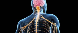
The central nervous system is the connecting link between all human organs and is responsible for their coordinated functioning. The central nervous system is responsible for the sensitivity and motor activity of the body. Problems in the functioning of the central nervous system cause disruption of the productive functioning of systems and organs. Most vascular diseases are associated with cerebrovascular accidents. They often lead to disability. This class of diseases is the greatest problem of modern medicine.
For the brain to function, it needs a lot of blood (up to 14% of all circulating in the body) and oxygen (up to 20%). Failure of blood supply provokes great difficulties in the functioning of the brain, and a complete break for 5-8 minutes. causes irreversible changes. At normal systolic pressure (60-180 mm Hg), blood flow is stable. With pathologies (high blood pressure, head injury, tumors) and other problems, problems with blood supply begin.
Vascular diseases of the brain include:
- Chronic cerebrovascular insufficiency, including encephalopathy.
- Acute cerebral circulatory disorders (transient, as well as two types of strokes).
- Anomalies of cerebral vessels (aneurysms, disorders of venous and spinal circulation).
Vascular diseases of the central nervous system can be provoked by diabetes mellitus, cervical osteochondrosis, hypertension, and atherosclerosis.
Diseases of the central nervous system
Meningitis - an inflammatory process of the membranes of the brain develops. General infectious symptoms are observed (chills, hyperthermia, changes in peripheral blood, and others) and meningeal syndrome, which includes general hyperesthesia, photo- and phonophobia, stiff neck (when flexing the neck), and specific neurological symptoms.
There is meningism - clinically manifested as meningitis, but irritation of the meninges is caused by non-infectious factors.
Encephalitis is a group of inflammatory diseases of the brain. Causes: viruses, other infectious agents (for example, mycoplasma, rickettsia), allergic factors. The clinical picture includes general infectious manifestations, focal neurological symptoms (loss of functions, paresis, aphasia and other manifestations).
Myelitis is an inflammatory lesion of the spinal cord. Specific symptoms of myelitis include motor and pelvic disorders, radicular pain, paresthesia in the limbs and body, and other sensory disorders.
Recently, laboratory tests for infections often reveal infection with Epstein-Barr viruses, cytomegaloviruses and other herpes viruses. The acute period of the disease can occur either without specific symptoms or in a mild form - fever, temperature, weakness, malaise, headaches, runny nose, skin rashes, swollen lymph nodes. That is, all the symptoms of acute respiratory viral infection (ARVI) are presented. In more severe cases, widespread damage to the nerves, brain and spinal cord, as well as internal organs develops, rashes on the lips, oral mucosa and genitals, on the skin, along the nerve trunks and roots. All this can be accompanied by severe pain, which lasts from several weeks to several months in the form of neuropathic pain.
There are many other infectious diseases of the central nervous system (poliomyelitis, neurosyphilis, toxoplasmosis, parasitic diseases of the central nervous system and others).
Autoimmune diseases of the central nervous system - arise as a result of improper functioning of the immune system, which begins to consider the cells of its own body as foreign. These include acute disseminated encephalomyelitis, neuromyelitis optica, multiple sclerosis, and optic neuritis. The most common of these is multiple sclerosis. A separate article on our website is dedicated to it.
Research in recent years has revealed a pattern between receiving certain vaccinations and increasing the likelihood of developing many autoimmune diseases.
Neurodegenerative diseases are slowly progressive diseases of the nervous system, accompanied by the slow death of nerve cells (neurodegeneration). These include Alzheimer's disease, Pick's disease, Parkinson's disease, dementia with Lewy bodies, multiple system atrophy, corticobasal degeneration and many others.
Prion diseases are a group of rare and severe neurodegenerative diseases (Creutzfeldt-Jakob disease, fatal familial insomnia, Gerstmann-Straussler-Scheinker syndrome and others). The causative agent is a pathologically altered protein capable of spontaneous aggregation. Aggregates of this protein accumulate inside and outside the neuron and cause tissue degeneration.
The clinical picture, depending on the characteristics of the lesion, may include a disorder of almost all functions of the nervous system - motor, sensory, cerebellar, extrapyramidal disorders, dementia (dementia).
Hereditary diseases of the central nervous system - many neurological diseases can be genetically determined or have a genetic predisposition. That is why our clinic sees a geneticist. You can read about neurogenetics in the section “Hereditary diseases of the nervous system - neurogenetics.”
Traumatic injuries to the spinal cord and brain - this section is described on our website under the title “consequences of injuries to the nervous system.”
How are NS pathologies diagnosed?
To identify and diagnose a vascular problem in the nervous system, a neurological examination is needed. The neurologist evaluates the following indicators: the patient’s intelligence and awareness, his ability to navigate reality and space, reflexes, etc.
Often, tests and hardware studies are necessary to establish and clarify the diagnosis. For this, patients are prescribed:
- CT;
- various types of ultrasound diagnostics;
- electroencephalography;
- X-ray examination;
- angiography;
- lumbar puncture;
- clinical blood tests;
- other diagnostic methods.
What methods are used?
By signing up for paid services in the neurology department, you get access to the most modern equipment and assistance from the highest class professionals.
Treatment can be carried out:
- medicinally;
- through lifestyle and nutrition correction;
- using physiotherapy methods;
- surgically.
Depending on what kind of disease the patient has, whether it is neurology, strokes, atherosclerosis or other malfunctions of the body, the most gentle and effective method is chosen.
The page is for informational purposes only. Find out the exact list of services provided and the specifics of the procedures by calling.
Symptoms of a stroke
- sudden numbness or weakening of the muscles of the face, arms or legs, often on one side of the body;
- sudden difficulties with articulation and perception of speech or text;
- a sharp violation of coordination of movements, the occurrence of unsteadiness of gait, dizziness;
- sudden deterioration of vision in one or both eyes;
- unexplained and sudden headache.
All these changes appear due to increased blood pressure. Therefore, call an ambulance immediately if you notice such symptoms.
Before the doctors arrive, put the patient to bed, give him an influx of fresh air and ensure complete rest.
Treatment of vegetative vascular dystonia
Physiotherapeutic methods are mainly used to treat the disease. This could be hydromassage, physical therapy. If the symptoms of vegetative-vascular dystonia are serious, acupuncture, manual therapy, and herbal medicine give good results. Vegetative-vascular dystonia responds well to treatment. The main thing is not to self-medicate. The choice of methods by which to treat vegetative vascular dystonia should be determined only by a doctor after conducting the necessary research. Often, VSD “masks” another disease, so its treatment should be aimed primarily at eliminating the underlying disease.
And, of course, first of all, don’t bring yourself to the point of needing to see a doctor: try to avoid stressful situations and emotional breakdowns, and, if possible, take a few days of rest and spend them as you would like for a long time.
Thus, treatment of vegetative vascular dystonia should be based on both drug therapy and regulation of the mental state.
Vascular diseases of the brain
– remain one of the most pressing health problems. In most industrialized countries, cerebrovascular diseases account for up to 15% of total mortality. Mortality from cerebrovascular accidents in Russia ranks third in the statistics of overall mortality. Mortality from cerebral strokes exceeds that from myocardial infarction by 2-3 times. They die more often in the acute period of stroke (up to 50% of patients). Only 20% of those who worked before the stroke remain able to work.
Classification of cerebrovascular disorders
■ A. Initial manifestations of circulatory failure:
- brain;
- spinal cord.
■ B. Transient cerebrovascular accidents:
- transient ischemic attacks (transient cerebrovascular accidents);
- hypertensive cerebral crises with cerebral and focal disorders;
■ B. Stroke:
- hemorrhagic stroke (in the brain and spinal cord): - subarachnoid hemorrhage (under the membranes of the brain and spinal cord); - parenchymal-subarachnoid hemorrhage; - ventricular-parenchymal-subarachnoid hemorrhage.
- ischemic stroke: - cerebral (with damage (occlusion) of the precerebral arteries, and with embolism of the cerebral arteries); - spinal (with compression of the vessels supplying the spinal cord in any part of the blood flow from the aorta to the intramedullary branches).
- stroke with restored neurological deficit - “minor stroke”;
- consequences of a previous stroke.
■ D. Progressive cerebrovascular accidents:
- chronic subdural hematoma;
- encephalopathy.
Etiology
. Cerebrovascular accidents can occur for many reasons. In 85-90% of cases, the main cause of their development is atherosclerosis, hypertension, or a combination of both. These disorders can also be caused by abnormalities of the cardiovascular system: saccular aneurysms and AVMs, aplasia and hypoplasia of cerebral vessels, Nishimoto-Takeuchi-Kudo disease (my-my), mitral valve prolapse, etc. A very important place in their occurrence belongs to arterial hypotension, lung diseases, infectious-allergic vasculitis, systemic connective tissue diseases, toxic lesions of cerebral vessels, and diabetes mellitus. Blood diseases, lesions and damage to brain vessels by bone formations and tumors play a certain role.
Pathogenesis
. The pathogenetic factors causing the development of ischemic strokes and transient cerebrovascular accidents are quite diverse. The main ones among them are the following.
Causes and types of stroke
Compression, blockage, or rupture of the blood vessels that supply blood to the brain cause a stroke. Most strokes are ischemic.
The mechanisms of development are similar to myocardial infarction. The atherosclerotic plaque, which is located in one of the arteries of the brain, is destroyed, and a blood clot appears in the area of damage - a thrombus that closes the lumen of the vessel.
Brain cells do not receive enough oxygen, and if neighboring arteries do not take over the blood supply to this area, then within a few minutes the cells die.
Less commonly, the lumen of the artery is blocked by drops of fat or air bubbles (with fat and air embolism), or blood clots pass from the left ventricle of the heart, or compression occurs from the outside due to injury or tumor.
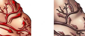
The cause of hemorrhagic stroke is hemorrhage. This form is less common, but is more life-threatening because the wall of the defective artery ruptures. The cause is an aneurysm, or the integrity of the vascular wall is disrupted, and these phenomena are provoked by an increase in blood pressure. Then the blood spills over the brain tissues, and the cells die from lack of oxygen, and the blood that spills begins to saturate and compress neighboring tissues, interfering with their vital functions.
Each area of the brain is responsible for a specific function - movement of the legs, arms, speech, vision, etc. Therefore, the outcome of a stroke depends on which area of the brain it occurs in. Perhaps this will be paralysis (the body is completely immobilized) or paresis (movements are partially impaired) of the leg or arm on the side opposite the lesion, serious impairment of speech and writing, memory impairment, and changes in sensitivity.
Neurology Clinic
Vascular diseases account for more than 40% of all pathologies of the nervous system; this is one of the most common diseases in neurology. Everyone is well aware of such terrible diseases as strokes and heart attacks, but these are only complications of long-term progressive vascular diseases of the brain. The most common causes of the development of progressive discirculatory encephalopathies are a combination of cerebral artery atherosclerosis and hypertension. Other possible causes are vasculitis in systemic inflammatory diseases, congenital anomalies of the structure of the heart, blood vessels, spine, venous disorders, pathology of the vertebral arteries, hereditary blood diseases, constitutional features of the autonomic nervous system with arterial hypotension and rarely other causes. Cerebrovascular insufficiency develops in stages. At stage 1
discirculatory encephalopathy is dominated by subjective unstable complaints of headaches, impaired performance, sometimes swaying and other mild nonspecific symptoms in the form of irritability, sleep disturbances.
At stage 2
symptoms become more pronounced and prolonged, objective symptoms in the form of crises are added, often against the background of an unstable increase in blood pressure, loss of coordination, changes in reflexes and other symptoms.
At stage 3
There may be persistent organic manifestations - memory impairment, impaired coordination, transient cerebrovascular accidents, complications in the form of strokes and their consequences are possible.
Diagnosis
Diagnosis of cerebrovascular insufficiency is carried out by a neurologist. Methods such as collecting anamnesis, assessing cardiac hemodynamics - blood pressure, pulse and other indicators are used; cognitive (mental) activity is checked both during a conversation with the patient and with the help of specially developed questionnaires and test tasks with a quantitative assessment of the points scored.
Laboratory tests - cholesterol content and its fractions, blood glucose levels and other biochemical indicators help in assessing the general condition.
An important method for assessing cerebral circulation is Doppler ultrasound with color Doppler mapping of the vessels of the neck and brain. This method allows you to identify atherosclerotic changes in the arteries and evaluate blood flow in all main arteries of the brain.
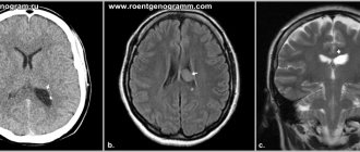
In modern conditions, it is also necessary to visualize the brain using magnetic resonance or computed tomography with angiography of the cerebral arteries, which will allow assessing structural changes in the brain and identifying the consequences of strokes.
At a late stage of the disease, multifocal vascular lesions of the brain are diagnosed on magnetic resonance imaging in the form of zones of atrophy and leukoaraiosis, which clinically is usually manifested by memory loss and impaired cognitive functions.
Treatment
Treatment methods for cerebrovascular insufficiency are varied; you just need to remember that vascular accidents are easier to prevent. Vascular, lipid-lowering, antihypertensive and metabolic therapy must be taken persistently and for a long time.
The most important role is played by lifestyle modification - quitting smoking, rational sports activities, diet and sleep.
Be healthy!
Symptoms of vascular diseases of the brain
Among the first signs of the disease are chronic headaches, memory loss, decreased hearing and vision, impaired movement, and dizziness.
Discirculatory encephalopathy, that is, a functional disorder of the brain that causes poor blood circulation, is characterized by special symptoms. In the first stages of the disease, the patient experiences emotional instability, chronic fatigue, irritation, dullness of attentiveness, headaches after overexertion (not only physical, but also mental). As a rule, symptoms are more pronounced in the evenings. In the later stages of dyscirculatory encephalopathy, the patient ceases to adequately assess his own condition, feels unsure of his abilities, and loses the ability to think in abstract concepts. At the same time, energy depletion occurs, depriving a person of the opportunity to independently cope with everyday tasks, opting for simpler actions. Hypochondria, a panicky fear of getting sick, is often noted.
Clinical manifestations of vascular diseases of the brain can be divided into the following forms:
- chronic cerebral ischemia stage I-II;
- cerebral circulatory disorder of a transient nature;
- cerebral infarction (ischemic stroke);
- bleeding in the brain (hemorrhagic stroke).
Cerebrovascular insufficiency: clinical picture, diagnosis and therapy
Stroke and chronic forms of cerebrovascular insufficiency represent one of the most pressing problems of modern neurology. According to epidemiological data, the incidence of stroke in the world is 150 cases per 100 thousand population per year. Chronic insufficiency of blood supply to the brain is also very widespread.
In the domestic literature, the term “dyscirculatory encephalopathy” (DE) is usually used to denote the clinical syndrome of brain damage resulting from insufficient blood supply to the brain. According to the classification of vascular diseases of the brain proposed by E.V. Schmidt (1985), dyscirculatory encephalopathy refers to chronic disorders of cerebral circulation.
Vascular diseases of the brain (E.V. Schmidt et al., 1985)
- Acute cerebrovascular accidents
*Stroke– Ischemic stroke (thrombotic, embolic, hemodynamic, lacunar)
– Hemorrhagic stroke (parenchymal hemorrhage, subarachnoid hemorrhage)
*Transient cerebrovascular accidents
– Transient ischemic attacks
– Hypertensive cerebral crises
- Chronic cerebrovascular accidents
*Initial manifestations of insufficient blood supply to the brain*Encephalopathy
However, as modern research shows, various cardiovascular diseases, as a rule, lead simultaneously to chronic cerebral ischemia and repeated acute cerebrovascular accidents. Therefore, it would be more correct to define discirculatory encephalopathy as a syndrome of chronic progressive brain damage, which is based on repeated strokes and/or chronic insufficiency of blood supply to the brain (N. N. Yakhno, I. V. Damulin, 2001).
Etiology and pathogenesis of DE
The most common causes of impaired blood supply to the brain are atherosclerosis of the main arteries of the head, heart disease with a high risk of thromboembolism in the brain, and hypertension. Less commonly, cerebrovascular accidents develop as a result of inflammatory changes in blood vessels (vasculitis), disorders of the blood coagulation system, abnormalities in vascular development, etc. In the vast majority of cases, cerebral vascular insufficiency develops in elderly people suffering from the above cardiovascular diseases.
As follows from the definition of DE, 2 main pathogenetic mechanisms play a role in the formation of this syndrome: stroke and chronic cerebral ischemia. Ischemic strokes of the brain develop as a result of thrombosis of cerebral arteries, thromboembolism in the brain, arteriolosclerosis, rheological and hemodynamic disorders.
Chronic cerebral ischemia is based on structural changes in the vascular wall, which arise as a consequence of prolonged arterial hypertension or an atherosclerotic process. It has been established that lipohyalinosis of small-caliber vessels penetrating the brain substance can lead to chronic ischemia of the deep white matter. A reflection of this process are changes in the white matter (leukoaraiosis), which are defined as focal or diffuse changes in signal intensity from deep cerebral structures on T2-weighted images of magnetic resonance imaging of the brain. These abnormalities are considered to be typical neuroimaging symptoms that develop in patients with long-term uncontrolled arterial hypertension.
Clinical manifestations of DE
The clinical picture of DE is highly variable. As mentioned above, most patients with chronic vascular diseases of the brain have a history of strokes, often repeated. The localization of strokes undoubtedly largely determines the characteristics of the clinic. However, in the overwhelming majority of cases with cerebrovascular pathology, along with the consequences of strokes, there are also neurological, emotional and cognitive symptoms of dysfunction of the frontal lobes of the brain. This symptomatology develops as a result of disruption of connections between the frontal cortex and the subcortical basal ganglia (the “disconnection” phenomenon). The reason for the “disconnection” lies in diffuse changes in the white matter of the brain, which, as mentioned above, are a consequence of the pathology of small-caliber cerebral vessels.
Depending on the severity of the disorders, it is customary to distinguish 3 stages of dyscirculatory encephalopathy. The first stage is characterized mainly by subjective neurological symptoms. Patients complain of headache, dizziness, heaviness or noise in the head, sleep disorders, increased fatigue during physical and mental stress. These symptoms are based on a mild to moderate decrease in mood associated with dysfunction of the frontal lobes of the brain. Objectively, mild impairments of memory and attention, as well as possibly other cognitive functions, are detected. There may be an asymmetrical increase in tendon reflexes, uncertainty when performing coordination tests, and slight changes in gait. Instrumental research methods that make it possible to detect the pathology of cerebral vessels are important in the diagnosis of cerebral vascular insufficiency at this stage of the pathological process.
The second stage of discirculatory encephalopathy is spoken of in cases where neurological or mental disorders form a clinically defined syndrome. For example, we may be talking about mild cognitive impairment syndrome. This diagnosis is legitimate in cases where impairments in memory and other cognitive functions clearly go beyond the age norm, but do not reach the severity of dementia. At the second stage of DE, neurological disorders such as pseudobulbar syndrome, central tetraparesis, usually asymmetrical, extrapyramidal disorders in the form of hypokinesia, mild or moderate increase in muscle tone of the plastic type, ataxic syndrome, neurological disorders of urination, etc. can also develop.
At the third stage of dyscirculatory encephalopathy, a combination of several of the above neurological syndromes is noted and, as a rule, vascular dementia is present. Vascular dementia is one of the most severe complications that develops with the unfavorable course of cerebrovascular insufficiency. According to statistics, vascular etiology underlies at least 10–15% of dementia in old age.
Vascular dementia, like DE in general, is a pathogenetically heterogeneous condition. Vascular dementia is possible after a single stroke in a strategic area of the brain for cognitive activity. For example, dementia can develop acutely as a result of a heart attack or hemorrhage in the thalamus. However, much more often vascular dementia is caused by repeated strokes (so-called multi-infarct dementia). Another pathogenetic mechanism of vascular dementia is chronic cerebral ischemia, reflected by changes in the white matter of the brain. Finally, in addition to cerebral ischemia and hypoxia, secondary neurodegenerative changes play an important role in the pathogenesis of dementia in cerebrovascular insufficiency, at least in some patients with DE. Modern research has convincingly proven that insufficient blood supply to the brain is a significant risk factor for the development of degenerative diseases of the central nervous system, in particular Alzheimer's disease. The addition of secondary neurodegenerative changes undoubtedly aggravates and modifies cognitive disorders in cerebrovascular insufficiency. In such cases, the diagnosis of mixed (vascular-degenerative) dementia is legitimate.
Clinical manifestations of vascular dementia in each specific case depend on the pathogenetic mechanisms determining the disease. In post-stroke and multi-infarction dementia, clinical features depend on the location of the strokes. Changes in the white matter of the deep lobes of the brain as a result of chronic ischemia lead to cognitive impairment of the “frontal” type. These disorders are characterized by emotional disorders in the form of decreased mood, depression or apathy, and loss of interest in the environment. Emotional lability, which is a rapid, sometimes causeless change of mood, tearfulness or increased irritability, is also very characteristic. In the cognitive sphere, memory and attention impairments, slowness of thinking, decreased intellectual flexibility, and difficulties associated with switching from one type of activity to another are determined. The behavior of patients changes: the ability for self-criticism and the sense of distance are reduced, increased impulsiveness and distractibility are noted, symptoms such as disregard for socially accepted rules of behavior, asociality, foolishness, flat and inappropriate humor, etc. may be present.
The presence of secondary neurodegenerative changes in vascular dementia is manifested primarily by progressive memory impairment. At the same time, to a greater extent, the patient forgets what happened recently, while memories of distant events are retained for quite a long time. The neurodegenerative process is also characterized by disturbances in spatial orientation and speech.
Diagnosis of dyscirculatory encephalopathy
To diagnose dyscirculatory encephalopathy syndrome, it is necessary to carefully study the medical history, assess the neurological status, and use neuropsychological and instrumental research methods. It is important to emphasize that the presence of cardiovascular diseases in an elderly person does not in itself serve as evidence of the presence of cerebral vascular insufficiency. A necessary condition for correct diagnosis is to obtain convincing evidence of a cause-and-effect relationship between neurological and cognitive symptoms and cerebrovascular pathology, which is reflected in the currently accepted diagnostic criteria for DE.
Diagnostic criteria for DE (N. N. Yakhno, I. V. Damulin, 2001)
- Presence of signs (clinical, anamnestic, instrumental) of brain damage.
- Presence of signs of acute or chronic cerebral dyscirculation (clinical, anamnestic, instrumental).
- The presence of a cause-and-effect relationship between hemodynamic disorders and the development of clinical, neuropsychological, and psychiatric symptoms.
- Clinical and paraclinical signs of progression of cerebrovascular insufficiency.
Evidence of a vascular etiology for symptoms would include the presence of focal neurological symptoms, a history of stroke, characteristic neuroimaging changes such as post-ischemic cysts or marked white matter changes.
Treatment of cerebrovascular insufficiency
Cerebrovascular insufficiency is a complication of various cardiovascular diseases. Therefore, etiotropic therapy for DE should be, first of all, aimed at the underlying pathological processes of cerebrovascular insufficiency, such as arterial hypertension, atherosclerosis of the main arteries of the head, heart disease, etc.
Antihypertensive therapy is an essential factor in the secondary prevention of the increase in mental and motor symptoms of cerebrovascular insufficiency. To date, however, the question of what blood pressure levels should be achieved in the treatment of hypertension has not been resolved. Most neurologists believe that complete normalization of blood pressure in elderly patients with a long history of hypertension, while reducing the risk of acute vascular episodes, can simultaneously contribute to the aggravation of chronic cerebral ischemia and an increase in the severity of cognitive dysfunction of the “frontal” type.
The presence of hemodynamically significant atherosclerosis of the main arteries of the head requires the prescription of antiplatelet agents. Drugs with proven antiplatelet activity include acetylsalicylic acid at doses of 75-300 mg per day and clopidogrel (Plavix) at a dose of 75 mg per day. The study showed that the use of these drugs reduces the risk of ischemic events (myocardial infarction, ischemic stroke, peripheral thrombosis) by 20-25%. The possibility of simultaneous use of these drugs has now been proven. Medicines with antiplatelet properties also include dipyridamole (chimes), which is used in doses of 25 mg three times a day. Monotherapy with this drug does not provide prevention of cerebral or other ischemia, however, with combined use, dipyridamole significantly increases the preventive effect of acetylsalicylic acid. In addition to the prescription of antiplatelet agents, the presence of atherosclerotic stenosis of the main arteries of the head requires referral of the patient for consultation with a vascular surgeon to decide on the advisability of surgical intervention.
If there is a high risk of thromboembolism in the brain, for example in cases of atrial fibrillation and valvular disease, antiplatelet agents may be ineffective. The listed conditions serve as an indication for the prescription of indirect anticoagulants. The drug of choice is warfarin. Therapy with indirect anticoagulants should be carried out under strict monitoring of coagulogram parameters.
The presence of hyperlipidemia that cannot be corrected by diet requires the prescription of lipid-lowering drugs. The most promising drugs are from the statin group (Zocor, Simvor, Simgal, Rovacor, Medostatin, Mevacor, etc.). According to some data, therapy with these drugs not only normalizes lipid metabolism, but also may have a preventive effect against the development of a secondary neurodegenerative process against the background of cerebrovascular insufficiency.
An important pathogenetic event is also the impact on other known risk factors for cerebral ischemia. These include smoking, diabetes, obesity, physical inactivity, etc.
In the presence of cerebrovascular insufficiency, the prescription of drugs that act primarily on the microvasculature is pathogenetically justified. These include:
- phosphodiesterase inhibitors: aminophylline, pentoxifylline, vinpocetine, tanakan, etc. The vasodilating effect of these drugs is associated with an increase in the cAMP content in the smooth muscle cells of the vascular wall, which leads to their relaxation and an increase in the lumen of blood vessels;
- calcium channel blockers: cinnarizine, flunarizine, nimodipine. They have a vasodilating effect due to a decrease in the intracellular calcium content in the smooth muscle cells of the vascular wall. Clinical experience suggests that calcium channel blockers, such as cinnarizine and flunarizine, may be more effective for vertebrobasilar circulatory failure; this is manifested by symptoms such as dizziness and unsteadiness when walking;
- α2-adrenergic receptor blockers: nicergoline. This drug eliminates the vasoconstrictor effect of the mediators of the sympathetic nervous system: adrenaline and norepinephrine.
Vasoactive drugs are among the most commonly prescribed drugs in neurological practice. In addition to the vasodilatory effect, many of them also have positive metabolic effects, which allows these drugs to be used as symptomatic nootropic therapy. Experimental data indicate that the vasoactive drug tanakan has the ability to deactivate free radicals, thereby reducing the processes of lipid peroxidation. The antioxidant properties of this drug also allow it to be used for secondary prevention of the increase in memory impairment and other cognitive functions in cases of secondary neurodegenerative changes.
In domestic practice, vasoactive drugs are usually prescribed in courses of 2-3 months, 1-2 times a year.
Metabolic therapy is widely used for cerebrovascular insufficiency, the purpose of which is to stimulate the reparative processes of the brain associated with neuronal plasticity. In addition, metabolic drugs have a symptomatic nootropic effect.
Piracetam was the first drug specifically synthesized to affect memory and other higher brain functions. In recent years, however, it has been possible to prove that in previously taken doses this drug has a relatively small clinical effect. Therefore, the use of piracetam in dosages of at least 4–12 g/day is currently recommended. Intravenous administration of this drug in saline solution is more appropriate: 20–60 ml of piracetam per 200 ml of saline solution intravenously, 10–20 infusions per course.
The peptidergic drug Cerebrolysin is no less successfully used for cerebrovascular insufficiency, as well as vascular and degenerative dementia. As in the case of piracetam, views on the dosage regimen of this drug have changed significantly in recent years. According to modern concepts, the clinical effect occurs in the case of intravenous administration of Cerebrolysin in doses of 30–60 ml intravenously in 200 ml of saline, 10–20 infusions per course.
Peptidergic drugs that have a beneficial effect on cerebral metabolism also include Actovegin. Actovegin is used in the form of intravenous infusions (250–500 ml per infusion, 10–20 infusions per course), or in the form of intravenous or intramuscular injections of 2–5 ml 10–20 injections, or orally 200–400 mg 3 times a day within 2-3 months.
Like vasoactive drugs, metabolic therapy is carried out in courses 1-2 times a year. Pathogenetically justified and appropriate is the combined implementation of vasoactive and metabolic therapy. Currently, the doctor has several combined dosage forms at his disposal, which include active substances with vasoactive and metabolic effects. These drugs include instenon, vinpotropil, fezam and some others.
The development of vascular dementia syndrome requires more intensive nootropic therapy. Among modern nootropic drugs, acetylcholinesterase inhibitors have the most powerful clinical effect on cognitive functions. Initially, drugs in this group were used in the treatment of mild and moderate dementia due to Alzheimer's disease. Today it has been proven that acetylcholinergic deficiency plays an important pathogenetic role not only in this disease, but also in vascular and mixed dementia. Therefore, cognitive disorders of vascular and mixed etiology are increasingly appearing among the indications for the prescription of acetylcholinesterase inhibitors.
In Russia, two drugs from the group of latest generation acetylcholinesterase inhibitors are currently available: Exelon and Reminyl. Exelon is prescribed at an initial dose of 1.5 mg 2 times a day, then the single dose is increased by 1.5 mg every 2 weeks. up to 6.0 mg 2 times daily or until side effects occur. Common side effects when using Exelon are nausea and vomiting. These phenomena do not pose a threat to the life or health of the patient, but may interfere with the achievement of a therapeutic effect. Reminyl is prescribed at 4 mg 2 times a day for the first 4 weeks, and then 8 mg 2 times a day. This drug is less likely to cause adverse events.
First generation acetylcholinesterase inhibitors include neuromidin. According to some data, this drug has a positive nootropic effect in both vascular and primary degenerative and mixed dementia. It is prescribed in a dose of 20–40 mg 2 times a day.
Therapy with acetylcholinesterase inhibitors should be carried out continuously. In this case, it is necessary to monitor the level of liver enzymes in the blood once every 3–6 months.
The use of akatinol memantine is also pathogenetically justified for vascular dementia. This drug is an inhibitor of NMDA glutamate receptors. Chronic intake of akatinol memantine has a symptomatic nootropic effect, and may also slow down the rate of increase in cognitive disorders. The effect of the drug was manifested in both mild and moderate and severe dementia. It should be noted that akatinol memantine is the only drug effective at the stage of severe dementia. It is prescribed during the first week 5 mg 1 time per day, during the second week - 5 mg 2 times a day, starting from the third week and then continuously - 10 mg 2 times a day.
In conclusion, it should be emphasized that a comprehensive assessment of the state of the cardiovascular system of patients with cerebrovascular insufficiency, as well as the impact on both the cause of the disorders and the main symptoms of DE, undoubtedly contribute to improving the quality of life of patients and preventing severe complications of cerebrovascular insufficiency, such as , such as vascular dementia and movement disorders.
V.V. Zakharov, Doctor of Medical Sciences Clinic of Nervous Diseases named after. A. Ya. Kozhevnikova, Moscow
Diagnosis of stroke
First, doctors determine the type of stroke. To do this, magnetic resonance imaging of the brain (MRI) is performed, which allows one to distinguish between hemorrhagic and ischemic strokes and exclude other diseases, and also establishes the exact location and size of the affected area of the brain.
Then an ultrasound of the vessels of the neck and brain is performed along with duplex scanning, as well as cerebral angiography and echocardiography. An additional examination method is Holter monitoring, that is, an electrocardiogram is recorded throughout the day.
In case of hemorrhagic stroke, in order to find out the causes of hemorrhage, it is necessary to perform cerebral angiography and Dopplerography of the cerebral vessels. If necessary, a consultation with a neurosurgeon is carried out.