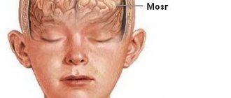
Intracranial hematoma is a pathological condition characterized by the accumulation of blood in the cranial cavity. Its danger lies in the fact that it can put pressure on, displace and damage nearby brain structures, causing them serious harm. The reasons for its appearance are traumatic injuries to the blood vessels of the brain or hemorrhages.
The Neurology Department of CELT invites you to undergo diagnostics and treatment of intracranial hematomas in Moscow. We employ leading domestic specialists who will accurately diagnose and select adequate treatment, which will certainly give the desired results. If you want to restore your health and get rid of severe symptoms, contact us and undergo treatment in accordance with international standards.
Causes
The main causes of this pathology are:
- stroke - in this case, rupture of blood vessels occurs due to increased intracranial pressure;
- head injuries;
- brain abscess - usually develops when an infection enters the meninges;
- vascular diseases;
- pathologies associated with blood clotting disorders;
- brain tumors;
- uncontrolled use of certain medications (for example, anticoagulants);
- addiction.
Symptoms of internal bleeding
How to recognize internal bleeding?
There are a number of signs that are characteristic of all types of bleeding in the early stages. This:
- general weakness;
- drowsiness;
- dizziness;
- cold sweat;
- pallor of the skin and mucous membranes;
- darkening of the eyes;
- thirst.
For low blood loss
Often there is a slight increase in heart rate and a slight decrease in blood pressure. However, such internal bleeding may sometimes not be accompanied by any symptoms at all.
For moderate internal bleeding
systolic pressure drops to 80-90 mmHg. Art., the pulse quickens to 90-100 beats per minute. The skin becomes pale, the extremities become cold, and increased breathing may occur. Also in such a situation, dry mouth, fainting and dizziness, adynamia, nausea, slow reaction, and weakness are possible.
In severe cases
systolic pressure drops to 80 mm Hg. Art. and even lower, the pulse increases to 110 beats per minute and above. Sticky cold sweat appears on the body, there is a strong increase in breathing, yawning, nausea and vomiting, apathy, hand tremors, increased drowsiness, painful thirst, a decrease in the amount of urine excreted, severe pallor of the skin and mucous membranes.
With massive internal bleeding
systolic pressure decreases to 60 mm Hg. Art., the pulse rate can reach 140-160 beats per minute. Characteristic features of massive internal bleeding are periodic breathing (Cheyne-Stokes breathing), delirium, confusion or lack thereof, cold sweat, severe pale skin. The gaze becomes indifferent, the facial features become sharpened, the eyes become sunken.
In case of fatal blood loss
systolic pressure decreases to 60 mm Hg. Art. or is not determined at all. Coma develops, breathing becomes agonal, severe bradycardia, convulsions, dilated pupils, and involuntary release of feces and urine are observed. The skin becomes dry and cold, acquiring a characteristic “marble” shade.
Signs of internal bleeding also depend on which cavity the blood is bleeding into. For example, nausea and vomiting of dark blood may indicate bleeding into the cavity of the stomach or esophagus; coughing with bright pulmonary blood is an unambiguous sign of pulmonary hemorrhage.
If you notice similar symptoms, consult a doctor immediately. It is easier to prevent a disease than to deal with the consequences.
Symptoms of intracranial hemorrhage
In most cases, cerebral hemorrhage occurs suddenly, often during the daytime. The clinical picture depends on many factors (how severe the hemorrhage was, in what part of the brain it occurred, how quickly the hematoma formed).
The patient complains of a sudden severe headache, his general condition deteriorates sharply, nausea appears, and then vomiting. One of the most common symptoms of intracranial hemorrhage is seizures.
If the hemorrhage occurs in the deep parts of the brain, the patient may suddenly lose consciousness. He experiences unilateral paralysis, his gaze deviates towards the affected hemisphere of the brain, his blood pressure decreases, his temperature rises, and vomiting occurs.
In severe cases, the pupils stop responding to light, breathing becomes impaired, a deep coma may occur, with fading reflexes, which can lead to death.
If hemorrhage occurs in the gray and white matter, the patient quickly experiences convulsions, followed by loss of consciousness.
If blood enters the cerebellum, the patient experiences pain in the back of the head, which is accompanied by severe, uncontrollable vomiting and dizziness.
The patient's respiratory function is impaired, and his gaze is directed in the direction opposite to the affected hemisphere. Coma may develop.
Intracranial and intravertebral phlebitis and thrombophlebitis
Thrombosis - in case of disruption of the outflow of venous blood from the cranial cavity, venous stagnation develops. There are a number of causes of venous stagnation. Such causes may include heart failure, pulmonary failure, brain tumors, traumatic brain injury, compression of extracranial veins, compression of intracranial veins in the case of craniostenosis and cerebral hydrops, as well as thrombosis of the cerebral veins and dural sinuses. Thrombosis of the cerebral veins can occur against the background of their previous inflammation - thrombophlebitis.
CLINICAL PICTURE AND SYMPTOMS
Thrombosis of superficial cerebral veins
Thrombosis of the superficial veins of the brain in most cases is a complication of various pathological processes in the body, such as inflammation, infectious diseases, surgical interventions, traumatic brain injuries, etc. Sometimes it can be a complication of pregnancy, childbirth, or abortion. The leading role in the pathogenesis of cerebral vein thrombosis is played by a pathological change in their walls, a violation of blood clotting, and a slowdown in blood flow, which leads to the formation of blood clots. Quite often, thrombosis of the cerebral veins is combined with thrombosis of the dural sinuses, as well as with thrombosis of the veins of the lower extremity.
Common infectious symptoms include increased body temperature, changes in blood tests such as increased ESR and neutrophilic leukocytosis. When examining the cerebrospinal fluid, an increase in the amount of protein, slight pleocytosis is noted, and in some cases blood is detected. Quite often, the first manifestations of the disease are headache, nausea, and vomiting. Impaired consciousness may occur in combination with psychomotor agitation. As the disease progresses, focal brain symptoms appear. Focal brain symptoms include epileptic seizures, aphasia, alexia, hemianopsia, paresis and paralysis of the limbs, etc.
The symptoms of superficial vein thrombosis are explained by the fact that against the background of impaired blood outflow, hemorrhagic infarctions develop, localized in the gray and white matter of the brain. In addition, intracerebral and subarachnoid hemorrhages are observed. In most cases, thrombophlebitis of the superficial veins of the brain develops in the postpartum period. In this case, when examining the cerebrospinal fluid, its hemorrhagic nature is noted.
Thrombosis of deep cerebral veins and vein of Galen (great cerebral vein)
Clinically, this disease is characterized by a particularly severe course; in typical cases, the patient’s condition is comatose. During the examination, pronounced cerebral symptoms are noted. The stem and subcortical structures turn out to be functionally incapable. Diagnosing this disease while the patient is alive is extremely difficult. When making a diagnosis, it is necessary to pay attention to the appearance of general local brain symptoms that occur against the background of thrombophlebitis of the upper extremities. Brain symptoms can also occur in the presence of foci of inflammation in the body, which occurs in the postpartum period, with diseases of the paranasal sinuses, ears, etc. In some cases, the disease is characterized by a chronic course over many years.
Thrombosis of the dural sinuses
Often this pathology is formed as a result of the ingress of an infectious agent from a nearby focus, which can be purulent processes in the orbit, paranasal sinuses, purulent otitis, mastoiditis, osteomyelitis of the skull bones, as well as purulent lesions of the scalp and face (boils, carbuncles). The spread of the infectious agent occurs through the diploic and cerebral veins. Thrombosis of the cerebral sinuses can develop against the background of thrombophlebitis of the veins of the upper and lower extremities, as well as the pelvis. In this case, there is a hematogenous mechanism for the development of the disease.
In some cases, thrombosis of the cerebral sinuses is accompanied by thrombophlebitis of the retinal veins, purulent meningitis, and brain abscess. Often, thrombosis of the cerebral sinuses develops in people suffering from tuberculosis, malignant neoplasms, and other diseases in which cachexia develops.
The clinical picture of venous sinus thrombosis is characterized by an increase in body temperature, a sharp headache, accompanied by vomiting. Blood changes in the form of leukocytosis are observed. Symptoms characteristic of increased intracranial pressure are noted. Symptoms of damage to the cranial nerves appear. Patients may be restless and may experience epileptic seizures. In some cases, patients, on the contrary, become drowsy, apathetic, lethargic. Focal symptoms in each specific case correspond to the location of the affected venous sinus.
Thrombosis of the sigmoid sinus
This pathology is the most common. Typically, thrombosis of this sinus develops as a complication of mastoiditis or purulent otitis. The appearance of headache and bradycardia is characteristic. Sometimes patients complain of diplopia. Body temperature rises to high levels, chills appear. The patient's condition may become soporous or comatose. Consciousness is impaired, which is manifested by delirium and agitation. On examination, there is a tilt of the head towards the pathological focus, as well as swelling of the tissue in the mastoid area. When palpated in this area, pain is determined. In some cases, the pathological process may spread to the jugular veins. In this case, symptoms of damage to the glossopharyngeal, vagus and hypoglossal nerves are added to the clinical picture.
Cavernous sinus thrombosis
Thrombosis of the cavernous sinus in most cases develops as a consequence of a septic condition with purulent diseases of the face, ear, paranasal sinuses and orbit. During examination, pay attention to emerging signs of venous stagnation, such as exophthalmos, venous hyperemia of the eyelids and their swelling, swelling of the periorbital tissues, chemosis, congestion in the fundus, and possible atrophy of the optic nerves. Symptoms of damage to the oculomotor nerves usually occur, which is manifested by external ophthalmoplegia. The superior branch of the trigeminal nerve is involved in the pathological process, which is clinically manifested by pain and hyperesthesia in the area of its innervation. Cavernous sinus thrombosis can be either unilateral or bilateral. With bilateral damage, the pathological process can spread to the adjacent sinus.
Thrombosis of the superior longitudinal sinus
The clinical manifestations of this disease depend on its etiology, on the rate of thrombus formation, on its localization in the superior longitudinal sinus, on the degree of involvement of the veins that flow into it into the pathological process. Thrombosis of this sinus can be septic and aseptic in nature. Thrombosis of the superior longitudinal sinus is most severe. When examining the patient, tortuosity of the veins of the temples, forehead, crown, root of the nose, as well as the eyelids is noted, which is associated with their overflow with blood. In addition to congestion and tortuosity of the veins, swelling of the above areas is noted. A characteristic feature of this pathology is the appearance of frequent nosebleeds. On percussion, pain is noted in the parasagittal region. There is an increase in intracranial pressure, and convulsive seizures develop. In some cases, lower paraplegia may occur in combination with urinary incontinence. The above symptoms add up to a general neurological syndrome.
TREATMENT
Treatment should be carried out exclusively by a neurologist. Self-medication is unacceptable.
A necessary condition is the sanitation of the source of infection, which led to the development of thrombosis of the cerebral veins and sinuses. In case of thrombosis or inflammation of the sigmoid sinus of a purulent nature, immediate surgical intervention is necessary. The scope of the operation includes opening and drainage of the area of the primary lesion, as well as opening of the sigmoid sinus and removal of the blood clot.
If thrombosis is complicated by the development of a brain abscess, which is most often localized in the temporal lobe and in the cerebellum, it is necessary to open and drain the abscess cavity. In the case of treatment of cavernous sinus thrombosis, in order to achieve a positive effect, it is necessary to open ulcers localized on the face, in the orbit, paranasal sinuses, etc.
Treatment
The main treatment for intracranial hemorrhage is surgery. The choice of surgical approach and surgical technique is made by the surgeon, depending on the nature, size and location of the hematoma.
After surgery, the patient is prescribed anticonvulsants to relieve or prevent post-traumatic seizures. Cases have been recorded when a patient's seizures began a year or more after the successful completion of the operation.
For some time after the operation, side effects such as impaired attention, headaches, and partial memory loss are possible. The rehabilitation period after intracranial hemorrhage is quite long. In adult patients, it is at least six months; in children, recovery is faster.
Good health to you!
- Non-traumatic extradural hemorrhage.
- Acute traumatic epidural hematoma.
- Subdural hemorrhage (acute) non-traumatic.
- Chronic traumatic subdural hematoma.
Treatment of internal bleeding
Treatment of internal bleeding is carried out in a hospital; the choice of department depends on the source of internal blood loss: it can be the department of general surgery, gynecology, neurosurgery, and so on.
In this case, specialists solve three problems:
- stopping internal bleeding;
- compensation for blood loss;
- improvement of microcirculation.
In some cases, in order to stop the bleeding, it is enough to cauterize the bleeding area, but most often surgical intervention under anesthesia is necessary. The specific actions of the surgeon, again, depend on the type of internal bleeding.
This article is posted for educational purposes only and does not constitute scientific material or professional medical advice.
Incidence (per 100,000 people)
| Men | Women | |||||||||||||
| Age, years | 0-1 | 1-3 | 3-14 | 14-25 | 25-40 | 40-60 | 60 + | 0-1 | 1-3 | 3-14 | 14-25 | 25-40 | 40-60 | 60 + |
| Number of sick people | 0 | 0 | 1.01 | 14.2 | 14.2 | 21.7 | 26.5 | 0 | 0 | 1.51 | 10.3 | 10.3 | 15.7 | 21.5 |
Symptoms
| Occurrence (how often a symptom occurs in a given disease) | |
| Sudden intense headache | 100% |
| Fainting (loss of consciousness) | 100% |
| Increased blood pressure (high blood pressure, hypertension, arterial hypertension) | 90% |
| Vomiting of various types, including indomitable | 90% |
| Weakness in one side of the body | 90% |
| Numbness of one half of the body | 70% |
| Restless behavior | 50% |
| Attacks of seizures with or without loss of consciousness (convulsions, convulsive seizures, convulsions, convulsions) | 50% |
| Memory loss (memory impairment, poor memory, memory impairment, forgetfulness) | 50% |
| Daytime sleepiness | 40% |
Etiology of intracranial hematomas
Intracranial hematomas are formed as a result of traumatic injuries or can be non-traumatic. Experts identify the following factors that can initiate their appearance:
- Ruptures of blood vessels due to traumatic brain injury;
- Bleeding that occurs after injury;
- Abnormal changes in blood characteristics during blood diseases;
- Diseases of the blood vessels of the brain;
- Destruction of the walls of blood vessels with a decrease in their plasticity, brain tumors, increased intravascular and blood pressure.
The hematoma itself can be formed by liquid or coagulated blood in a volume of one to one hundred milliliters. It puts pressure on the structures located around it, damages them and can cause them to die, and can also lead to swelling of the brain.