Benign intracranial hypertension syndrome in children: diagnosis and treatment
A lot of controversy arises around the so-called intracranial hypertension syndrome.
Someone says that such a condition does not exist and doctors are allegedly engaged in pseudo-treatment, deceiving parents. Most often, this misinformation is carried out by doctors of related specialties themselves, and sometimes even directly by some pediatric neurologists. The purpose of this short article is to explain in an accessible form what this mysterious, ambiguous condition is - intracranial hypertension syndrome? To do this, I will rely primarily on scientific articles, the results of various studies, as well as my personal experience as a practicing neurologist at a medical center with almost 10 years of medical experience.
Symptoms of pathology
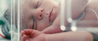
We often have to listen to parents complain that their child has such unhealthy signs as:
- tearfulness;
- restless sleep;
- decreased appetite;
- irritability.
Of course, these manifestations are not characteristic only of intracranial hypertension syndrome (ICH). Almost any illness can cause these symptoms. One can say even more, it is impossible to reliably diagnose ICH syndrome only on the basis of patient complaints. Any pain syndromes in children tend to generalize. Also, headaches in children, especially in the first years of life, are manifested by general reactions similar to the complaints described above, which complicates topical diagnosis.
Examination of the child is very important for a high-quality diagnosis of pathology. First of all, you should pay attention to such additional signs as:
- Enlarged, rounded head shape.
- Swelling and pulsation of the large fontanelle.
- Expansion of the vascular network at the temples, bridge of the nose, and around the eyes.
- Neck muscle tension.
- Graefe's symptom (manifestation of sclera between the upper eyelid and the iris of the eye, exophthalmos).
The baby's behavior also changes. He has a desire to lie on a cold surface, putting his head against it, he can touch, hit his head with his hand or a toy, both his own and someone else’s, hit his head on the floor or wall, or simply scratch behind the ear. Often, intracranial hypertension syndrome can be accompanied by nosebleeds.
Features of the disease
Information about the child’s life history, illness, and heredity is important for a specialist. The main cause of the development of ICH syndrome is perinatal hypoxic-ischemic damage to the central nervous system (CNS). Children with a burdened obstetric history are at risk for the development of intracranial hypertension. The course of ICH syndrome is chronic, with periods of exacerbation followed by periods of remission. Like any chronic disease, there is no point in trying to cure it completely. The main task of the doctor is to transfer the course of the disease to the stage of remission.
It should be understood that in the remission stage such a child is no different from a healthy peer. He is also well developed in psycho-speech and motor development. If this is not the case, then it is a different condition, but it is important to know that ICH syndrome does not cause delays in the development of the child. In medical practice, there are cases of the spread of ICH syndrome in one family (among siblings and cousins).
Diagnosis of the disease
In terms of instrumental confirmation of ICH in children, nowadays there are many neuroimaging methods, in particular:
- neurosonography (number 1 for children of the first year of life);
- computed tomography (CT);
- magnetic resonance imaging (MRI).
It is worth noting that none of these methods can directly determine the amount of intracranial pressure, but they can show indirect signs (expansion of the liquor spaces, dilation of the veins and venous sinuses of the brain). Most authors agree that for a reliable diagnosis of benign intracranial hypertension, a child needs a combination of three of the following four criteria:
- Increase in head circumference.
- The presence of ventriculomegaly (enlargement of the ventricles of the brain).
- Headaches or their equivalent in children under 3 years of age.
- Complicated obstetric history.
There are other syndromes with similar manifestations that require a different approach in terms of treatment and diagnosis. These include: the consequences of traumatic brain injury or neuroinfections, intracranial tumors and hematomas, the initial stage of occlusive hydrocephalus, which we will not dwell on in this article.
Treatment methods
So we found out that the child has ICH syndrome. What to do? First of all, don't panic! As noted above, developmental delays do not develop in children with this syndrome. The main problem is pain. To eliminate it, you should not use painkillers. It is necessary to dehydrate the body with diuretics. In particular, the drug Diacarb, by inhibiting the enzyme carbonic anhydrase, not only accelerates the excretion of cerebrospinal fluid, but also reduces its production. Do not try to prescribe medications to your child yourself! This should only be done by a qualified doctor. The inclusion of nootropic and vasoactive drugs in therapy increased the percentage of positive treatment outcomes in various studies.
Preventive measures
In addition to drug treatment, it is necessary to follow certain rules necessary to prevent exacerbation of ICH syndrome:
- Any blows, even minor ones, to the child’s head are undesirable.
- Correct sleep and wakefulness patterns.
- Do not allow your child to stay in noisy rooms for a long time.
- Limit water load in the evening.
- Do not walk for a long time in the sun, especially without a hat.
And, perhaps, the best information: the manifestations of ICH syndrome become less frequent with age and can often gradually disappear forever. That is why the incidence of benign intracranial hypertension in adults is extremely low.
Kabardiev Alimkhan Arslanovich, pediatric neurologist.
Works in a clinic of a medical network in the city. Khasavyurt at the address: st. Abubakarova, 9a.
Opening hours: daily, Monday to Saturday - from 13.00 to 17.00.
Phone numbers for inquiries and appointments: +7
The size of the ventricles is normal
In infants, the ventricles are often dilated. This condition does not at all mean that the child is seriously ill. The dimensions of each ventricle have specific values. These indicators are shown in the table.
First and second ventricles (lateral)
To assess normal indicators, the determination of all structural elements of the lateral ventricles is also used. The lateral cisterns should be less than 4 mm deep, the anterior horns between 2 and 4 mm, and the occipital horns between 10 and 15 mm.
Basic Concepts
Functional asymmetry is a property of nervous tissue, characterized by the distribution of mental functions between the hemispheres of the telencephalon. One of the main properties is not only the specialization of the right or left hemisphere, but their careful and precise interaction. This phenomenon is considered as a fundamental law of the functioning of the higher nervous system. Lateralization is inherent not only in humans, but also in animals.
At a time when neuroscience was underdeveloped, researchers believed that the two parts of the brain were identical. However, further study of the structure showed that there are many anatomical differences.
The phenomenon of asymmetry is explained by one genetic model, which states that there is a certain gene that is a factor in the “right shift”. If this gene is in a dominant position, then most likely the person will be born right-handed, and vice versa: the recessive position of the protein determines the left-handedness of the individual.
Provoking diseases
The main disease causing this pathology is hydrocephalus. It can interfere with the absorption of cerebrospinal fluid. This leads to its accumulation in the lateral ventricles.
Excessive formation of cerebrospinal fluid is also observed with serious lesions of the central nervous system. Poor circulation is also associated with the formation of cysts, tumors and other neoplasms.
A common cause of hydrocephalus is a defect of the Sylvian aqueduct. If this defect was discovered during the prenatal period, termination of pregnancy is recommended. At the birth of a child, complex systematic treatment will be required.
Another cause is aneurysm of the vein of Galev and Arnold-Chiari syndrome. However, in children, the disease can be caused by rickets or due to the specific structure of the skull, so observation by a specialist is important if there is a predisposition to the disease.
Types of lateroventriculoasymmetry
Cerebral asymmetry of the lateral ventricles is classified into two types:
- symptomatic - the development of pathology occurs due to a specific cause;
- idiopathic - the etiology of the disease is unknown.
In addition to types, the disease varies in severity:
- mild asymmetry - no therapy is required, since no serious deviations are observed;
- moderate lateroventriculoasymmetry - determined by a slight disturbance in the outflow of cerebrospinal fluid, a slight asymmetry of up to 15-17 mm is noticeable;
- severe (or dangerous) form - there is maximum dilatation of the ventricles on the left or right, as well as significant dysfunction of the outflow of cerebrospinal fluid.
With a strong degree of severity, complex symptoms, anomalies and pathology of the brain and nervous system are added to the asymmetry.
Lateroventriculoasymmetry is divided into types:
- isolated type - an increase in the volume of only the lateral ventricles;
- extensive - dilatation of different groups, for example, asymmetry of the anterior horns of the lateral ventricles (ALV);
- hereditary predisposition.
The pathology forecast is made after analyzing all types, forms, types, as well as measuring several altered parameters of the ventricles.
What it is
Ventriculomegaly is a change characterized by an increase in the size of the ventricles in the brain. As a result of the progression of this pathology, various brain ailments arise and neurological abnormalities appear.
Important! A timely undetected pathology, in addition to a negative effect on the central nervous system, rapidly worsens the functioning of the heart and provokes the development of abnormalities in the musculoskeletal system. There are 4 ventricles in the brain, 2 of them are located in the white matter, and are called lateral
The interventricular foramina serve as peculiar connecting elements between the third ventricle (located between the visual tuberosities), which is connected to the fourth via the cerebral aqueduct. The location of the latter is the bottom of the diamond-shaped dimple. It is attached to the central canal of the spinal cord
There are 4 ventricles in the brain, 2 of them are located in the white matter and are called lateral. The interventricular foramina serve as peculiar connecting elements between the third ventricle (located between the visual tuberosities), which is connected to the fourth via the cerebral aqueduct. The location of the latter is the bottom of the diamond-shaped dimple. It is attached to the central canal of the spinal cord.
The ventricular system is necessary for the production of cerebrospinal fluid, which, when pathology occurs, must enter the spinal canal. When its outflow is disrupted, ventriculomegaly develops. At the same time, the ventricles expand. Normally, the size of one of them should be 1-4 mm, and with the development of diseases - 12-20 mm. Find out what happens in the body of the pregnant woman and the fetus on 1, 2, 3, 4, 5, 6, 7, 8, 9 , 10, 11, 12, 13, 14, 15, 16, 17, 18, 19, 20, 21, 22, 23, 24, 25, 26, 27, 28, 29, 30, 31, 32, 33, 34 , 35, 36, 37, 38, 39, 40 weeks of pregnancy.
There is a treatment
Therapy is prescribed only for severe clinical signs. Only the doctor decides how to treat, based on the conclusion, as well as the individual characteristics of the patient.
A specific treatment regimen is selected, aimed directly at eliminating the cause of the pathology.
What is used:
- diuretics;
- nootropics;
- NSAIDs (anti-inflammatory);
- vasoactive substances;
- neuroprotectors;
- sedatives;
- in the presence of infectious diseases, antibacterial agents are used.
Additionally, massage, therapeutic exercises and physiotherapy are prescribed, which have a positive effect on the dynamics of recovery.
Some people prefer to see an osteopath - a specialist who treats without medication using palpation.
Dr. Komarovsky spoke in detail about the effectiveness of this technique, who devoted a series of programs to this topic.
Treatment methods
The main principle of treating this disease is timely identification and elimination of the cause that provoked this disease. When an infection occurs inside the womb, doctors are faced with the task of killing this infection with the help of drug therapy.
The goal of treatment is to eliminate the provoking factor and prevent possible complications and consequences that may appear during the course of the disease and cause irreparable harm, including death.
Since the pathology appears in utero, pharmaceutical treatment is intended for the expectant mother. Mild forms of the disease can be managed without medications; regular monitoring of the dynamics of development and constant monitoring by a doctor are sufficient.
For moderate and severe forms, treatment is prescribed immediately, including the following groups of drugs:
- vitamin complexes and minerals;
- antihypoxants in order to increase tissue resistance to lack of oxygen in the body;
- diuretics to remove excess fluid;
- nootropics that improve blood circulation;
- diuretics;
- To prevent neurological disorders, special agents are used that prevent potassium from being washed out of the body.
In combination with medications, a course of massage and physical therapy classes are prescribed. These measures are also prescribed for children who were born with pathology in order to strengthen their muscles. In any case, with timely measures taken, there is a great chance for a small child to gain a normal, full life.
According to statistics, more than 75% of children outgrow this condition and develop further without deviations from the normal indicators of their peers.
However, there are situations in which drug therapy does not give the desired results either in the womb or at birth. Such pathologies can be solved exclusively by surgical methods. During the operation, a special drainage tube is installed in the child’s skull, which will help drain excess fluid from the brain.