The main task of the human body is to maintain a constant internal environment (i.e. homeostasis), as well as adaptation (i.e. adaptation) to the environment. These tasks are performed by 3 body systems: 1. The immune system, which is responsible for protecting against foreign genetic information. 2. The humoral (endocrine) system is responsible for the slow regulation of the activity of individual organs and tissues of the body. 3. The nervous system, which appeared later than the previous two; is responsible for the rapid and precise regulation of the activity of individual organs and tissues.
Content
- Functions of the nervous system
- NS departments
- Reflex as the basic principle of the nervous system
Neurophysiology views the nervous system as part of a living system that specializes in the transmission, analysis and synthesis of information, and neuropsychology as the material substrate of complex forms of mental activity, formed on the basis of the integration of various parts of the brain into functional systems.
The nervous system (NS) is a set of anatomically and functionally interconnected nervous structures that provide regulation and coordination of the activities of the human body and its interaction with the environment.
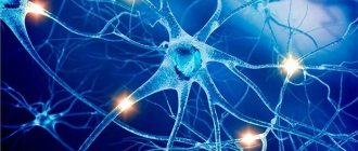
The structural unit of the NS is a cell with a process ( neuron , or neurocyte). The nervous system is a collection of neurons that communicate with each other through synapses.
Neuron
All organs of the nervous system are built from nerve cells called neurons. A neuron is capable of receiving and transmitting information in the form of a nerve impulse.
Rice. 1. Structure of a neuron.
The body of a neuron has processes with which it communicates with other cells. The short processes are called dendrites, the long ones are called axons.
Functions of the nervous system
The nervous system occupies a special position among other body systems. It ensures the relationship of the organism with the environment . Receptors respond to any signals from the external and internal environment, converting them into streams of nerve impulses that enter the central nervous system. Based on the analysis of the flow of nerve impulses encoding information, the brain forms an adequate response.
Together with the endocrine glands, the nervous system regulates the functioning of all organs . This regulation is carried out due to the fact that the spinal cord and brain are connected by nerves to all organs through bilateral connections. The central nervous system receives signals from the organs about their functional state, and the nervous system, in turn, sends signals to the organs, correcting their functions and ensuring all vital processes - movement, nutrition, excretion, etc. The nervous system ensures coordination of the activities of cells, tissues, organs , organ systems. In this case, the body functions as a single whole.
The nervous system is the material basis of mental processes : attention, memory, speech, thinking, etc., with the help of which a person not only learns about the environment, but can also actively change it.
Thus, several functions of the nervous system can be distinguished: 1. Connects the body with the environment (perception and transmission).
2. Provides interaction between tissues of organs and body systems and their regulation.
NS departments
Central nervous system and peripheral nervous system
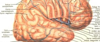
According to topographical principles, the nervous system is divided into central and peripheral. The central nervous system (CNS) includes those parts that are contained in the cranial cavity and the spinal canal, i.e. brain and spinal cord. The spinal cord is a tube with a small canal in the middle, surrounded by neurons and their processes. The brain is an extension of the spinal cord. The topographic boundary with the spinal cord is a plane passing through the lower edge of the foramen magnum. The average brain weight is 1400 g with individual variations from 1100 to 2000 g.
The peripheral nervous system (PNS) includes all the nerve structures located outside of it. These are nodes and bundles of fibers that connect the central nervous system with sensory organs and various effectors (muscles, glands, etc.), i.e. ganglia and nerves. The peripheral nervous system connects the spinal cord and brain with receptors and effectors. It consists of 12 pairs of cranial and 31-33 pairs of spinal (spinal) nerves.
Somatic nervous system and autonomic nervous system
According to the functional classification, the nervous system is divided into somatic nervous system and autonomic nervous system. Both somatic and autonomic nervous systems include central and peripheral parts.
The somatic nervous system includes the parts of the nervous system that regulate the functioning of skeletal muscles. Responsible for connecting the body with the external environment, providing sensitivity and movement, causing contraction of skeletal muscles. It regulates primarily the functions of voluntary movement. Its neurons are located in the anterior horns of the spinal cord, and their axons through the anterior roots of the spinal cord are directed to the skeletal (striated) muscles.
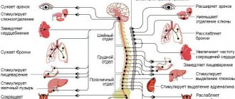
The autonomic (autonomic) nervous system is a collection of nerves and ganglia through which the heart, blood vessels, internal organs, glands, etc. are regulated. Internal organs receive double innervation (supply of organs and tissues with nerves, which ensures their connection with the central nervous system) - from the sympathetic and parasympathetic divisions of the autonomic nervous system. These two departments have excitatory and inhibitory influences, determining the level of activity of the organs.
The vegetative nervous system provides metabolism, respiration, and excretion. Influencing the metabolic activity in various organs and tissues in accordance with the changing conditions of their functioning and the external environment, it carries out an adaptive-professional function.
The autonomic nervous system innervates smooth muscles; it is also called the visceral nervous system. The division of the peripheral nervous system into somatic and autonomic is quite arbitrary, since in the central nervous system there is a significant overlap of projections of both, and somatic and autonomic reactions are equal components of any behavioral reaction.
In the autonomic nervous system, there are 2 divisions that are functional antagonists : sympathetic and parasympathetic . They differ in the localization of centers in the brain and peripheral nodes, as well as the nature of the effect on internal organs.
Differences between sympathetic and parasympathetic nervous system
1. The fibers of the sympathetic nervous system emerge from the thoracic and lumbar spinal cord, where the first sympathetic neuron lies. They then converge on the sympathetic ganglia located along the spine, where the second sympathetic neuron is located. The fibers of the parasympathetic nervous system begin in the spinal cord above or below the place where the sympathetic nerves exit the cranial and sacral regions, and then converge in ganglia located not along the spinal column, but close to the innervated organ.
2. The peculiarities of the location of the ganglia of these two systems suggest a difference in the effect they have. The action of the sympathetic nervous system is more diffuse, while the parasympathetic nervous system is more specific, since it is associated only with changes in the organ next to which the ganglion is located.
3. These systems also differ in the mediators involved in synaptic transmission. The main transmitter for the sympathetic nervous system is adrenaline, and for the parasympathetic nervous system it is acetylcholine.
4. The results of the activity of these two systems are largely opposite. While the main function of the sympathetic nervous system is to mobilize the body for fight or flight, the parasympathetic nervous system primarily ensures the maintenance of homeostasis.
5. Activation of the sympathetic nervous system underlies the behavior of a person eager to fight. The stimulation of the parasympathetic nervous system ensures digestion in a person lying on the couch after a hearty lunch.
The sympathetic nervous system excites, and the parasympathetic nervous system inhibits the activity of the heart, the first weakens the motor activity of the intestines, the second strengthens it. At the same time, they can act at the same time: together they increase the motor activity of the salivary and gastric glands, although the composition of the secreted juice varies depending on the share of participation of each system.
Structure, properties and age-related changes in nerve fibers
A nerve fiber is a process of a nerve cell covered with membranes. The central part of any process of a nerve cell (axon or dendrite) is called the axial cylinder. The axial cylinder is located in the axoplasm and consists of the finest fibers - neurofibrils and is covered with a membrane - the axolemma.
When examined under an electron microscope, it was found that each neurofibril consists of even thinner fibers of different diameters, having a tubular structure. Tubes with a diameter of up to 0.03 µm are called neurotubules, and those with a diameter of up to 0.01 µm are called neurofilaments. Neurotubules and neurofilaments carry substances that are formed in the cell body and serve to transmit nerve impulses to the nerve endings.
The axoplasm contains mitochondria, the number of which is especially high at the ends of the fibers, which is associated with the transfer of excitation from the axon to other cellular structures. There are few ribosomes and RNA in the axoplasm, which explains the low level of metabolism in the nerve fiber.
The axon is covered with a myelin sheath up to the point of its branching at the innervated organ, which is located along the axial cylinder not in a continuous line, but in segments 0.5-2 mm long. The space between the segments (1-2 µm) is called the node of Ranvier. The myelin sheath is formed by Schwann cells by repeatedly wrapping them around an axial cylinder. Each segment is formed by one Schwann cell, twisted into a continuous spiral.
In the region of the nodes of Ranvier, the myelin sheath is absent, and the ends of the Schwann cells are tightly adjacent to the axolemma. The outer membrane of Schwann cells covering myelin forms the outermost sheath of the nerve fiber, which is called the Schwann sheath or neurilemma. Schwann cells are given special importance; they are considered satellite cells, which additionally ensure metabolism in the nerve fiber. They take part in the process of regeneration of nerve fibers.
There are pulpy, or myelinated, and non-myelinated, or non-myelinated, nerve fibers. Myelin fibers include fibers of the somatic nervous system and some fibers of the autonomic nervous system. Non-pulp fibers are distinguished by the fact that they do not develop a myelin sheath and their axial cylinders are covered only with Schwann cells (Schwann sheath). These include most fibers of the autonomic nervous system.
Properties of nerve fibers. In the body, excitation is carried out through nerves, which include a large number of nerve fibers of different structure and function.
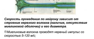
The main properties of nerve fibers are as follows: connection with the cell body, high excitability and lability, low level of metabolism, relative fatigue, high speed of excitation (up to 120 m/s). Myelination of nerve fibers occurs in a centrifugal direction, retreating a few microns from the cell body to the periphery of the nerve fiber. The absence of the myelin sheath limits the functionality of the nerve fiber. Reactions are possible, but they are diffuse and poorly coordinated.
As the myelin sheath develops, the excitability of the nerve fiber gradually increases. The peripheral nerves begin to myelinate earlier than others, then the fibers of the spinal cord, the brainstem, the cerebellum, and later the cerebral hemispheres. Myelination of the spinal and cranial nerves begins in the fourth month of intrauterine development. Motor fibers are covered with myelin at birth. Most mixed and centripetal nerves are myelinated by three months after birth, some by three years.
The spinal cord pathways are well developed at birth and almost all are myelinated. Myelination of only the pyramidal tracts does not end. The rate of myelination of cranial nerves varies; most of them myelinate by 1.5-2 years. Myelination of nerve fibers in the brain begins in utero and ends after birth. Despite the fact that by the age of three the myelination of nerve fibers basically ends, the growth in length of the myelin sheath and the axial cylinder continues even after the age of three.
Reflex as the basic principle of the nervous system
I. M. Sechenov in 1863 in his work “Reflexes of the Brain” he developed the idea that the reflex is the basic principle of the functioning of not only the spinal cord, but also the brain.
A reflex is the body’s response to irritation with the participation of the central nervous system. Reflexes are divided into: 1) unconditioned reflexes: innate (hereditary) reactions of the body to stimuli carried out with the participation of the spinal cord or brain stem; 2) conditioned reflexes: temporary reactions of the body acquired on the basis of unconditioned reflexes, carried out with the obligatory participation of the cerebral cortex, which form the basis of higher nervous activity.
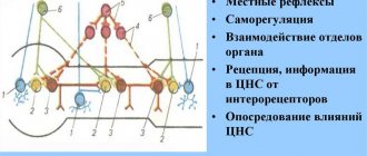
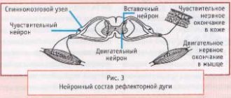
Each reflex has its own reflex arc - this is the path along which excitation passes from the receptor to the effector (executive organ).
The reflex arc is represented by a chain of neurons that provide the perception of irritation, the transformation of the energy of irritation into a nerve impulse, the conduction of a nerve impulse to the nerve centers, the processing of incoming information and the implementation of a response.
Any reflex arc consists of 5 components
1. A receptor is a specialized cell designed to perceive a stimulus (sound, light, chemical, etc.). 2. Afferent pathway, which is represented by afferent neurons. 3. Part of the central nervous system, represented by the spinal cord or brain; 4. The efferent pathway consists of the axons of efferent neurons extending beyond the CNS. 5. An effector is a working organ (muscle, gland, etc.).
The simplest reflex arc includes 2 neurons and is called monosynaptic (based on the number of synapses). A more complex one is represented by 3 neurons and is called three-neuron or disynaptic. However, most reflex arcs include a large number of interneurons and are called polysynaptic.
Reflex arcs can pass through the spinal cord only (for example, withdrawing a hand when touching a hot object) or through the brain only (for example, closing the eyelids when a stream of air is directed at the face), or through both the spinal cord and the brain.
Reflex arcs are closed into reflex rings using feedback connections. The concept of feedback and its functional role was indicated by Bell in 1826. He wrote that two-way connections are established between the muscle and the central nervous system. With the help of feedback, signals about the functional state of the effector are sent to the central nervous system.
The morphological basis of feedback is the receptors located in the effector and the afferent neurons associated with them. Thanks to feedback afferent connections, fine regulation of the effector’s work and an adequate response of the body to environmental changes are carried out.
Structure of the spinal cord
The spinal cord is actually an extension of the brain, surrounded by the same membranes and cerebrospinal fluid. It makes up two-thirds of the central nervous system and is a kind of conduction system for nerve impulses.
Figure 4. The structure of the vertebra and the location of the spinal cord in it
The spinal cord makes up two-thirds of the central nervous system and is a kind of conduction system for nerve impulses. Sensory information (touch sensations, temperature, pressure, pain) goes through it to the brain, and motor commands (motor function) and reflexes pass from the brain through the spinal cord to all parts of the body. The flexible, boney vertebral column protects the spinal cord from external influences. The bones that make up the spine are called vertebrae ; their protruding parts can be felt along the back and back of the neck. The different parts of the spine are called departments (levels), there are five in total: cervical ( C
), thoracic (
Th
), lumbar (
L
), sacral (
S
) and coccygeal[1].
[1] The sections of the spine are designated by Latin symbols based on the initial letters of the corresponding Latin names.
Within each section, the vertebrae are numbered.
Figure 5. Sections of the spine
A tumor of the spinal cord can form in any part - for example, they say that the tumor is found at the C1-C3
or at level
L5
. Along the entire spinal column, 31 pairs of spinal nerves extend from the spinal cord. They are connected to the spinal cord through nerve roots and pass through openings in the vertebrae to various parts of the body.
Tumors can develop inside the spinal cord (intramedullary tumors) or on the outside of the spinal cord (extramedullary tumors). Since there is very little room inside the spine for the tumor to grow, it begins to compress the spinal cord, and this is what causes the observed symptoms.
With spinal cord tumors, two types of disorders occur. Local (focal) symptoms - pain, weakness or sensory disturbances - are associated with tumor growth in a specific area, when this growth affects the bone and/or spinal nerve roots. More common disorders are associated with disruption of the transmission of nerve impulses through the part of the spinal cord affected by the tumor. Weakness and loss of sensation or muscle control in the area of the body controlled by the spinal cord below the level of the tumor (paralysis or paresis) may occur. Possible problems with urination and defecation (bowel movements).
During surgery to remove a tumor, the surgeon sometimes has to remove a piece of outer bone (the vertebral arch plate, or arch) to get to the tumor.
Important!
This can subsequently provoke curvature of the spine, so such a child should be observed by an orthopedist
.