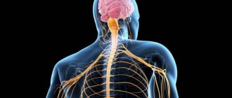
General information
All nerves include a huge number of fibers that are surrounded by connective tissue.
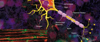
The fiber itself consists of a special process - an axon, which is covered with an ectodermal membrane. They are collected in certain bundles, thus creating tracts in the brain, spinal cord and peripheral nervous system. It is worth noting that the processes are either pulpy or non-pulpous (for example, the nerve endings of the skin). They all differ in the nature of their coverage, as well as in their belonging to a particular nervous system. They are divided into two main groups: those covered with myelin and those without it. In general, the first group predominates in the human body.
Unmyelinated fibers are located in the sympathetic autonomic nervous system.
Let's take a closer look at the structure of the myelin fiber.
Its main components are:
- a cylinder that runs along the central axis;
- directly the sheath of myelin nature, which covers the axial cylinder;
- Schwann membrane.
The main components of the cylinder are neurofibrils. Due to the presence of myelin components in the sheath, the impulse reaction passes through the nerve fiber faster. It is important to note that the cylinder is not completely covered with a shell; there are separate areas of Ranvier on it. It is at this point that the cylinder comes into contact with the Schwann shell. Its cells are of ectodermic origin. It is important to note that these types of membranes are present only in the peripheral nervous system. In the case of complete absence of a shell, the cylinders are called “naked”.
Classification
Among the existing nerve cells, experts traditionally distinguish the following units, according to the number of processes and functional purpose:
Based on the number of endings:
- unipolar - with a single process;
- pseudounipolar - from two branches of the same dendrite;
- bipolar – there is 1 dendrite and 1 axon;
- multipolar - several dendrites, but 1 axon.
By functional responsibilities:
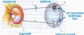
- perceivers - for receiving and transmitting signals from the outside, as well as from internal tissues;
- contact – intermediate, which provide processing and transmission of information to motor neurons;
- motor - generate control signals and then transmit them to other organs.
Additional units of the peripheral nerve regulatory system are lemmocytes. They envelop the processes of neurons and form the unmyelinated/myelin sheath. They are also called Schwann cells in honor of their discoverer. It is the Schwann cell membrane that, as the axon encircles and the membrane forms, helps to improve the conductivity of the nerve impulse.
It’s important to “take it”: reviews from doctors and patients, instructions
Experts must identify special contacts of neurons in the brain tissue, their synapses, the classification of which depends on the form of signal transmission:
- electrical – are important in the embryonic period of human development for the process of interneuron interactions;
- chemical - widely represented in adults; they use mediators to transmit a nerve impulse, for example, in motor cells for unidirectional excitation along the fiber.
Such a classification gives a complete picture of the complex structure of the brain tissue of humans, as representatives of a subclass of mammals.
Classification of nerve fibers
All of them are classified into three main groups:
- by impulse transmission speed;
- by transverse diameter;
- by the duration of the action potential.
It is worth noting that the larger their diameter and myelination, the faster the impulse travels through it. There are three varieties:
- Group A. All of them are covered with a membrane, their action potential is the lowest. In turn, they are divided into 4 subspecies: alpha, beta, gamma and delta. These include all receptors of the somatic nervous system, sensory fibers of the skin, thermoregulation, and proprioceptors. All these processes are responsible for human tactile senses.
- Group B. The processes are not completely covered with a myelin sheath; these include components of the autonomic nervous system. These include pain mediators and indicators of the functioning of internal organs.
- Group C. The membrane is completely absent, the speed of impulse conduction is low. These include ANS cells, as well as somatic pain and temperature cells.
Myelin contains phospholipids, cholesterol, basic protein and other useful components.
Thus, the membrane is a unique membrane, thanks to which the nervous system becomes able to quickly transmit impulses. All nerve processes are divided into two main groups: afferent (conduct impulses from tissues to the central nervous system) and efferent (act vice versa).
Some things we control, some things we cannot control.
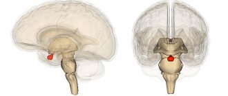
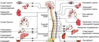
The properties of nervous tissue explain the fact that the somatic nervous system obeys the will of a person, innervating the work of the support system. Motor centers are located in the cerebral cortex. Autonomous, which is also called vegetative, does not depend on the will of a person. Based on your own requests, it is impossible to speed up or slow down your heartbeat or intestinal motility. Since the location of the autonomic centers is the hypothalamus, the autonomic nervous system controls the functioning of the heart and blood vessels, endocrine apparatus, and abdominal organs.
The nervous tissue, the photo of which you can see above, forms the sympathetic and parasympathetic divisions of the autonomic nervous system, which allow them to act as antagonists, producing a mutually opposite effect. Excitation in one organ causes inhibition processes in another. For example, sympathetic neurons cause strong and frequent contractions of the heart chambers, vasoconstriction, and surges in blood pressure, as norepinephrine is released. Parasympathetic activity, releasing acetylcholine, helps to weaken heart rhythms, increase the lumen of arteries, and lower blood pressure. Balancing these groups of mediators normalizes heart rhythm.
The sympathetic nervous system operates during times of intense tension such as fear or stress. Signals arise in the area of the thoracic and lumbar vertebrae. The parasympathetic system is activated when resting and digesting food, during sleep. The cell bodies of neurons are in the trunk and sacrum.
By studying in more detail the features of Purkinje cells, which are pear-shaped with many branching dendrites, one can see how impulse transmission occurs and reveal the mechanism of successive stages of the process.
Myelination of nerve fibers and demyelination
As described above, each process contains an axial cylinder, which is covered with a special myelin sheath. This process is called myelination. Due to the presence of sites of Ranvier, impulse is transferred from one to another. This is what ensures high transmission of excitation along the process towards the nerve.
Areas that are covered with the myelin sheath (pulpous) take part in the metabolic processes of muscle muscles; they have a high level of resistance to the effects of currents of a bioelectric nature.
In Ranvier gaps, impulse reactions are generated and accelerated. Their functions in the autonomic nervous system are assumed by oligodendroglia.
Non-pulpless tissues do not have a myelin sheath in their composition, therefore they are characterized by low insulating ability. In this case, the speed of impulse transmission is significantly reduced due to the fact that when it is transmitted from neurons, it comes into direct contact with the environment. The transmission of impulses for them requires large energy expenditures from the body (unlike pulp-type fibers).
From these two groups of fibers, a large nerve is subsequently formed, which has small bundles at its end. They differ in their main functions. It is important to note that these areas are the final ones in the formation of the interneuronal system.
When the functioning of the myelin sheath is disrupted or damaged, the process of demyelination occurs. This pathology can be caused by the presence of an inflammatory or infectious process in the body, metabolic disorders, ischemic processes in tissues, or the spread of neuroinfection. As a result of this process, the myelin in the sheath is replaced by fibrous plaques. The conductivity of impulse reactions in this case is significantly reduced.
There are two types of dimyelination:
- myelinopathy, which is the result of autoimmune disorders in the body;
- myelinoclasty appears with a genetic predisposition to the process of demyelination.
This process is considered quite dangerous, as it causes serious disturbances in the functioning of the central nervous system. It is very important to diagnose the disease at an early stage in order to provide effective therapy.
Treatment
Since many factors lead to damage to the optic nerve, therapy is prescribed only after a final diagnosis has been made. In most cases, the fight against the disease is carried out in a hospital.
Ischemic neuropathy is a very dangerous pathology that requires emergency care. Therapy must be started within the first twenty-four hours from the onset of the attack. Delaying treatment increases the risk of a severe and irreversible drop in visual acuity. Treatment of the disease includes taking corticosteroids, diuretics, and angioprotectors.
- Structure and morphological characteristics of tissue
- Nerve fibers and their endings
- Transport function of nerve fibers
- Some things we control, some things we cannot control.
- Classification
- The structure of nerve fibers
- Nerve fibers (meaty and non-meaty) - structure and functions. Conduction of excitation along a nerve
- Structure and properties of nerve fibers.
Traumatic abnormalities of the optic nerve can lead to serious vision problems. First of all, it is necessary to eliminate the pressure on the chiasm. For this purpose, forced diuresis is used and craniotomy is performed. The prognosis for such injuries is ambiguous. Sometimes vision can be preserved completely, and sometimes the patient goes blind.
| Retrobulbar and bulbar neuritis in most cases signal the development of multiple sclerosis. The second most common cause of pathologies is infections (influenza, rubella, measles). Therapy is aimed at eliminating swelling and inflammation of the nerve. Corticosteroids, antibacterial and antiviral agents are used. |
Benign tumors are diagnosed in children in 90% of cases. Glioma is located inside the optic canal and is prone to growth. The disease cannot be treated, and the baby may go blind.
The main symptoms of the pathology:
- On the damaged side, visual acuity drops very quickly, up to its complete loss.
- Exophthalmos develops. Bug-eye affects the eye whose nerve is affected by the neoplasm.
Most often, glioma damages the fibers of the optic nerve, in rare cases the optic-chiasmal area. The tumor in the latter is difficult to diagnose at an early stage and can lead to spread to the second eye.
Optic nerve atrophy is treated in courses. Therapy is carried out twice a year to maintain the patient's optimal condition. It includes taking medications (Mexidol, Retinalamine) and physiotherapy (electrical stimulation, magnetophoresis).
Functions of nerve fibers
The main function of nerve processes is the transmission of impulse response from neuron to neuron. There are two types of such transfer:
- impulse. It is based on electrolyte and neutrotransmitter mechanisms. As described above, in fibers covered with a myelin sheath the transmission rate is much higher;
- non-pulse. All reactions occur due to axoplasmic flow using axon microtubules. The latter contain a special substance that has a trophic effect on the innervating organ.
During the transmission of an impulse, a transformation of electrical potentials occurs, as a result of which unique molecules are formed - neurotransmitters.
All this formation has unique properties:
- lability (a limited number of impulses can be carried out over a certain time);
- excitability;
- conductivity.
It is believed that the nerve fiber is tireless. This is due to the low cost of ATP during the transmission of the impulse reaction. In the case of unmyelinated fibers, energy is required many times more, and therefore the transmission speed is significantly reduced.
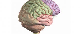
Structure and morphological characteristics of tissue
The main component of the brain is nervous tissue, which has a cellular structure. It is based on neurons, as well as neuroglia, an intercellular substance. A similar structure of nervous tissue ensures its physiological parameters - tissue irritation, subsequent excitation, as well as the production and transmission of signals.
Neurons are large functional units. They consist of the following elements:
- core;
- dendrites;
- body;
- axon.
Neuroglia contain auxiliary cells - for example, plasma astrocytes, oligodendrites, Schwann cells. A neuron, as a basic morpho-functional unit, usually consists of several dendrites, but always one axon - the action potential moves along it from one cell to neighboring ones. It is with the help of these endings that the human body communicates between the internal organs and the brain.
In their mass, the processes of neurons form fibers in which the axial cylinder breaks up into sensory and motor endings. On top they are surrounded by many myelin and non-myelin cells of the protective sheath.
Nerve fibers (meaty and non-meaty) - structure and functions. Conduction of excitation along a nerve
Previous12345678910111213141516Next
The function of rapid transmission of excitation to and from a nerve cell is performed by its processes - dendrites and axons, i.e. nerve fibers. Depending on their structure, they are divided into pulpy, having a myelin sheath, and non-pulpy. This membrane is formed by Schwann cells, which are modified glial cells. They contain myelin, which is mainly composed of lipids. It performs isolating and trophic functions. One Schwann cell forms the sheath per 1 mm of nerve fiber. Areas where the shell is discontinuous, i.e. not covered with myelin, called nodes of Ranvier. Interception width 1 µm.
Functionally, all nerve fibers are divided into 3 groups:
1. Type A fibers are thick fibers that have a myelin sheath.
This group includes 4 subtypes:
• And alpha - motor fibers of skeletal muscles and afferent nerves coming from muscle spindles - stretch receptors. Conduction speed 70-120 m/s.
• And beta - afferent fibers coming from the pressure and touch receptors of the skin. Speed 30-70 m/s.
• And gamma – efferent fibers going to muscle spindles (15-30 m/s).
• And delta – afferent fibers from temperature and pain receptors of the skin (12-30 m/s).
2. Group B fibers are thin myelinated fibers, which are preganglionic fibers of the autonomic efferent pathways. Conduction speed 3-18 m/s.
3. Group C fibers are non-myelinated postganglionic fibers of the autonomic nervous system. Speed 0.5-3 m/s.
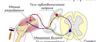
The conduction of excitation along the nerves is subject to the following laws:
1. The law of anatomical and physiological integrity of nerves, i.e. the nerve is able to perform its function only under both of these conditions. The first violations occur during cutting, the second - due to the action of substances that block conduction, for example, novocaine.
2. The law of bilateral excitation. It spreads in both directions from the site of irritation. In the body, excitation most often goes through afferent pathways to the neuron, and through efferent pathways from the neuron. This type of distribution is called orthodromic. Very rarely, reverse or antidromic propagation of excitation occurs.
Important How to use the drug Tebantine correctly?
3. Law of isolated conduction. Excitation is transmitted from one nerve fiber to another fiber, which is part of the same nerve trunk.
4. Law without decremental implementation. Excitation is carried out along the nerves without decrement, i.e. without attenuation. Therefore, nerve impulses are not weakened as they pass through the nerves.
5. The speed of conduction is directly proportional to the diameter of the nerves.
Nerve fibers have the properties of an electrical cable, which is not very well insulated. The mechanism of excitation is based on the occurrence of local current. As a result of the generation of an action potential in the axon hillock and reversal of the membrane potential, the axon membrane acquires a positive charge. Outside it becomes negative, inside positive. The membrane of the underlying unexcited axon is charged in the opposite way. Therefore, local currents begin to pass between these areas along the outer and inner surfaces of the membranes. These currents depolarize the membrane of the underlying unexcited portion of the nerve to a critical level, and an action potential is also generated in it. Then the process is repeated and a more distant part of the nerve is excited, etc.
Since local currents flow through the membrane of the pulpless fiber without interruption, such conduction is called continuous. With continuous conduction, local currents cover a large surface area of the fiber, so they require a long time to pass through a section of the fiber. As a result, the range and speed of transmission along the non-pulp fiber is small.
In the pulp fibers, areas covered with myelin have high electrical resistance. Therefore, continuous conduction of the action potential is impossible. When an action potential is generated, local currents flow only between adjacent nodes. According to the “all or nothing” law, the node of Ranvier closest to the axon hillock is excited, then the adjacent underlying node, etc.
This is called a saltatory jump. With this mechanism, local currents do not weaken, and nerve impulses travel over a greater distance and at high speed.
Previous12345678910111213141516Next
Date: 2015-07-02; view: 551; Copyright infringement
| Did you like the page? Like for friends: |