: 30 Oct 2021, The Fellowship of the Ring, volume 88, no. 3
Since childhood, we have heard that nerve cells do not recover. And although the question of the possibility of the formation of new neurons in the adult brain is still open, there is already evidence that the process of neurogenesis in humans continues into old age. Any disturbances in the development of nerve cells can lead to serious, sometimes irreversible pathologies. One of these disorders is defects in the protective insulating sheath (myelin) of nerve cell processes, which can form in a person even before birth. They are almost impossible to diagnose using traditional imaging methods
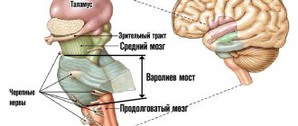
The average human brain contains about 100 billion neurons that receive, store, process and transmit information using electrical and chemical signals. Interaction between a neuron and other nerve cells and organs occurs through short ( dendrites)
) and long (
axon
) processes.
Each axon, like a wire, is covered with an insulating material - the myelin sheath
, which provides a higher speed of nerve impulses and protects nerve fibers from damage. In addition, this shell has a supporting function, and also, according to the latest data, serves as a kind of “refueling station” for the axon, which needs a large amount of energy.
All damage to the myelin sheath or defects that occur during its formation lead to serious, sometimes incurable diseases. Among them, the most famous is multiple sclerosis.
is a chronic autoimmune disease that primarily affects young people.
Myelin is also destroyed during strokes
, which occur not only in adults (primarily, as is commonly believed, in older people), but also in children, including the unborn.
Intrauterine stroke most often occurs after the 28th week of pregnancy, in children - a month after birth. Stroke in a fetus leads to the development of brain defects, and in children it can cause cerebral palsy
at an early age.
At the same time, today we judge the “quality” of myelination of the brain of a particular person only by indirect clinical symptoms or magnetic resonance imaging
(MRI), which can usually detect myelin defects at a late, often irreversible stage.
Principles of treatment of shell defects
Diseases associated with the destruction of the pulp membrane are very difficult to treat. Therapy is aimed mainly at relieving symptoms and stopping destruction processes. The earlier the disease is diagnosed, the greater the chances of stopping its progression.
Possibilities of myelin restoration
Thanks to timely treatment, the process of myelin restoration can be started. However, the new myelin sheath will not perform its functions as well. In addition, the disease may enter a chronic stage, and the symptoms will persist, only slightly smoothing out. But even minor remyelination can stop the course of the disease and partially restore lost functions.
Modern drugs aimed at myelin regeneration are more effective, but are very expensive.
- What are these areas of the brain and what is known about them?
- What does it consist of?
- Functions
- Biotin in myelin repair in MS
- Molecular organization of myelin
- Learning activates the brain
- Lecithin delivery
- Compound
- The role of microglia in the destruction of myelin structure
- Conclusion
- What did you find and what conclusions did you draw?
Therapy
The following drugs and procedures are used to treat diseases caused by the destruction of the myelin sheath:
- beta interferons (stop the course of the disease, reduce the risk of relapses and disability);
- immunomodulators (affect the activity of the immune system);
- muscle relaxants (help restore motor functions);
- nootropics (restore conductive activity);
- anti-inflammatory (relieve the inflammatory process that caused the destruction of myelin);
- neuroprotectors (prevent damage to brain neurons);
- painkillers and anticonvulsants;
- vitamins and antidepressants;
- cerebrospinal fluid filtration (a procedure aimed at cleansing cerebrospinal fluid).
Disease forecast
Currently, the treatment of demyelination does not give a 100% result, but scientists are actively developing drugs aimed at restoring the pulp membrane. Research is carried out in the following areas:
- Stimulation of oligodendrocytes . These are the cells that produce myelin. They do not work in a body affected by demyelination. Artificial stimulation of these cells will help start the process of restoring damaged areas of the myelin sheath.
- Stem cell stimulation . Stem cells can develop into full-fledged tissue. There is a possibility that they can also fill the pulpy shell.
- Regeneration of the blood-brain barrier . During demyelination, this barrier is destroyed and allows lymphocytes to negatively affect myelin. Its restoration protects the myelin layer from attack by the immune system.
Perhaps soon diseases associated with the destruction of myelin will no longer be incurable.
What are these areas of the brain and what is known about them?
The nucleus accumbens is part of the interconnected ventral area-nucleus accumbens (VTA-NAc) system. This system is critical for obtaining “rewards” through the release of dopamine. VTA dopaminergic neurons also innervate several areas of the prefrontal cortex (PFC), central amygdala, basolateral amygdala (BLA), and hippocampus, as well as other areas (Fig. 2). All of these so-called reward regions of the brain are interconnected in complex ways: for example, the NAc receives dense glutamatergic innervation from the PFC, amygdala, and hippocampus; The PFC, amygdala, and hippocampus form reciprocal glutamatergic connections with each other. The functional output of each of these areas is modulated by several types of GABAergic interneurons.
Figure 2. Diagram of the ventral area-nucleus accumbens (VTA-NAc) reward system. Simplified diagram of the major dopaminergic, glutamatergic, and GABAergic connections around the ventral area (VTA) and nucleus accumbens (NAc) in the rodent brain. The primary circuitry of the reward system involves dopaminergic connections from the VTA to the NAc, which release dopamine in response to reward-associated stimuli (and in some cases aversive-associated stimuli). There are also GABAergic projections from the NAc to the VTA; The NAc also receives dense innervation from glutamatergic monosynaptic circuits of the medial prefrontal cortex (mPFC), hippocampus (Hipp), and amygdala (Amy), as well as other regions. The VTA receives glutamatergic stimuli from the lateral dorsal segment (LDTg), the lateral leash of the epithalamus (LHb), and the lateral hypothalamus (LH). These various glutamatergic inputs control reward-related aspects of perception and memory. Dashed lines show internal inhibitory projections. Red lines—glutamatergic connections; green - dopaminergic; blue - GABAergic.
The release of dopamine into the nucleus accumbens is the way the brain receives pleasure signals
Feelings of pleasure motivate us to repeat behaviors that are vital for survival, and disruptions in the acquisition of dopamine rewards are associated with symptoms such as anhedonia and impaired reward-related perception and memory
The medial prefrontal cortex is the part of the prefrontal cortex that is also associated with the reward system, as shown above. Research has shown that patients with depression have less cortical volume, including less white matter in this area, which leads to impaired perception of events that previously brought joy.
What does it consist of?
Such an amazing biological insulating function of myelin turned out to be possible due to its structure. Don’t think that myelin is just an insulating layer wrapped around a neuron. Let us remember that in nature everything consists of cells, and the myelin of a peripheral nerve is simply an overgrown Schwann cell that has wrapped its cytoplasm around the axial cylinder of the neuron several times. It is myelin that gives the white color to nerve fibers, hence the concept of “white matter of the brain.” These are nothing more than bundles of nerve fibers that contain a lot of myelin. Their function is to be conductors of current. The pons, brain stem, midbrain are all areas consisting of an unimaginably large number of conductive bundles.
Therefore, myelin consists mostly of lipids, which repel water, and proteins. Myelin contains about 75% lipids, which is much higher than in most membranes. It's clear why this happens. After all, a membrane consisting of a bilipid layer should not only delimit the internal environment of the cell. This is a complex transport system that occurs with the help of carrier proteins. As for the myelin “wrappings” of the nerve, their task is very simple - to isolate the nerve fiber as much as possible. That’s why myelin is so “fat.” In the area of nodes of Ranvier, ions can enter the cytoplasm of the neuron, causing depolarization of the membrane, but not in the myelin areas. Thanks to this, the uninterrupted passage of impulses is ensured.
But there are situations in which myelin begins to break down. This process is called demyelination, and it manifests itself in a whole group of diseases of the same name. Why does this happen and how does it manifest itself?
Demyelination and its manifestations
Defects in the myelination of nerve fibers are called demyelination. This can occur due to genetic defects (called myelinopathy). Sometimes myelin is synthesized normally, but the physiological restoration of myelin occurs slowly or with damage. Demyelination is the process by which nervous tissue responds to pathological influences. Sometimes demyelination is secondary, that is, first the nerve is destroyed, and then its sheath. But still, most often myelin is lost first, and only then the process of the nerve cell is damaged.
Most often, immune inflammation is to blame for the primary destruction of myelin. Nerve insulation is destroyed by cytokines, enzymes and other active substances that are synthesized by plasma cells and macrophages. Antimyelin antibodies cause significant damage.
Important Cards pecs
The most common causes of demyelination are the following processes:
intoxication (alcoholism, radiation, increased glucose levels in diabetes mellitus);
cerebrovascular diseases, strokes, atherosclerosis;
vasculitis and systemic collagenosis;
autoimmune post-vaccination and post-infectious reactions.
The most well-known disease from this group is multiple sclerosis, which can occur with very diverse clinical symptoms (paralysis, paresis, dysfunction of the pelvic organs, tremor, ophthalmoplegia, extinction of reflexes, impaired coordination of movements). In multiple sclerosis, symptoms depend on where the lesion is located and the severity of demyelination.
Demyelination also occurs from the action of physical factors. Very serious exacerbations of multiple sclerosis can occur accidentally if you do not follow the rules of behavior. It has long been established that myelin is destroyed by thermal procedures. Thus, patients are strictly prohibited from:
take a steam bath;
take hot baths and showers;
sunbathing and being in the sun with exposed parts of the body.
Also, serious exacerbations occur after ARVI, influenza, and other diseases that occur with fever syndrome. An increase in temperature in multiple sclerosis and similar diseases stimulates the breakdown of myelin.
Myelination defects
Destruction of the myelin layer of the nervous system is a very serious pathology, due to which the transmission of nerve impulses is disrupted. It causes dangerous diseases, often incompatible with life. There are two types of factors influencing the occurrence of demyelination:
- genetic predisposition to myelin destruction;
- impact on myelin of internal or external factors.
- Demyelization is divided into three types:
- acute;
- relapsing;
- acute monophasic.
Why does destruction occur?
The most common causes of destruction of the pulp membrane are:
- rheumatic diseases;
- significant predominance of proteins and fats in the diet;
- genetic predisposition;
- bacterial infections;
- heavy metal poisoning;
- tumors and metastases;
- prolonged severe stress;
- bad ecology;
- pathologies of the immune system;
- long-term use of antipsychotics.
Diseases due to demyelination
Demyelinating diseases of the central nervous system:
- Canavan disease is a genetic disorder that occurs early in life. It is characterized by blindness, problems with swallowing and eating, and motor and developmental disorders. Epilepsy, macrocephaly and muscle hypotonia are also consequences of this disease.
- Binswanger's disease. Most often caused by arterial hypertension. Patients are expected to have thinking disorders, dementia, as well as walking and pelvic organ dysfunction.
- Multiple sclerosis. May cause damage to several parts of the central nervous system. It is accompanied by paresis, paralysis, convulsions and impaired motor skills. Also, symptoms of multiple sclerosis include behavioral disorders, weakening of the facial muscles and vocal cords, and sensory disturbances. Vision is impaired, the perception of color and brightness changes. Multiple sclerosis is also characterized by disorders of the pelvic organs and dystrophy of the brain stem, cerebellum and cranial nerves.
- Devic's disease is demyelination in the optic nerve and spinal cord. The disease is characterized by disturbances in coordination, sensitivity and functions of the pelvic organs. It is characterized by severe visual impairment and even blindness. The clinical picture also includes paresis, muscle weakness and autonomic dysfunction.
- Osmotic demyelination syndrome . Occurs due to a lack of sodium in the cells. Symptoms include convulsions, personality disorders, loss of consciousness, even coma and death. The consequences of the disease are cerebral edema, hypothalamic infarction and brainstem herniation.
- Myelopathies are various dystrophic changes in the spinal cord. They are characterized by muscle disorders, sensory disorders and pelvic organ dysfunction.
- Leukoencephalopathy is the destruction of the myelin sheath in the subcortex of the brain. Patients suffer from constant headaches and epileptic seizures. Impairments in vision, speech, coordination and walking are also observed. Sensitivity decreases, personality and consciousness disorders are observed, and dementia progresses.
- Leukodystrophy is a genetic metabolic disorder that causes the destruction of myelin. The course of the disease is accompanied by muscle and movement disorders, paralysis, visual and hearing impairment, and progressive dementia.
Demyelinating diseases of the peripheral nervous system:
- Guillain-Barré syndrome is an acute inflammatory demyelination. It is characterized by muscle and movement disorders, respiratory failure, partial or complete absence of tendon reflexes. Patients suffer from heart disease, disruption of the digestive system and pelvic organs. Paresis and sensory disturbances are also signs of this syndrome.
- Neural amyotrophy of Charcot-Marie-Tooth is a hereditary pathology of the myelin sheath. It is distinguished by sensory disturbances, limb dystrophy, spinal deformity and tremor.
These are only some of the diseases that arise due to the destruction of the myelin layer. The symptoms are similar in most cases. An accurate diagnosis can be made only after a computer or magnetic resonance imaging scan. An important role in making a diagnosis is played by the level of qualifications of the doctor.
Functions
The main functions of the myelin sheath are:
- axon isolation;
- acceleration of impulse conduction;
- energy savings by maintaining ion flows;
- nerve fiber support;
- axon nutrition.
How pulses work
Nerve cells are isolated due to their membrane, but are still interconnected. The areas where cells touch are called synapses. It is where the axon of one cell and the soma or dendrite of another meet.
An electrical impulse can be transmitted within a single cell or from neuron to neuron. This is a complex electrochemical process that is based on the movement of ions through the membrane of the nerve cell.
In a calm state, only potassium ions enter the neuron, and sodium ions remain outside. At the moment of excitement they begin to change places. The axon is positively charged from within. Then sodium stops flowing through the membrane, but the outflow of potassium does not stop.
The change in voltage due to the movement of potassium and sodium ions is called the "action potential". It spreads slowly, but the myelin sheath that envelops the axon speeds up this process by preventing the outflow and influx of potassium and sodium ions from the axon body.
Inflammation of the oral cavity - stages of treatment of the mucous membrane and methods of eliminating inflammation
Passing through the node of Ranvier, the impulse jumps from one part of the axon to another, which allows it to move faster.
Once the action potential crosses the break in the myelin, the impulse stops and the resting state returns.
This method of energy transfer is characteristic of the central nervous system. As for the autonomic nervous system, it often contains axons covered with little or no myelin. There are no jumps between Schwann cells, and the impulse travels much more slowly.
Functions of myelin in the nervous system
It is known that in addition to neurons in the central nervous system, there are glial cells that help neurons and serve them, performing a supporting and trophic function. In the central nervous system, the role of “insulating” nerve fibers is played by oligodendrocytes, and in the peripheral nervous system, by Schwann cells, which form the myelin substance.
If you cut a thick nerve, it can be compared to a cable, which consists of individual nerve bundles. The nerve bundles can be split until we get to a very thin extension of just one neuron. And every axon of every cell is protected by a myelin sheath. Myelin fibers are tightly wrapped around the nerve fiber, with virtually no gaps. It looks a little like a cylindrical roll of toilet paper with a pencil stuck in the center. The paper will rather roughly but faithfully imitate the myelin layers.
About jumps and interceptions
Electric current is known to travel at the speed of light when it comes to the flow of electrons in an ideal conductor, for example, in metals or under superconducting conditions. But the process of conducting an impulse in neurons is called electrochemical. Therefore, it takes a very short but finite time to “recharge” the membrane. This occurs at specific sites where the myelin protein is located.
There is then a bottleneck in the nerve where the myelin sheath is interrupted. This area is called the node of Ranvier. They are located at a distance of 1-2 mm, and between them there is a myelin sheath, “wrapped” onto the nerve. Therefore, the current moves in “jumps”, from interception to interception. The interception "interrupts" the potential, and then it accumulates on the other side of the conductor. The thicker the shell, the more perfect the impulse conduction function .
There are fibers poor in myelin, and axons in general, devoid of myelin, in which the impulse conduction speed is only 1-2 m/s, that is, 100 times slower. They are contained in the autonomic nervous system, where increased impulse speed is not very important, but slow and thorough work is required, for example, in the innervation of vasomotor-trophic reactions. It is precisely in such areas that there is a continuous conduction of the impulse, without “jumps” between the insulator - myelin.
Biotin in myelin repair in MS
Biotin is a vitamin involved in both energy metabolism and the creation of fatty acids in the body. It is found in dietary supplements for hair, skin and nail growth, as well as in multivitamins and prenatal vitamins.
Because the myelin sheath is a fatty sheath, researchers hypothesize that by giving people large doses of biotin (for example, 300 mg per day), the myelin sheath can be repaired.
In addition to restoring myelin (fatty covering), some scientists believe that biotin may reduce axonal degeneration by increasing energy production. Axonal degeneration occurs predominantly in progressive types of MS and refers to the loss of nerve fibers and ultimately the death of nerve cells.
The big picture is that experts suggest biotin may provide therapeutic benefits in two ways—double bonus. However, to date, scientific evidence supporting the role of biotin in the treatment of MS is sparse and inconclusive.
Let's take a closer look at a few studies showing these mixed results.
Thumbs up for biotin
In a small study called Multiple Sclerosis and Related Disorders, 23 people with primary or secondary progressive MS were given high doses of biotin and improvements were noted in a range of symptoms, particularly visual acuity, spinal cord problems and fatigue.
Mixed Results for Biotin
A large study in Multiple Sclerosis found that high doses of biotin improved MS-related disability in about 12 percent of participants with progressive MS. However, the fact that only 12 percent showed improvement suggests that potentially only a proportion of people with MS may benefit from biotin.
Also of concern is the fact that the researchers in this study noted that those taking biotin had more new or enlarged brain lesions on their MRI compared to those in the placebo group. The researchers questioned whether biotin causes inflammation answer (that wouldn't be good).
Thumbs down for biotin
The third study discusses the issue of biotin. In this study, there was no improvement in MS-related disability in people with progressive MS. In fact, about one-third of participants experienced worsening of their disease, particularly with greater leg weakness, poorer balance, and more falls.
Of course, the worsening of their disease may not be due to biotin and due to the natural progression of multiple sclerosis. However, the study researchers wondered whether high-dose biotin had anything to do with it. It is possible that biotin shifted the body's metabolic needs away from protecting the brain and spinal cord, allowing the immune system to wreak havoc.
Important Why do you feel dizzy after quitting smoking?
On a regular tomograph
Any pathology of the fetal brain that doctors suspect during an ultrasound examination of a pregnant woman is usually an indication for an MRI; Similar studies have been carried out at the ITC SB RAS for more than ten years. MRI results can confirm, clarify, refute, or even change the preliminary diagnosis and, accordingly, pregnancy management tactics.
The fact is that the amount of myelin and the size of individual brain structures in the embryo are so small that any measurements are very complex and time-consuming. In addition, the fetus is constantly moving, which makes it very difficult to obtain high-quality images and reliable quantitative data. Therefore, we need technology that allows us to obtain images quickly and with high resolution even on small objects.
This is exactly what the method for fast mapping of the macromolecular proton fraction
(MPF) is a biophysical parameter that describes the proportion of protons in tissue macromolecules involved in the formation of the MRI signal, whereas the signal source is usually protons contained in water (Yarnykh, 2012; Yarnykh et
al
., 2015).
The method is based on a specialized procedure for mathematical processing of MRI images, which makes it possible to isolate signal components associated with the MPF of cell membranes. And in the brain of humans and animals, the main part of them is contained in myelin. MPF maps are reconstructed based on initial data, which can be obtained on almost any clinical tomograph.
To reconstruct the MPF maps, four source images obtained by various traditional MRI methods are used. The correctness of this approach was confirmed by the results of its testing on laboratory animals at Tomsk State University: in mice that were injected with a solution that causes myelin destruction, the results of MPF mapping coincided with the data of histological examination of tissues (Khodanovich et al
., 2017).
Molecular organization of myelin
A unique feature of myelin is its formation as a result of the spiral entanglement of glial cell processes around axons, so dense that there is practically no cytoplasm left between the two layers of the membrane. Myelin is this double membrane, meaning it consists of a lipid bilayer and proteins associated with it.
Myelin proteins include so-called intrinsic and extrinsic proteins. The internal ones are integrated into the membrane, the external ones are located superficially and therefore are weaker connected to it. Myelin also contains glycoproteins and glycolipids.
Proteins make up 25-30% of the dry matter mass of the myelin sheath of neurons in the central nervous system of mammals. Lipids account for approximately 70-75% of the dry mass of the brain. Spinal cord myelin has a higher percentage of lipids than brain myelin. Most of the lipids are phospholipids (43%), the rest are cholesterol and galactolipids in approximately equal proportions.
Myelin function and structure
Before discussing the processes of normal myelination in the human brain, it is necessary to understand the structure of myelin and its functions in the central nervous system.
Myelin is found in both the central and peripheral nervous systems. In the central nervous system, it is found predominantly in the white matter (although some amounts are also present in the gray matter), giving it its color. Myelin acts as an electrical insulator: the speed of action potential increases 10-100 times compared to unmyelinated fibers. Julia Edgar and Jessica Garburn (2004) showed that the absence of myelin major protein (PLP/DM20) in oligodendrocytes leads to severe impairment of axonal transport in a mouse model of hereditary spastic paraplegia. It was also found that myelin likely plays a role in regulating both the ion composition and the fluid volume around the axon. Myelination is the process of formation of a bilipid myelin layer around the axon. This process further ensures the rapid transfer of information necessary for cognitive, behavioral, and emotional functions. Myelination begins during the embryonic period and continues after birth.
Myelin is a modified extension of the processes of oligodendroglial cells. The oligodendrocyte is a key cell in myelination of the CNS and is the predominant type of neuroglia in the white matter. The myelin sheath consists of multiple segments of myelin that wrap around the axon. This membrane is capable of conducting an action potential with its increase along the axon thanks to nodes of Ranvier, which are unmyelinated areas containing sodium ion channels - they are responsible for accelerating the conduction of action potentials along the axon. Myelin also has some symbiosis with the axon. Myelin is involved in the metabolic transformations of its components and contains a large number of myelin enzymes. Myelin also plays a role in ion transport, which helps buffer ions around the axon.
One oligodendrocyte can provide the myelin sheath for up to 40 fibers. Myelin is approximately 70% lipid and 30% protein. Myelin basic protein (MBP) makes up 30% of the total protein fraction, proteolipid protein (PLP) makes up 50%, and cyclic nucleotide phosphodiesterase makes up 4%. Myelin lipids contain cholesterol, phospholipids, and glycosphingolipids.
Myelin and MRI
There is no MRI technique or technology that can directly image the myelin bilayer. Myelin is assessed qualitatively on the basis of T1- and T2-weighted images, MR spectroscopy, and diffusion tensor imaging (DTI). In clinical practice, traditional anatomical imaging is the mainstay due to its ease of performance. Quantification of myelin can be obtained in a multicomponent relaxation (MCR or multicomponent relaxation) assay. MCR analysis is a volume-weighted summation of microscopic compartments of water. Using this method, two water domains are determined: a slow-relaxing domain, including free intra- and extracellular water, and a fast-relaxing domain, including water contained in the myelin lipid bilayer.
Currently, standard MRI sequences are not able to specifically quantify the amount of myelin. They allow you to evaluate changes in the density and size of axons, changes in membrane structure, namely the content of proteins, lipids and water. DTI is an unreliable measure of the total amount of myelin, but does provide information about possible changes in myelin.
James Barkovich identifies two distinct populations of water molecules that play a major role in generating the myelin signal on MRI—those molecules found within the myelin sheath and those outside the myelin sheath. On anatomical images, myelin is hyperintense relative to gray matter on T1 and hypointense on T2. On T1-weighted images, hyperintensity relative to the cortex is most likely determined by the presence of a large amount of glycolipids (especially galactocerebrosides) and cholesterol in the myelin sheath. On T2-weighted images, hypointensity is due to decreased water content.
Learning activates the brain
When we learn something new, whether programming in Ruby on Rails, consulting on the phone, playing chess, or making a cart wheel, our brains begin to function at a higher level.
Any new experience, be it playing the guitar or even computer games, promotes the production of myelin in the brain, which leads to strengthening mental abilities
Science has long proven that our brain is very mobile, that is, it does not stop developing even at 25. Of course, many things, especially languages, are easier for children than for adults. But there are many examples in the world of how older people learn something new.
But how does this happen? To perform any task, we must activate different parts of the brain. For example, to explain something, our brain coordinates a series of actions that include motor function, visual and auditory processing, speech, and more.
At first our explanation will be terse and chaotic.
We may forget to say something important. But with practice, our speech becomes smoother, more natural and softer.
Practice helps the brain optimize and coordinate all its actions through a process called myelination.
Lecithin delivery
Lecithin, also known as phophatidylcholine, is a fatty substance made up of choline, fatty acids and other lipid molecules. Lecithin is important for nerve transmission and can serve as a source of choline for myelin production. Dietary sources of lecithin include egg yolks, soybeans, wheat germ and liver. Brussels sprouts, shrimp, peanut butter and chocolate also contain significant levels of lecithin. If you follow a vegetarian diet that excludes dairy or eggs, you risk becoming deficient in lecithin and choline. Taking a vegetarian lecithin supplement can help ensure you're getting enough of these important nutrients.
Compound
The myelin layer consists of two layers of lipids and three layers of protein. There are much more lipids in it (70-75%):
- phospholipids (up to 50%);
- cholesterol (25%);
- glactocerebroside (20%), etc.
The high fat content causes the myelin sheath to be white, which is why the neurons covered by it are called “white matter.”
Protein layers are thinner than lipid layers. The protein content in myelin is 25-30%:
- proteolipid (35-50%);
- myelin basic protein (30%);
- Wolfgram proteins (20%).
There are simple and complex proteins of nervous tissue.
The role of lipids in the structure of the shell
Lipids play a key role in the structure of the pulp membrane. They are the structural material of nervous tissue and protect the axon from energy loss and ion flows. Lipid molecules have the ability to restore brain tissue after damage. Myelin lipids are responsible for adaptation of the mature nervous system. They act as hormone receptors and communicate between cells.
The role of proteins
Protein molecules are of no small importance in the structure of the myelin layer. They, along with lipids, act as building materials for nervous tissue.
Their main task is to transport nutrients into the axon. They also decipher signals entering the nerve cell and speed up reactions in it. Participation in metabolism is an important function of the myelin sheath protein molecules.
The role of microglia in the destruction of myelin structure
Microglia are macrophages capable of phagocytosis and able to recognize various pathogenic particles - antigens. Thanks to membrane receptors, these glial cells produce enzymes - proteases, as well as cytokines, for example, interleukin 1. It is a mediator of the inflammatory process and immunity.
The myelin sheath, whose functions are to insulate the axial cylinder and improve the conduction of nerve impulses, can be damaged by interleukin. As a result of this, the nerve is “exposed” and the speed of excitation is sharply reduced.
Moreover, cytokines, by activating receptors, provoke excessive transport of calcium ions into the neuron body. Proteases and phospholipases begin to break down the organelles and processes of nerve cells, which leads to apoptosis - the death of this structure.
It is destroyed, breaking up into particles, which are devoured by macrophages. This phenomenon is called excitotoxicity. It causes degeneration of neurons and their endings, leading to diseases such as Alzheimer's disease and Parkinson's disease.
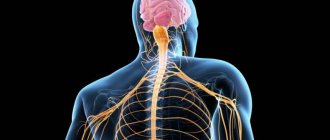
Nervous tissue
Nervous tissue is the main tissue that forms the nervous system and creates the conditions for the implementation of its many functions. Nervous tissue is of ectodermal origin; it is not customary to divide nervous tissue into any types of tissue. It has two main properties: excitability and conductivity.
Neuron
The structural and functional unit of nervous tissue is a neuron (from ancient Greek νεῦρον - fiber, nerve) - a cell with one long process - axon (Greek axis - axis), and one / several short ones - dendrites (Greek dendros - tree ).
I hasten to inform you that the idea that a short process of a neuron is always a dendrite, and a long process is always an axon, is fundamentally wrong. From a physiological point of view, it is more correct to give the following definitions: dendrite - a process of a neuron along which a nerve impulse moves to the body of a neuron, axon - a process of a neuron along which an impulse moves from the body of a neuron.
Neurons have 4 properties:
- Reception (lat. receptio - acceptance) - capable of perceiving incoming signals (dendrites)
- In response to signals, they are able to switch to a state of excitation or inhibition
- Conduction of excitation (from the dendrite to the neuron body, then to the end of the axon)
- Transmission of a signal to other objects - a neuron or an effector organ
In physiology, an effector organ (from the Latin efferes - efferent) is often called an executive organ or a target organ (muscles, glands). The effector organ carries out certain “orders” of the central nervous system (CNS) or endocrine glands
The processes of neurons conduct nerve impulses and transmit them to other neurons, effectors, due to which muscles contract or relax, and the secretion of glands increases or decreases.
Myelin sheath
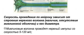
Nerve fibers are divided into myelinated and unmyelinated. A nerve fiber is one or more processes of neurons (can be either axons or dendrites) with a surrounding sheath.
Unmyelinated nerve fibers are found predominantly in the autonomic nervous system (conduction speed 1-2 m/s). Myelin - form the white matter of the brain and spinal cord, nerve fibers of the somatic nervous system (5-120 m/s).
In myelinated nerve fibers, the processes of neurons are covered with a myelin sheath (70-75% composed of lipids (fats)), which ensures isolated transmission of the nerve impulse along the nerve. If there were no myelin sheath (imagine!), nerve impulses would propagate chaotically, and when we wanted to move our arm, our leg would move along with our arm.
There is a disease in which one’s own antibodies destroy the myelin sheath of the nerve fibers of the brain and spinal cord (such malfunctions of the body also occur). This disease - multiple sclerosis, as it progresses, leads to the destruction of not only the myelin sheath, but also the nerves - which means muscle atrophy occurs and the person gradually becomes immobilized.
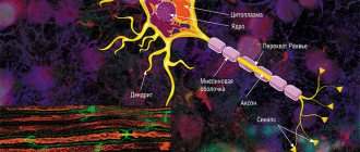
The myelin layer is represented by several layers of glial cell membrane (lemmocyte, Schwann cell), which twist around the axial cylinder (neuron process). This twisting is clearly visible in the picture of a healthy nerve, just above 
The myelin layer of the fiber sheath is regularly interrupted at the junction of neighboring lemmocytes - nodes of Ranvier. The myelin sheath provides isolated and faster conduction of excitation (saltatory type, Latin salto - I gallop, jump).
Neuroglia (Greek νεῦρον - fiber, nerve + γλία - glue)
You have already seen how important neurons are; their high specialization leads to the emergence of a special environment - neuroglia. Neuroglia (glial cells, gliocytes) are an auxiliary part of the nervous system that performs a number of important functions:
- Supporting - supports neurons in a certain position
- Regenerative (Latin regeneratio - rebirth) - in case of damage to nerve structures, neuroglia promotes regeneration
- Trophic (Greek trophe - nutrition) - with the help of neuroglia, neurons are nourished: neurons do not come into direct contact with blood
- Electrical insulating - lemmocytes (Schwann cells) curl around the processes of neurons and form the myelin sheath
- Barrier and protective - isolate neurons from the tissues of the internal environment of the body
- Some gliocytes secrete cerebrospinal fluid - cerebrospinal fluid (from the Latin liquor - liquid)
Neuroglia consists of different cells; there are tens of times more of them than neurons themselves. In the peripheral part of the nervous system, the myelin sheath, which we studied, is formed precisely from neuroglia - Schwann cells (lemmocytes). Between them, the nodes of Ranvier are clearly visible - areas devoid of the myelin sheath between two adjacent Schwann cells.
Classification of neurons
Neurons are functionally divided into sensory, motor and intercalary.
Sensory neurons are also called afferent, centripetal, sensory, perceptive - they perceive irritations, convert them into nerve impulses and transmit them to the central nervous system. A receptor is the terminal ending of sensory nerve fibers that perceive a stimulus.
Interneurons are also called intermediate, associative - they provide communication between sensory and motor neurons, transmit excitation to various parts of the central nervous system, and participate in information processing and the generation of commands.
Motor neurons are also called efferent, centrifugal, or motor neurons - they transmit a nerve impulse (excitation) to an effector (working organ). The simplest example of the interaction of neurons is the knee reflex (however, there is no interneuron in this diagram). We will study reflex arcs and their types in more detail in the section on the nervous system.
Synapse
In the diagram above, you probably noticed a new term - synapse (Greek sýnapsis - connection). A synapse is the point of contact between two neurons or between a neuron and an effector (target organ). At the synapse, the nerve impulse is “transformed” into a chemical one: special substances are released - neurotransmitters (the most famous is acetylcholine) into the synaptic cleft.
Let's look at the structure of a synapse in the diagram. It is made up of the presynaptic membrane of the axon, next to which there are vesicles (Latin vesicula - bubble) with a neurotransmitter inside (acetylcholine). If a nerve impulse reaches the terminal (end) of the axon, then the vesicles begin to merge with the presynaptic membrane: acetylcholine flows out into the synaptic cleft.
Once in the synaptic cleft, acetylcholine binds to receptors on the postsynaptic membrane, thus excitation (nerve impulse) is transmitted to another neuron. This is how the nervous system works: the electrical transmission path is replaced by a chemical one (at the synapse).
Poison curare
It is much more interesting to study any subject with examples, so I will try to please you with them as often as possible. I cannot hide the story about the poison curare, which the Indians have used for hunting since ancient times.
This poison blocks acetylcholine receptors on the postsynaptic membrane, and, as a result, the chemical transfer of excitation from one neuron to another becomes impossible. This leads to the fact that nerve impulses cease to flow to the effectors, including the respiratory muscles (intercostal muscles, diaphragm), as a result of which breathing stops and the death of the animal occurs.
Nerves and ganglia
When the processes of neurons (nerve fibers) come together, they form bundles of nerve fibers. Nerve bundles unite into nerves, which are covered with a connective tissue sheath. If the bodies of neurons are concentrated in one place outside the central nervous system, their clusters are called a nerve ganglion - or ganglion (from the ancient Greek γάγγλιον - node).
In the case of complex connections between nerve fibers, they speak of nerve plexuses. One of the most famous is the brachial plexus.
Nervous system diseases
Neurological diseases can develop anywhere in the nervous system: the clinical picture will depend on this. If the sensitive pathway is damaged, the patient ceases to feel pain, cold, heat and other irritants in the area of innervation of the affected nerve, while movements are fully preserved.
If the motor link is damaged, movement in the affected limb will be impossible: paralysis occurs, but sensitivity may remain.
There is a severe muscle disease - myasthenia gravis (from the ancient Greek μῦς - “muscle” and ἀσθένεια - “powerlessness, weakness”), in which one’s own antibodies destroy motor neurons (motor neurons).
Gradually, any muscle movements become more and more difficult for the patient, it becomes difficult to speak for a long time, and fatigue increases. A characteristic symptom is observed - drooping of the upper eyelid. The disease can lead to weakness of the diaphragm and breathing muscles, making breathing impossible.
© Bellevich Yuri Sergeevich 2018-2021
This article was written by Yuri Sergeevich Bellevich and is his intellectual property. Copying, distribution (including by copying to other sites and resources on the Internet) or any other use of information and objects without the prior consent of the copyright holder is punishable by law. To obtain article materials and permission to use them, please contact Yuri Bellevich
.