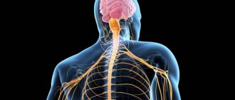
Somatic nervous system
(from the Greek soma - body) - part of the human nervous system, which is a set of afferent (sensitive) and efferent (motor) nerve fibers innervating muscles (in vertebrates - skeletal), skin, joints. The somatic system is the part of the peripheral nervous system that carries motor (motor) and sensory (feeling) information to and from the central nervous system. This system consists of nerves attached to the skin, sensory organs and all the muscles of the skeleton. It is responsible for almost all conscious muscle movements, as well as for processing sensory information received through external stimuli: vision, hearing and touch. The name somatic nervous system comes from the Greek word “soma” (body). The somatic nervous system contains two main types of neurons: sensory (afferent) neurons, which carry information from the nerves to the central nervous system, and motor (efferent) neurons, which carry information throughout the body from the brain and spinal cord to muscle tissue.
The neurons of the somatic nervous system extend from the central nervous system directly to the muscles and receptors. The body of the neuron is located in the central nervous system, and the axons extend further until they reach the skin, sensory organs, or muscles. Electrochemical impulses travel through axons from the brain to the spinal cord. The somatic nervous system also includes reflex arcs that are responsible for unconscious actions (reflexes). With the help of reflex arcs, muscles move without signals from the brain. This happens when nerve pathways connect directly to the spinal cord. Some examples of reflex arcs are when you quickly remove your hand from a hot pan or unconsciously raise your leg when the doctor taps your knee.
Stages of nervous system development
In evolution, the nervous system has undergone several stages of development, which became turning points in the qualitative organization of its activities.
These stages differ in the number and types of neuronal formations, synapses, signs of their functional specialization, and in the formation of groups of neurons interconnected by common functions. There are three main stages of the structural organization of the nervous system: diffuse, nodular, tubular. The diffuse nervous system is the most ancient, found in coelenterates (hydra). Such a nervous system is characterized by a multiplicity of connections between neighboring elements, which allows excitation to freely spread throughout the nervous network in all directions.
This type of nervous system provides wide interchangeability and thereby greater reliability of functioning, but these reactions are imprecise and vague.
The nodal type of nervous system is typical for worms, mollusks, and crustaceans.
It is characterized by the fact that the connections of nerve cells are organized in a certain way, excitation passes along strictly defined paths. This organization of the nervous system turns out to be more vulnerable. Damage to one node causes dysfunction of the entire organism as a whole, but its qualities are faster and more accurate.
The tubular nervous system is characteristic of chordates; it includes features of the diffuse and nodular types. The nervous system of higher animals took all the best: high reliability of the diffuse type, accuracy, locality, speed of organization of nodal type reactions.
Role
It is difficult to overestimate the role of the somatic system in the full functioning of the human body. After all, it regulates a lot of conscious muscle contractions - from facial expressions to fine motor skills of the fingers.
Due to the fact that the somatic system does not respond in a timely manner to external irritating factors, a person gets the opportunity to control his body and keep it intact. In general, this structure, with the help of reflex arcs, regulates the work of every, even the smallest, skeletal muscle - through impulses from motor neurons.
You can imagine the role of the somatic system using the example of any extreme situation:
- sense organs perceive and transmit information about changes in the external environment;
- the corresponding neuron of the nerve fiber transmits the signal to the sensory neuron in the spinal ganglia;
- the impulse travels through the entire chain of neurons to the motor neuron;
- the required muscle group is activated.
It is the speed of reaction to the threat that directly determines the preservation of human bodies as units of living matter. The role of somatic regulation is not simply limited to processing the information received and reacting to it - the system is involved in the regulation of voluntary movements by the consciousness. For example, move your foot from a broken board slightly to the side to avoid falling, or move your body higher/lower and regain your balance.
The leading role of the nervous system
At the first stage of the development of the world of living beings, interaction between the simplest organisms was carried out through the aquatic environment of the primitive ocean, into which the chemical substances released by them entered. The first oldest form of interaction between the cells of a multicellular organism is chemical interaction through metabolic products entering the body fluids. Such metabolic products, or metabolites, are the breakdown products of proteins, carbon dioxide, etc. This is the humoral transmission of influences, the humoral mechanism of correlation, or connections between organs.
The humoral connection is characterized by the following features:
- lack of an exact address to which a chemical substance entering the blood or other body fluids is sent;
- the chemical spreads slowly;
- the chemical acts in minute quantities and is usually quickly broken down or eliminated from the body.
Humoral connections are common to both the animal and plant worlds. At a certain stage of development of the animal world, in connection with the appearance of the nervous system, a new, nervous form of connections and regulation is formed, which qualitatively distinguishes the animal world from the plant world. The higher the development of an animal’s organism, the greater the role played by the interaction of organs through the nervous system, which is designated as reflex. In higher living organisms, the nervous system regulates humoral connections. Unlike the humoral connection, the nervous connection has a precise direction to a specific organ and even a group of cells; communication is carried out hundreds of times faster than the speed of distribution of chemicals. The transition from a humoral connection to a nervous connection was not accompanied by the destruction of the humoral connection between the cells of the body, but by the subordination of nervous connections and the emergence of neurohumoral connections.
At the next stage of development of living beings, special organs appear - glands, in which hormones are produced, formed from food substances entering the body. The main function of the nervous system is both to regulate the activity of individual organs among themselves, and in the interaction of the body as a whole with its external environment. Any impact of the external environment on the body appears, first of all, on receptors (sensory organs) and is carried out through changes caused by the external environment and the nervous system. As the nervous system develops, its highest department—the cerebral hemispheres—becomes “the manager and distributor of all the activities of the body.”
Introduction
First of all, it is worth informing that the nervous system is designed to transmit information and commands to our body. The main functions of the human nervous system are the perception of changes within the body and the space surrounding it, the interpretation of these changes and the response to them in the form of a certain form (including muscle contraction).
The nervous system is a set of different nervous structures interacting with each other, which, along with the endocrine system, provides coordinated regulation of the work of most of the body's systems, as well as a response to changing conditions of the external and internal environment. This system combines sensitization, motor activity and the correct functioning of systems such as endocrine, immune and more.
Structure of the nervous system
The nervous system is formed by nervous tissue, which consists of a huge number of neurons - a nerve cell with processes.
The nervous system is conventionally divided into central and peripheral.
The central nervous system includes the brain and spinal cord, and the peripheral nervous system includes the nerves that arise from them.
The brain and spinal cord are a collection of neurons. In a cross section of the brain, white and gray matter are distinguished. Gray matter consists of nerve cells, and white matter consists of nerve fibers, which are processes of nerve cells. In different parts of the central nervous system, the location of white and gray matter is different. In the spinal cord, gray matter is located inside, and white matter is outside, but in the brain (cerebral hemispheres, cerebellum), on the contrary, gray matter is outside, white matter is inside. In various parts of the brain there are separate clusters of nerve cells (gray matter) located inside the white matter - the nuclei. Clusters of nerve cells are also located outside the central nervous system. They are called nodes and belong to the peripheral nervous system.
review
The central nervous system can be considered as a system of coordination and integration within organisms. The central nervous system receives signals from the peripheral nervous system, processes them, then creates new signals to coordinate the actions of various body systems. For example, long- and short-term metabolism and homeostasis are regulated through the close interaction between the central nervous system and the endocrine system.
While the CNS is functionally composed of neurons, other cell types such as glial cells play important supporting roles. Some cranial nerves, such as the optic and olfactory nerves, are also considered part of the central nervous system. All other nerves are part of the peripheral nervous system, but they still connect to the central nervous system. Thus, the central nervous system is the body's coordinated processing center.
Based on these stimuli, the CNS modifies skeletal muscle contraction. Once children learn to walk, it happens involuntarily, no longer requiring conscious thought or concentration. This process of receiving complex stimuli and generating a coordinated response is required for a wide variety of activities—whether balancing a bicycle, holding a conversation, or enhancing an immune response.
Thought and Processing
The CNS, especially the brain, is considered the physical site for most higher-order mental functions. Neural connections form the basis for thinking and memory. The brain plays an important role in the development of speech, language and communication. These tasks involve associating abstract symbols and sounds with concrete objects and emotions. Motivation, ambition, reward and satisfaction are also mediated through neural connections in the central nervous system.
At the same time, the limbic system of the brain also controls the most basic emotions and drives, such as pleasure, fear, anger, hunger, thirst, sleepiness and sexual desire. Additionally, involuntary reflexes are mediated by the spinal cord, providing protection and quickly preventing injury.
The CNS directly or indirectly affects almost every internal organ system whether related to respiration, digestion, excretion, circulation or reproduction.
Reflex activity of the nervous system
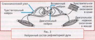
The main form of activity of the nervous system is the reflex. Reflex is the body’s reaction to changes in the internal or external environment, carried out with the participation of the central nervous system in response to irritation of receptors.
With any irritation, excitation from the receptors is transmitted along centripetal nerve fibers to the central nervous system, from where, through the interneuron along centrifugal fibers, it goes to the periphery to one or another organ, the activity of which changes. This entire path through the central nervous system to the working organ, called the reflex arc, is usually formed by three neurons: sensory, intercalary and motor. A reflex is a complex act in which a significantly larger number of neurons take part. Excitation, entering the central nervous system, spreads to many parts of the spinal cord and reaches the brain. As a result of the interaction of many neurons, the body responds to irritation.
Sensory neurons
Afferent sensory neurons of the somatic nervous system provide the CNS with information about joint angle, muscle length, muscle tension, and the presence of noxious stimuli.
proprioceptors
In addition to the typical extrafusal muscle fibers, the muscle body also contains muscle spindles. These small sensory organs contain specialized muscle fibers that have a central non-contractile segment. Afferent neurons of the somatic nervous system have their sensory dendrites in this area. These dendrites contain ion channels that open in response to mechanical stimuli on the cell. When the muscle spindle is stretched, the opening of ion channels generates an action potential in these sensory neurons. The presence of mechanically closed ion channels allows these neurons to carry detailed information about the state of the muscle and its contractile activity.
Nociceptors
Nociceptors are pain receptors found throughout the body and are an important part of injury prevention, especially in muscle fibers. These neurons fire in response to potentially harmful stimuli, such as heat, cold, or extreme forces. The presence of nociceptors prevents us from overextending our joints, straining our muscles, and protects us from a wide range of injuries.
- Alpha Motor Neurons – Lower motor neurons of the brainstem and spinal cord are associated with striated muscle fibers and are directly responsible for their contraction.
- Extrafusal muscle fiber Skeletal striated muscle fibers are innervated by alpha motor neurons and are attached to bone through tendons. The main function is the formation of skeletal movements as a result of contractile tension. They make up the majority of striated muscles in the human body.
- Lower motor neurons - Effector neurons of voluntary muscle contraction, usually synapse at the neuromuscular junction with striatal muscle fibers. The cell body is located either in the medullary pyramids of the brain stem or in the anterior horn of the spinal cord.
- Upper motor neurons - Neurons that originate in the brainstem or motor cortex and carry information to the lower motor neurons of the spinal cord or medullary pyramids.
Spinal cord
The spinal cord is a cord about 45 cm long, 1 cm in diameter, located in the spinal canal, covered with three meninges: dura, arachnoid and soft (vascular).
The spinal cord is located in the spinal canal and is a cord that at the top passes into the medulla oblongata and at the bottom ends at the level of the second lumbar vertebra. The spinal cord consists of gray matter containing nerve cells and white matter consisting of nerve fibers. Gray matter is located inside the spinal cord and is surrounded on all sides by white matter.
In a cross section, the gray matter resembles the letter H. It distinguishes the anterior and posterior horns, as well as the connecting crossbar, in the center of which there is a narrow canal of the spinal cord containing cerebrospinal fluid. In the thoracic region there are lateral horns. They contain the bodies of neurons that innervate internal organs. The white matter of the spinal cord is formed by nerve processes. Short processes connect sections of the spinal cord, and long ones make up the conductive apparatus of bilateral connections with the brain.
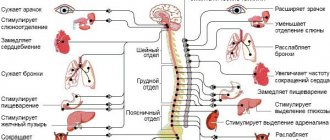
The spinal cord has two thickenings - cervical and lumbar, from which nerves extend to the upper and lower extremities. 31 pairs of spinal nerves arise from the spinal cord. Each nerve begins from the spinal cord with two roots - anterior and posterior. The dorsal roots are sensitive and consist of processes of centripetal neurons. Their bodies are located in the spinal ganglia. The anterior roots - motor - are processes of centrifugal neurons located in the gray matter of the spinal cord. As a result of the fusion of the anterior and posterior roots, a mixed spinal nerve is formed. The spinal cord contains centers that regulate the simplest reflex acts. The main functions of the spinal cord are reflex activity and conduction of excitation.
The human spinal cord contains reflex centers for the muscles of the upper and lower extremities, sweating and urination. The function of excitation is that impulses from the brain to all areas of the body and back pass through the spinal cord. Centrifugal impulses from organs (skin, muscles) are transmitted through ascending pathways to the brain. Along descending pathways, centrifugal impulses are transmitted from the brain to the spinal cord, then to the periphery, to the organs. When the pathways are damaged, there is a loss of sensitivity in various parts of the body, a violation of voluntary muscle contractions and the ability to move.
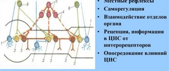
Characteristics of the reflex arc
The functioning of the nervous system is the continuous processing of information coming from outside and reaction to it. The somatic section in this chain is responsible for motor reflexes - in most cases, even without control from the brain.
Anatomically, the path of impulse transmission is described by a reflex arc. Among its links are several neurons that are interconnected by synapses. It is through them that the unidirectional movement of an information impulse occurs - in most cases through a chemical intermediary. It is also called a mediator.
Structures of the somatic arc:
- sensory neuron - localized in the spinal ganglion or in the zone of sensory ganglia inside the brain;
- intercalary, peripheral neuron - located in the nuclei of the dorsal horns of the spinal cord or the nuclei of the subcortical brainstem;
- The motor neuron lies in the nuclei of the ventral horns of the spinal substance, as well as in the nuclei of the central brain stem.
The originating nerve impulse travels all the way from the sensory neuron to the muscle bundle, which innervates the motor neuron as part of the spinal fiber. For example, from the receptor on the skin of the finger, with which a person checks the heating of the stove, to the muscles of the same finger, if it is necessary to withdraw the hand to avoid a burn.
Evolution of the vertebrate brain
The formation of the central nervous system in the form of a neural tube first appears in chordates. In lower chordates, the neural tube is preserved throughout life; in higher chordates, vertebrates, a neural plate is formed on the dorsal side during the embryonic stage, which is immersed under the skin and folded into a tube. In the embryonic stage of development, the neural tube forms three swellings in the anterior part - three brain vesicles, from which parts of the brain develop: the anterior vesicle gives rise to the forebrain and diencephalon, the middle vesicle turns into the midbrain, the posterior vesicle forms the cerebellum and medulla oblongata. These five brain regions are characteristic of all vertebrates.
Lower vertebrates - fish and amphibians - are characterized by a predominance of the midbrain over other parts. In amphibians, the forebrain somewhat enlarges and a thin layer of nerve cells is formed in the roof of the hemispheres - the primary medullary vault, the ancient cortex. In reptiles, the forebrain increases significantly due to accumulations of nerve cells. Most of the roof of the hemispheres is occupied by the ancient cortex. For the first time in reptiles, the rudiment of a new cortex appears. The hemispheres of the forebrain creep onto other parts, as a result of which a bend is formed in the region of the diencephalon. Beginning with ancient reptiles, the cerebral hemispheres became the largest part of the brain.
The structure of the brain of birds and reptiles has much in common. On the roof of the brain is the primary cortex, the midbrain is well developed. However, in birds, compared to reptiles, the total brain mass and the relative size of the forebrain increase. The cerebellum is large and has a folded structure. In mammals, the forebrain reaches its greatest size and complexity. Most of the brain matter is made up of the neocortex, which serves as the center of higher nervous activity. The intermediate and middle parts of the brain in mammals are small. The expanding hemispheres of the forebrain cover them and crush them under themselves. Some mammals have a smooth brain without grooves or convolutions, but most mammals have grooves and convolutions in the cerebral cortex. The appearance of grooves and convolutions occurs due to the growth of the brain with limited dimensions of the skull. Further growth of the cortex leads to the appearance of folding in the form of grooves and convolutions.
Distinctive features
Each structural unit of the human nervous system has its own functional purposes. However, together they form a complex whole. However, in the somatic part of the nervous system, neurons are more stable, and the impulse reaches its target faster. After all, its speed can reach 100–120 m/s. This significantly increases the chances of people surviving in dramatically changed external conditions - for example, when attacked by a predator during a hunt.
It is in the somatic department that information processing will occur more slowly than in the autonomic nervous system. Since they are faced with different functional tasks - the first must respond to information from motor neurons and carry out a reaction to them. Whereas vegetatives is nutrition, respiration, and reproduction.
Another significant difference is that there is no division in the somatic department. Most often it is represented by standard reflex arcs. Whereas in the autonomic system it is customary to distinguish the sympathetic as well as the parasympathetic departments. This makes it easier to control different muscle groups on an unconscious level, which takes some of the load off the cerebral cortex.
In general, all departments of the system cannot exist without close interaction with each other. This is why a person gets the opportunity to survive even in the most difficult conditions.
Brain
If the spinal cord in all vertebrates is developed more or less equally, then the brain differs significantly in size and complexity of structure in different animals. The forebrain undergoes particularly dramatic changes during evolution. In lower vertebrates, the forebrain is poorly developed. In fish, it is represented by the olfactory lobes and nuclei of gray matter in the thickness of the brain. The intensive development of the forebrain is associated with the emergence of animals onto land. It differentiates into the diencephalon and two symmetrical hemispheres, which are called the telencephalon. Gray matter on the surface of the forebrain (cortex) first appears in reptiles, developing further in birds and especially in mammals. Truly large forebrain hemispheres become only in birds and mammals. In the latter, they cover almost all other parts of the brain.
The brain is located in the cranial cavity. It includes the brainstem and telencephalon (cerebral cortex).
The brainstem consists of the medulla oblongata, pons, midbrain and diencephalon.
The medulla oblongata is a direct continuation of the spinal cord and, expanding, passes into the hindbrain. It basically retains the shape and structure of the spinal cord. In the thickness of the medulla oblongata there are accumulations of gray matter - the nuclei of the cranial nerves. The posterior pons includes the cerebellum and the pons. The cerebellum is located above the medulla oblongata and has a complex structure. On the surface of the cerebellar hemispheres, gray matter forms the cortex, and inside the cerebellum - its nuclei. Like the spinal medulla oblongata, it performs two functions: reflex and conductive. However, the reflexes of the medulla oblongata are more complex. This is reflected in its importance in the regulation of cardiac activity, the condition of blood vessels, respiration, and sweating. The centers of all these functions are located in the medulla oblongata. Here are the centers for chewing, sucking, swallowing, saliva and gastric juice. Despite its small size (2.5–3 cm), the medulla oblongata is a vital part of the central nervous system. Damage to it can cause death due to cessation of breathing and heart activity. The conductor function of the medulla oblongata and the pons is to transmit impulses from the spinal cord to the brain and back.
In the midbrain there are primary (subcortical) centers of vision and hearing, which carry out reflexive orienting reactions to light and sound stimuli. These reactions are expressed in various movements of the torso, head and eyes towards the stimuli. The midbrain consists of the cerebral peduncles and quadrigeminalis. The midbrain regulates and distributes the tone (tension) of skeletal muscles.
The diencephalon consists of two sections - the thalamus and hypothalamus, each of which consists of a large number of nuclei of the visual thalamus and subthalamic region. Through the visual thalamus, centripetal impulses are transmitted to the cerebral cortex from all receptors of the body. Not a single centripetal impulse, no matter where it comes from, can pass to the cortex, bypassing the visual hillocks. Thus, through the diencephalon, all receptors communicate with the cerebral cortex. In the subtubercular region there are centers that influence metabolism, thermoregulation and endocrine glands.
The cerebellum is located behind the medulla oblongata. It consists of gray and white matter. However, unlike the spinal cord and brainstem, the gray matter - the cortex - is located on the surface of the cerebellum, and the white matter is located inside, under the cortex. The cerebellum coordinates movements, makes them clear and smooth, plays an important role in maintaining the balance of the body in space, and also influences muscle tone. When the cerebellum is damaged, a person experiences a decrease in muscle tone, movement disorders and changes in gait, speech slows down, etc. However, after some time, movement and muscle tone are restored due to the fact that the intact parts of the central nervous system take over the functions of the cerebellum.
The cerebral hemispheres are the largest and most developed part of the brain. In humans, they form the bulk of the brain and are covered with cortex over their entire surface. Gray matter covers the outside of the hemispheres and forms the cerebral cortex. The human cerebral cortex has a thickness of 2 to 4 mm and is composed of 6–8 layers formed by 14–16 billion cells, different in shape, size and functions. Under the cortex is a white substance. It consists of nerve fibers connecting the cortex with the lower parts of the central nervous system and the individual lobes of the hemispheres with each other.
The cerebral cortex has convolutions separated by grooves, which significantly increase its surface. The three deepest grooves divide the hemispheres into lobes. In each hemisphere there are four lobes: frontal, parietal, temporal, occipital. The excitation of different receptors enters the corresponding receptive areas of the cortex, called zones, and from here they are transmitted to a specific organ, prompting it to action. The following zones are distinguished in the cortex. The auditory zone is located in the temporal lobe and receives impulses from auditory receptors.
The visual zone lies in the occipital region. Impulses from the eye receptors arrive here.
The olfactory zone is located on the inner surface of the temporal lobe and is connected to the receptors of the nasal cavity.
The sensory-motor zone is located in the frontal and parietal lobes. This zone contains the main centers of movement of the legs, torso, arms, neck, tongue and lips. This is also where the center of speech lies.
The cerebral hemispheres are the highest division of the central nervous system, controlling the functioning of all organs in mammals. The importance of the cerebral hemispheres in humans also lies in the fact that they represent the material basis of mental activity. I.P. Pavlov showed that mental activity is based on physiological processes occurring in the cerebral cortex. Thinking is associated with the activity of the entire cerebral cortex, and not just with the function of its individual areas.
| Brain department | Functions | |
| Medulla | Conductor | Connection between the spinal and overlying parts of the brain. |
| Reflex | Regulation of the activity of the respiratory, cardiovascular, digestive systems:
| |
| Pons | Conductor | Connects the cerebellar hemispheres to each other and to the cerebral cortex. |
| Cerebellum | Coordination | Coordination of voluntary movements and maintaining body position in space. Regulation of muscle tone and balance |
| Midbrain | Conductor | Approximate reflexes to visual and sound stimuli (turns of the head and torso). |
| Reflex |
| |
| Diencephalon | thalamus
hypothalamus
| |
The structural and functional unit of the animal nervous system is the neuron, which is a nerve cell with all its processes and terminal devices, and the main, elementary nervous act is the reflex.
In the nervous system, therefore, one can distinguish: 1) nerve centers, or ganglia, in which the switching of nerve impulses occurs; 2) conductors connecting the centers with each other and 3) peripheral nerves with their terminal devices.
The central nervous system consists of the brain and spinal cord. An intermediate position is occupied between them by the medulla oblongata, which is similar in structure to the spinal cord and retains the segmental apparatus in the form of cranial nerves and the conduction apparatus in the form of motor and sensory pathways.
In accordance with the pronounced physiological function in the brain, the forebrain, or cerebrum, the diencephalon (visual thalamus and their region), the midbrain (the quadrigeminal dorsal side and the cap (cerebellum and pons)) are distinguished.
The spinal cord is divided into cervical, thoracic and lumbar sections.
Under the influence of intermediate connections, the central nervous system functions as a single organ. All functions of the nervous system can be reduced to two main aspects of its activity: as an organ in which excitations are automatically generated, and as an organ that transfers impulses from a sensory neuron to a motor one and maintains communication between the periphery and the center. Both of these aspects of brain activity are closely intertwined.
The forebrain consists of two hemispheres and has a complex structure. The bulk of the hemispheres is white matter, consisting of pathways and subcortical nodes. The white matter is covered by an even layer of gray matter—the cerebral cortex. It is generally accepted that the cerebral cortex consists of six cellular layers arranged horizontally. Numerous processes of cortical cells penetrate all layers and form a dense network. Some fibers rise from the white matter to the cortex, others run parallel to the surface of the cortex and lie in the intermediate layers between the pyramids. Nerve fibers provide multilateral communication between individual parts of the brain.
The main function of the cerebral cortex is higher nervous activity aimed at regulating all functions of the animal’s body.
Higher nervous activity consists of the activity of the cerebral cortex and the subcortex closest to it. The cortex subordinates the subcortex and regulates its activity, and the subcortex, in turn, is a source of energy for the cortex.
The subcortex is the center of unconditioned reflexes, such as digestive, sexual, defensive, etc., while the cerebral cortex is the center of conditioned reflexes. In animals, conditioned reflex activity is determined by the direct effect of stimuli coming from the external or internal environment on the sense organs and their receptors located in the cerebral cortex.
Perception, higher analysis and synthesis, as well as consciousness, thinking and memory, i.e. the so-called mental manifestations, are closely related to the activity of the cerebral cortex. It has been experimentally proven that after removal of the bark, the dog is not able to assess the situation, does not recognize the owner, and becomes aggressive (Goltz, cited by Viktorov).
It has been experimentally established that the cerebral cortex can be divided according to the localization of specific functions into motor and sensory spheres. It has also been established that the motor and sensitivity of the body are controlled crosswise. The right hemisphere controls the left half of the body, and the left, vice versa, controls the right.
Motor spheres Control such volitional movements as flexion and extension of limbs, turning the neck and head, etc. The term “volitional movements” in animals has a relative meaning, since most of them are performed automatically, but some movements undoubtedly have elements conscious nature. Such movements include, for example, jumping over an obstacle, which is overcome by estimating the height and calculating the tension.
Motor centers do not represent sharply defined and isolated foci; zones of neighboring centers move towards each other.
Sphere of vision Located in the occipital lobe in the area of cunens et fissura, calcarina, the destruction of which leads to hemianopia, i.e., loss of the ability to see objects and phenomena in the field of vision. Reflexes related to the visual organs, such as blinking and turning the eyes, are preserved.
The sphere of hearing is located in the anterior part of the first gyrus of the temporal lobe of the hemisphere. Destruction of this sphere leads to deafness, and partial destruction leads to decreased hearing on the opposite side.
Sphere of smell Located under the Sylvian fissure of the ulcus gyri hippocampi and connected with the horn of ammonium. The nerve pathways from the tractus olfactorius go to the hemisphere of both its own and the opposite side. The destruction leads to a decrease in the sense of smell.
The sphere of general sensitivity (pain, tactile, thermal) is located throughout the cortex, but is more closely associated with the central gyri, where the motor sphere is localized.
Sphere of associations. The entire space of the cerebral cortex not occupied by motor and sensory areas belongs to the area of higher nervous activity.
Pavlov's teaching on conditioned reflexes showed that even internal organs, which were considered in many respects autonomous from the influence of the cerebral cortex, not only obey the direct influence of the cortex, but also send afferent excitations to the cortex, informing it of one or another of their states.
The study of conditioned reflexes has shown that the cerebral cortex has the ability to organize temporary connections, some of which can disappear, while others, with appropriate reinforcement, become stable and necessary.
The diencephalon is anatomically and functionally directly connected with the cerebral cortex and forms the “proximal subcortex”, which contains the visual thalamus, the hypothalamic region and the striatum.
The visual tubercle is the main subcortical node of all sensory conductors - visual, olfactory and general sensitivity. All sensory pathways gather here to switch to the last neurons that conduct sensitivity to the cerebral cortex. The main mass of the optic nerve passes through the diencephalon to the optic thalamus and further to the sphere of vision in the cerebral cortex. Here exteroceptive impulses are combined with interoceptive impulses.
In the hypothalamic region there are vegetative centers: metabolism, digestion, respiration, blood circulation, etc. Here are the basic mechanisms by which the cerebral cortex adapts the body to environmental conditions.
The striatum is located adjacent to the optic thalamus and is closely connected with it. The main function of the striatum is to maintain a certain muscle tone and ensure their readiness to contract.
The midbrain consists of the quadrigeminal, central gray matter, red nuclei and cerebral peduncles. The main function of the midbrain is the mobilization of motor apparatus and regulation of muscle tone.
The quadrigeminal structure is extremely complex. It contains sensory and motor neurons, as well as gray matter nuclei that serve as intermediate points.
The quadrigeminal consists of two cranial and two caudal colliculi. The former are related to vision, and the latter to hearing. Both of these senses take part in the animal’s orientation when moving. When the quadrigeminal dysfunction occurs in animals, hearing and vision deficiency occurs with a simultaneous disturbance in the correctness of movements.
Red core Located under the water supply in the cap. The red nucleus is the most responsible center, which perceives pathways from the cerebral cortex and bundles from the cerebellum. The red nucleus controls muscle tension, and together with the Deiters nucleus, nuclei of the cerebellum and medulla oblongata, the state of muscle tension in the body, which determines its position in space, controls the position of the head and the direction of the eyes according to the position of the body.
Transection of the brain stem along the quadrigeminal causes the appearance of rigor with a predominance of extensors. The animal stretches its legs and throws its head back. If the red nucleus is left with the spinal cord, then muscle tone will not be disturbed.
The hindbrain includes the pons and cerebellum.
The pons is a continuation of the nuclei of the medulla oblongata and contains transmission points for the cerebellum. It contains centripetal and centrifugal paths.
The cerebellum is similar in structure to the brain. The white matter is located in the center, while the gray matter forms the cortex, but is included in the nuclei and in the white matter. The nerve fibers of the white matter diverge towards the periphery in a fan-shaped manner.
The function of the cerebellum is reduced to receiving stimulation from sensory nerve endings and coordinating contractions of tonic muscle tension aimed at maintaining body balance.
Destruction of the cerebellum leads to impaired coordination of movements, collectively called cerebellar or cerebellar ataxia. When the function of the cerebellum is impaired, there is a weakening of muscle tone (atonia), a disturbance in the sense of strength (asthenia) and a violation of the correctness of movements (asthesia).
The loss of the coordinating influences of the cerebellum on the autonomic nervous system, mainly the sympathetic one, leads to severe disturbances in the motor and secretory functions of the digestive apparatus, the activity of the cardiovascular system, etc. (Orbeli, Astratyan). The regulation of the movements of the forelimbs comes from the anterior parts of the cerebellar hemispheres, and the hind limbs from the posterior parts.
The medulla oblongata can be considered as the anterior part of the spinal cord. The function of the medulla oblongata is quite complex, as is its structure. Both white and gray matter, when moving into the medulla oblongata, move and mix with each other, forming the so-called nuclei. These include the nuclei of the dorsal bundles of the spinal cord. From the accumulation of these nerve cells, secondary neurons of the secondary sensory pathways begin, heading towards the visual thalamus. The centripetal nerves of the brain end above them.
The automatic centers of the medulla oblongata include: a) the breathing center—lies at the height of the nuclei of the 9-12th nerve; b) the center of inhibition of the heart - connected with the nucleus of the recurrent nerve, it also works as a reflex center; c) vasomotor center—lies at the bottom of the fourth ventricle at the height of the nucleus of the facial nerve, it is connected with centers in the spinal cord and with the vascular center in the hypothalamic region; also works as a reflex center; d) sugar metabolism center - lies in the region of the nucleus of the recurrent nerve. Associated with metabolism. An injection into its area causes the development of glucosuria.
Spinal cord. The continuation of the medulla oblongata is the spinal cord, located in the spinal canal and suspended in the cerebrospinal fluid. In the area of the lumbar vertebrae, the spinal cord forms a filum terminale, which contains almost no brain matter. Along the entire length of the intervertebral foramina, spinal nerves arise from the spinal cord, forming the spinal ganglia as they exit the spine.
The gray matter of the spinal cord in a cross section occupies the central part and has the shape of the letter H, that is, it consists of a central wide part and four projections—two dorsal and two ventral horns. The lateral horns also protrude on the neck and lumbar region.
The white matter of the spinal cord occupies the periphery of the transverse section and is divided by gray matter into three main bundles (on each side): dorsal, lateral and ventral.
Each part of the spinal cord has a specialized purpose, its own function. To understand pathological conditions, it is very important to represent the function of the spinal cord through its cross section.
The neurites of the dorsal ganglion cells enter the dorsal horns of the spinal cord in the form of dorsal roots and are divided here into collaterals. Some of them are directed cranially, others caudally. Cranially they reach the medulla oblongata. All these numerous branches enable the spread of sensitive stimulation of the skin, mucous membranes and from the sensory endings inside the muscles, joints, periosteum and tendons. The ventral horns contain groups of large nerve cells with short neurites and dendrites.
Spinal nerves arise from two roots: ventral and dorsal. The dorsal ones carry centripetal nerve fibers, while the ventral ones carry centrifugal nerve fibers. Cutting the dorsal roots leads to loss of sensitivity, and cutting the ventral roots leads to the development of paralysis of the corresponding muscle groups. The centrifugal and centripetal nerves on each side cover specific areas of the body corresponding to a specific segment of the spinal cord. The dorsal bundle of white matter of the spinal cord, located between the roots and the middle plate, belongs to the sensitive ones and transmits deep sensitivity. This bundle transmits excitation from the periphery and from the spinal cord to the medulla oblongata, where it passes to the other side and reaches the visual thalamus.
The ventral fascicle, located between the ventral roots and the middle part of the gray matter of the spinal cord, is motor. Impulses pass through its fibers, originating in the cerebral cortex and regulating the movements of the animal. At the periphery of this bundle is the motor bundle, which begins in the medulla oblongata and ends above the motor cells of the ventral horns. These fibers carry impulses that regulate the balance of the body during movement.
The lateral fascicle, occupying the space between the dorsal and ventral roots, contains both centripetal and centrifugal pathways. In the lateral column there are fibers that carry impulses to the cerebellum from sensory endings in the muscles and blood vessels (deep sensitivity), fibers for conducting skin sensitivity, as well as motor nerves from the respiratory center, secretory nerves for the sweat glands and numerous nerves to the blood vessels.
In all three bundles of white matter there are numerous nerve fibers for the transmission of reflexes, intercentral communication and communication between segments of the spinal cord. Complete transection of the spinal cord causes sensory and motor paralysis of the entire part of the body below the transection. When half of the brain is cut, motor paralysis occurs on one side, sensory paralysis on the other, and weakening of tactile sensitivity on both sides.
The spinal cord contains numerous centers. Reflex centers are located along the length of the spinal cord and are intended for the motor system. Motor centers react to skin irritation by organizing protective movements, determining the coordination of movements and performing the function of muscle tone. Along the spinal cord there are numerous vascular-motor centers that work under the influence of the blood reaction and under the influence of sensitive irritations from the skin and organs. In the lumbosacral part of the spinal cord there are centers that regulate the activity of autonomic functions. This includes the center that controls the act of defecation, the center for the act of urination, the center for erection of the penis, and the center for ejaculation. These centers cause effects under the influence of excitations coming from the periphery on the one hand and the brain on the other. It must, however, be borne in mind that these centers influence the functions of internal organs through the sympathetic and parasympathetic nerves, which are independent to a certain extent.
Cerebral cortex
The surface of the human cerebral cortex is about 1500 cm2, which is many times greater than the inner surface of the skull. This large surface of the cortex was formed due to the development of a large number of grooves and convolutions, as a result of which most of the cortex (about 70%) is concentrated in the grooves. The largest grooves of the cerebral hemispheres are the central one, which runs across both hemispheres, and the temporal one, which separates the temporal lobe from the rest. The cerebral cortex, despite its small thickness (1.5–3 mm), has a very complex structure. It has six main layers, which differ in the structure, shape and size of neurons and connections. The cortex contains the centers of all sensory (receptor) systems, representatives of all organs and parts of the body. In this regard, centripetal nerve impulses from all internal organs or parts of the body approach the cortex, and it can control their work. Through the cerebral cortex, conditioned reflexes are closed, through which the body constantly, throughout life, very accurately adapts to the changing conditions of existence, to the environment.
Functions
Experts traditionally include the regulation of activity and body position in direct dependence on external stimulation as the functions of the somatic nervous system. A person “consciously” controls the parts of his body.
Since the motor neurons of the somatic nervous system innervate muscle structures on the periphery - limbs, internal organs, its functions will be as follows:
- processing information that comes from the senses;
- quick reactions to changing environmental conditions;
- body orientation in space;
- purposefulness of movements of all parts of the body.
Thanks to the motor neurons of the department, a person carries out conscious muscle movements - touching things, distinguishing tastes, assessing the position of his limbs in space and changing it as needed. At the same time, it regulates activity in the somatic nervous system, mainly its spinal part. Also called primitive. Despite the fact that in the process of evolution, people gained the ability to perform complex movements that obey the central structures of the cerebral cortex.
What is he responsible for?
The functional division of the nervous system during the evolution of man as a biological unit gave him special advantages in the struggle for existence. So, if the somatic department began to specialize in the perception of information from the outside - temperature, the presence of danger, then the autonomous, or as it is also called, the vegetative department completely switched to managing internal organs.
A functional comparison of the parts of the nervous system allows us to see the complexity of their activities:
- building a home, searching for food, hunting and fishing required coordination of the muscular frame of the human body - the somatic system began to be responsible for this;
- establishment of the correct heart rate, depth of breathing, blood pressure on the wall of blood vessels, movement of the food bolus through the intestines and metabolic processes - the organization of the “internal economy” came under the control of the autonomous system.
However, both departments are subordinate to the brain - each at its own level. The regulatory role comes down to correcting possible failures so that the functioning of the body as a whole corresponds to the current situation. After all, despite the fact that people's goals are much more complex than those of animals, they ultimately come down to satisfying their needs.
So, having set a task for himself, a person thinks about the best ways to implement it. He subordinates his movements to her - moving the body in space, fine motor skills. At the same time, at an unconscious level, changes occur in the internal environment - pulse rate, breathing, release of hormones and neurotransmitters. All changes occur, both automatically and arbitrarily.