Cervicocranialgia; Barre-Lieu syndrome; spinal nerve syndrome; Cervical migraine, vertebral artery syndrome; Neuritis of the sympathetic vertebral nerve - under such names can be found in foreign and domestic medical literature - Posterior cervical sympathetic syndrome.
The syndrome described above is generally considered to be a functional stage of vertebral artery syndrome. Vertebral artery syndrome refers to cerebrovascular diseases that lead to vascular disorders of cerebral circulation. Brain strokes are the main cause of disability and mortality among the adult population of planet Earth!! This problem, which is extremely unfavorable in epidemiological terms, also exists in Ukraine.
- The reasons are impaired blood circulation in the vertebral arteries, compression of blood vessels in the cervical spine.
Clinical picture
Headache in the cervico-occipital region, radiating to the parietal, temporal, fronto-orbital and ear regions, more often on one side. The headache is throbbing, aching, burning in nature and is provoked by head movements. Often, cervicocranialgia is combined with short-term vestibulo-cochlear disorders (balance disorders), sometimes with transient visual (blurred vision, darkening of the eyes). Physical and mental performance decreases sharply.
Vertebral artery syndrome. Pain, a feeling of numbness, a “crawling sensation” in the cervical-occipital region with irradiation to the anterior parts of the head, parietal, temporal, and postauricular regions. There is a sensation of a foreign body in the eye on the affected side, dizziness, nausea (or vomiting), congestion in the ear, tinnitus. There may be sensations of falling through, surrounding objects falling on the patient, a transient decrease in visual acuity, the patient falling without loss of consciousness - falls of the dropp attack type. Unsteadiness when walking. Possible increase in body temperature. General weakness, increased irritability, mood instability, feeling of heaviness in the head, sleep disturbances, memory problems.
What leads to all these reasons?
1. working at a computer for a long time, especially a laptop
2. Frequent work with gadgets (mobile phone, tablet)
3. incorrect sleeping place (pillow too high, mattress too soft, sleeping place does not correspond to the posture and weight of the sleeping person)
4. Frequent driving.
5. incorrect organization of the workplace (the height of the table and chair does not correspond to the person’s height)
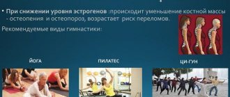
6. lack of regular dynamic physical activity (swimming, aerobics, dancing, gym)
Treatment of ShCS
Therapy for compression radicular syndromes is traditionally aimed at eliminating negative pathogenetic effects such as ischemia, edema, hypoxia and oxidative stress. The priority is to relieve pain in order to prevent chronicity of the disease. Short courses of painkillers are recommended, due to the risk of developing “silent” addiction and side effects that may go unnoticed by the doctor and the patient for a long time. Various non-opioid analgesics are used to treat low-intensity pain [4]. The prescription of non-steroidal anti-inflammatory drugs (NSAIDs) is also justified, the mechanism of action of which is associated with inhibition of prostaglandin synthesis (increasing the threshold of pain sensitivity). Since pain during NK compression has multiple causes, it is advisable to use drugs that improve the rheological properties of blood and endothelium-dependent reactions of the vessel walls. These are derivatives of succinic acid, vinpocetine (Cavinton), etc.
In complex treatment, vitamin-containing complexes with a multimodal mechanism of action are traditionally used. The main points of application of their pharmacological effects are neurological diseases, which are accompanied by pain, disturbances in the trophism of peripheral nerves and require their enhanced regeneration [3]. The need to use B vitamins is dictated by their high metabolic activity in the cells of the peripheral nervous system. Taking part in numerous biochemical processes, they act as coenzymes for a number of important reactions. Thus, thiamine is part of key enzyme systems that provide oxidative decarboxylation of pyruvic and α-ketoglutaric acids. An important feature of its action is the ability to form reserves of plastic substrates for the synthesis of nucleic acids. In addition, thiamine is directly involved in the synthesis of ATP and thereby increases the energy potential of the injured cervical root. Thiamine, pyridoxine and cyanocobalamin also have an antinociceptive effect [10, 13]. Possible points of their application are pain receptors, the sensitivity of which varies as a result of the influence of various tissue hormones (for example, bradykinin) and neuropeptides [7]. Sensitization of pain receptors is manifested, for example, by inflammatory hyperalgesia (increased pain sensitivity). In addition, there are several areas in the brain stem that, through descending pathways in the spinal cord, exert an inhibitory effect on the secondary neuron and thus cause a decrease in pain sensitivity. This mechanism of action is the rationale for the use of B vitamins in the treatment of compression radicular syndromes, the main manifestation of which is pain [8]. Apparently, the mediator in this case is serotonin. While the active metabolite of vitamin B6 is involved in the synthesis of serotonin as a coenzyme, thiamine performs an important function in its storage and transport [13]. It is here that, perhaps, the point of realization of the analgesic effect of pharmacological doses of thiamine and pyridoxine is located. Cyanocobalamin promotes remyelination through activation of transmethylation due to its effect on the synthesis of phospholipids and proteins of the myelin sheaths of peripheral nerves. The simultaneous use of thiamine, pyridoxine and cyanocobalamin helps stimulate the axoplasmic part of the transport of structural elements of the membrane or myelin sheath, such as choline. The combination of vitamins B1, B6 and B12 provides inhibition of nociceptive neurons in the dorsal nuclei of the spinal cord and thalamic nuclei due to stimulation of noradrenergic and serotonergic antinociceptive systems [3, 11]. High doses of neurotropic B vitamins have a positive effect on nerve regeneration [9].
In neurological practice, the drug of this group of drugs, Milgamma, is successfully used. The drug has a direct neurotropic effect in combination with an indirect analgesic effect. Milgamma solution for intramuscular injection (2 ml) contains thiamine hydrochloride - 100 mg, pyridoxine hydrochloride - 100 mg, cyanocobalamin hydrochloride - 1000 mcg, as well as lidocaine, which allows for painless injections. Due to its composition, Milgamma is comparable in speed and duration of analgesic effect after completion of a course of therapy for back pain to the standard drug of the NSAID group - diclofenac [11]. A number of studies confirm the high effectiveness of combination therapy with Milgamma + diclofenac compared with diclofenac monotherapy [3]. In case of severe pain syndrome, when NSAIDs cannot be prescribed, Milgamma allows you to significantly reduce the dose and duration of taking these drugs, which reduces the risk of complications from the gastrointestinal tract and increases the safety of therapy. At the same time, restoration of metabolism in nerve fibers helps to achieve a long-term therapeutic effect and prolong the period of remission to three or more months. Intramuscular administration of Milgamma allows for relief of acute pain syndrome within 2 days. After 10 daily injections, it is possible to switch to more infrequent injections - 2-3 times a week (for 2-3 weeks) or taking the drug Milgamma compositum, which makes it possible to achieve a lasting positive result and reduce the frequency and severity of relapses of pain.
The drug Milgamma compositum is advisable to use for long-term and preventive treatment. It contains benfotiamine and pyridoxine 100 mg each. The lipophilic analog of thiamine benfotiamine has high bioavailability and is not destroyed by intestinal thiaminases [12]. Since only treatment with sufficiently large doses of thiamine to achieve its high concentration in the blood and cytoplasm of cells is effective, benfotiamine has undoubted advantages over water-soluble thiamine. Oral therapy with Milgamma compositum is carried out for 4–6 weeks, 3 tablets per day. The two-stage course of therapy with Milgamma and Milgamma compositum has a pathogenetic basis: a 10-day course of intramuscular injections allows you to quickly achieve target concentrations of thiamine in the blood plasma and relieve pain symptoms; oral administration of Milgamma compositum increases the effectiveness of the injection course and prolongs the therapeutic effect. In the future, it is recommended to conduct courses of Milgamma compositum 2 times a year. The optimal course of treatment for neck pain includes 10 injections of the drug Milgamma and a 14-day intake of the drug Milgamma compositum, 1 tablet 3 times a day. Research results confirm that this treatment regimen allows prolonging the therapeutic effect for up to 3 months.
Thus, the modern strategy for treating patients with radicular compression syndromes should be the optimal selection of medications as part of a comprehensive program, including the use of neurotropic B vitamins and NSAIDs. An important aspect of the management of patients with compression radicular syndromes, including ShCS, is early clinical and radiological diagnosis. It prevents severe neurological complications and the unreasonable prescription of certain types of treatment.
Vertebral artery syndrome caused by abnormal structure of the cervical spine
Petryanina E.L., Ismagilov M.F.
Department of Neuropathology, Neurosurgery and Medical Genetics (Head of the Department, Prof. M.F. Ismagilov) Kazan State Medical University
In the clinic of vertebrogenic diseases of the nervous system, posterior cervical sympathetic syndrome (Barre-Leu syndrome, or cervical migraine) occupies a significant place. The disease manifests itself more often in the third decade of life in the form of unilateral or bilateral headaches, spreading from the occipital to the parietotemporal region, and cochleovestibular disorders.
In the overwhelming majority of observations, according to I.R. Schmidt and A.A. Lutsik [3], Barré-Lieu syndrome is caused by uncovertebral exostoses and extensor subluxation of the vertebrae. Ya.Yu. Popelyansky and I.R. Schmidt [2], based on an analysis of extensive clinical and radiological material, conclude that vertebral artery syndrome is the leading neurological manifestation of congenital blocks of the cervical vertebrae. According to their observations, the CII-CIII vertebrae are more often blocked, combined with other anomalies and remaining clinically latent for a long time. Painful symptoms are associated with earlier development of osteochondrosis and pathological mobility of the vertebrae.
Blocked vertebrae are classified as developmental defects [1]. Blocks (concrescence, synostoses) of the vertebrae - complete or partial fusion of the bodies, arches, articular or spinous processes of adjacent vertebrae - are associated with impaired segmentation in the early or late prenatal period.
Congenital blocks of the cervical spine are considered a variant of Klippel-Feil syndrome. The population frequency of the syndrome is 1:120,000. The clinic is varied and not fully studied. Characteristic: short neck, low hair growth, limited range of motion, minor developmental anomalies. The first clinical descriptions of the syndrome were given by Klippel er Feil in 1912 [4]. A valuable method for diagnosing abnormalities is radiography. Treatment is mainly symptomatic.
In connection with the above, it is of particular interest to describe the early clinical manifestation of the abnormal structure of the cervical spine (an additional hypoplastic vertebra with a bone block in CIII - bodies, arches, articular and spinous processes) in the form of posterior cervical sympathetic syndrome against the background of congenital cerebral insufficiency, minor developmental anomalies .
Here is a brief extract from the medical history.
Patient V., 17 years old, a vocational school student, was admitted to the clinic with complaints of constant headaches starting in the occipital region and spreading to the parietotemporal region on the left. The latter intensify with sudden movements of the head, usually in the morning, and are sometimes accompanied by noise, ringing in the ear, systemic (proprioceptive) dizziness, photopsia (at the height of headaches). Considers himself sick for about 3 years. The deterioration is associated with a head and neck injury in 1991, accompanied by short-term stupefaction.
Life history: mother, 44 years old, only child, moderate blue asphyxia during labor. An allergic history without any peculiarities or hereditary diseases cannot be identified.
Objectively: the consciousness is clear, speech contact is accessible, the physique is asthenic, height is above average, nutrition is reduced. The cervical lordosis is straightened, movements are carried out due to the lower part: tilt forward - 45°, backward - 30°, right - 30°, left - 45°, turn - 45° in both directions. Palpation pain in the projection of the spinous processes of the CII, CIII intervertebral joints of this level on the left, the point of the vertebral artery on the left with vibration recoil syndrome. Minor developmental anomalies: low hair growth, Michaelis rhombus hair growth, high palate, attached earlobes.
The neurological status reveals a horizontal small-scale nystagmus of the first degree, which intensifies when the neck is extended and turned to the right. Slight smoothing of the left nasolabial fold and slight deviation of the tongue to the right. Proprioceptive reflexes are live without a clear difference between sides, Rossolimo's sign on both sides, rough axial signs.
Fundus of the eye: on the right - pink, the boundaries of the optic nerve nipple are clear, on the left - pink, the boundaries of the nipple are blurred in the lower part of the temporal half, the arteries are narrowed, the veins are not changed.
X-ray of the skull: slight digital impressions, the sella turcica is normal. Radiographs of the cervical spine in 4 projections and in a state of maximum flexion and extension reveal an additional hypoplastic vertebra in a complete bone block with CIII (body, arches, articular and spinous processes), decreased height of the discs of CIV-CV, CV-CVI vertebrae, anterosuperior angle CV is beveled. Tchaikovsky index 0.85.
Echoencephaloscopy: signs of moderate intracranial hypertension on both sides, width of the third ventricle - 7 - 8 mm, IMP = 2.0, CC = 140. M - midline echo.
Rheoencephalography: the intensity of pulse blood filling of the carotid and vertebral arteries is sufficient, with dystonic signs of mixed type. In functional tests, blood flow in the left vertebral artery worsened by 30%.
Clinical diagnosis: moderately severe vertebrogenic cervicocranialgia with frequent cochleovestibular paroxysms (posterior cervical sympathetic syndrome, compression-irritative form), caused by an anomaly of the cervical spine (an additional vertebra with a complete bone block in the cervical spine); initial phenomena of osteochondrosis (according to Zecker grades I-II) in the vertebral segments CIV-CV, CV-CVI against the background of residual microfocal neurological symptoms and intracranial (moderate) hypertension. Recurrent course, exacerbation.
Dehydration, vascular, vegetotropic, and restorative therapy was carried out. There was an improvement in general condition, a decrease in headaches, and a decrease in paroxysms. Follow-up observation after discharge for 3 months did not reveal any deterioration of the condition. The patient receives basic therapy under the supervision of a neurologist.
Thus, we observed a patient with a rare anomaly of the cervical spine (an additional vertebra with a complete bone block in CIII), which caused the development of posterior cervical sympathetic syndrome in adolescence against the background of congenital cerebral insufficiency and minor developmental anomalies.
Literature
1. Dyachenko V.A. Anomalies of the development of the spine in x-ray anatomical lighting. - M., 1949.
2. Popelyansky Y.Yu., Shmidt I.R. Neurological manifestations of congenital blocks of the cervical vertebrae // Vertebrogenic diseases of the nervous system. - Novokuznetsk, 1969. - P. 80-107.
3. Shmidt I.R., Lutsik A.A. Some pathogenetic mechanisms of damage to the vertebral artery in connection with cervical osteochondrosis // Ibid. — P. 58-67.
Petryanina E.L., Ismagilov M.F. Vertebral artery syndrome caused by abnormal tension of the cervical spine // Neurological Bulletin. - 1994. - T. XXVI, issue. 3-4. — P.58-59
Diagnosis of cervical compression radicular symptoms
Diagnosing compression radicular symptoms is quite a difficult task. Only after a thorough examination to identify various pathologies (somatic, oncological, etc.) can treatment be prescribed. The examination of the patient must begin with a thorough examination and study of the neurological status. When diagnosing the level of the lesion, it should be taken into account that weakness of the extensors and flexors of the hand does not allow differentiating lesions of the C6, C7 and C8 NKs. The dorsal roots of adjacent cervical segments are often connected by intradural communication fibers. The connections between the dorsal root and the overlying cervical segment are especially constant. In this regard, erroneous localization of the level of the lesion one segment higher is possible. Patients with ShCS often experience a variety of vertebral, muscular, and neurovascular symptoms [2]. When palpating paravertebral points and interspinous ligaments, pain and limited mobility of individual vertebral segments are almost always noted. When the nodes of the borderline sympathetic trunk are affected, paresthesia, burning pain spreading to half the body, and itching in the area of innervation of the affected ganglion are often observed. Possible muscle wasting, redness or pallor of the skin in the area of innervation of the affected vegetative formations, regional disorders of thermoregulation and sweating, swelling or pastiness of tissues and other skin-trophic disorders.
Autonomic disorders can be confirmed using skin thermometry, reflex dermographism, pilomotor reflex, and adrenaline skin test. To determine the cause of the infringement (tumor, violation of the intervertebral discs in the form of protrusion or hernia), an X-ray examination or magnetic resonance imaging is performed [5]. To determine the degree of involvement of the spinal cord, NK and peripheral nerves in the pathological process, electromyographic studies are used. For differential diagnosis with dissection of the vertebral arteries, the patient is prescribed duplex or triplex scanning of the vertebral arteries, and if vascular pathology is suspected, magnetic resonance angiography is prescribed.