Definition
Neuroepithelial cysts (NCs) , also called neuroglial or glioependymal cysts, are developmental anomalies resulting from the absorption of part of the developing neuroectoderm, from leptomeningeal neuroglial heterotopia, or from folds of the choroidal pia mater in the case of choroidal sulcal cysts. NCs can be classified according to location into intraventricular, choroidal sulcus, and intraparenchymal cysts.
Morphology
Intraventricular neuroepithelial cyst
Intraventricular neuroepithelial cysts arise from the choroid plexus, usually at the level of the glomerulus in the triangle of the lateral ventricle. In such a location, they are often bilateral, and usually do not require any treatment. However, they can increase due to the secretive activity of the hair epithelium.
Fig. 1 Bichat's fissure cyst (choroidal fissure cyst)
Due to the relationship between the choroidal fissure and the choroid plexus, cysts associated with the choroidal fissure are considered to be of neuroepithelial origin, although they have also been classified among arachnoid cysts. Because they are located close to the hippocampus, which is often compressed, they can cause complex partial seizures.
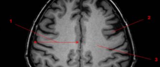
Fig.2 Intraparenchymal NC
Intraparenchymal neuroepithelial cysts are very rare. Differentiation from dilated perivascular spaces (see below) may not be possible on imaging, although location in the pericommissural or periventricular regions and multiplicity strongly favor dilated perivascular spaces. Sometimes, high-resolution MRI can detect a tiny band corresponding to the vessel's entry space. In the absence of such functions, they may be considered intraparenchymal NCs. Several histologically proven cases showed glio-ependymal elements along the cyst wall
Liquid collectors for anatomical variants
Fluid reservoirs, sometimes cystic in appearance, may result from accumulation of CSF in non-arachnoid cavities, which represent paraphysiologic developmental variations. These volumes usually cause slight deformation of adjacent structures, and, as a rule, are found in oligosymptomatic or completely asymptomatic children. These conditions include pseudocystic dilatation of the perivascular (ie, Virchow-Robin) spaces, cavities of the septum pellucida, cavities of Verges, and the spaces of the velum intermedius.
Expansion of perivascular spaces (PVS or Virchow-Robin spaces)
Perivascular spaces are tiny spaces that surround the arteries, arterioles, veins, and venules that penetrate the brain parenchyma. Contrary to what was commonly thought, the SVCs are not continuous with the subarachnoid space. Rather, they are separated by a single (cortex) or double (basal ganglia) layer of pia mater that invaginates with penetrating arteries.
Thus, the pia mater separates the PVC from the subarachnoid space, and the PVC is not filled with CSF, but rather with interstitial fluid, with the pia mater acting as a regulatory interface between the two compartments. PVAs are also functionally important in the elimination of high molecular weight substances from the brain. Thus, blocking such pathways can lead to the accumulation of these substances in the extracellular spaces of the brain.
On imaging, PVP appears as smoothly demarcated, circular, linear parallel areas that are similar in signal intensity to CSF. So they are ↓ on CT, ↓ on T1, ↑ on T2, and ↓ on FLAIR. There is no perifocal gliosis. They tend to cluster around the anterior commissure and inferior parts of the basal ganglia, as well as in the external capsule.
Fig.3
Periventricular regions, especially posteriorly around the triangles, are another typical location of prominent VVP. Unlike other regions, the periventricular VVP may be surrounded by a hyperintense rim on FLAIR in the pediatric age group. This may reflect incomplete myelination of the white matter (ie, terminal zones) or possibly spongy changes. In white matter, their orientation is typically radial, reflecting the course of penetrating arteries within the brain parenchyma.
The normal diameter of PVP does not exceed 5 mm under normal conditions. However, there are known cases of frank dilatation of the RVP, which can occur in the absence of any pathological condition as an anatomical variant. “Giant” PVS can be up to 3 cm in diameter and may be difficult to distinguish from neuroepithelial cysts on imaging. Recognition of radial orientation or direct central visualization on high-resolution MRI is helpful for correct diagnosis. Occasionally, expansion of the PVP may be widespread and involve both hemispheres of the brain.
Several pathological conditions can be characterized by dilatation of the PVP. In the pediatric age group, this includes storage diseases such as mucopolysaccharidoses (ie types I and II). In these disorders, blockage of the PVP is thought to result in the accumulation of abnormal materials in the PVP. In such cases, the PVP expansion noticeably includes, in addition to the periventricular white matter, the corpus callosum. Thus, the discovery of several callosal PVAs should prompt metabolic studies. In addition, VVP can provide a route for the spread of a number of diseases, including infections, inflammation, demyelination and tumors.
Cavity of the septum pellucida and Verge's cavity
The septum pellucidum is a thin two-layer curtain located between the anterior parts of the corpus callosum above, and the anterior columns of the fornix and anterior commissure below.
The septum pellucidum cavity (SCP) is a virtual midline. The cavity between the two layers of the septum under the corpus callosum. The continuation of the cavity into the space between the hippocampal commissure below, and the callosal isthmus and splenium above, is called Verge's cavity (VC).
PPP is present in the majority (i.e. 97%) of individuals, in 85% at 2 months and 41% at 3 months postnatal age, while it is found in 12%-20% of mature brains. CV is found in 100% of fetuses at 6 months of pregnancy but only in 30% of individuals subsequently; the incidence in mature brains is 1%-3%. Frank dilatation of the PPP and CV often represents an incidental finding in neuroimaging performed for a number of reasons.
Headaches are a common concern, although it is difficult to correlate headaches with dilated PPP/PV. An increase in the prevalence of abnormal PPP/PV has been noted in patients with schizophrenia and other affective disorders since childhood. An increased prevalence of abnormal PPP has also been implicated as a marker of neurodevelopmental delay.
In the photographs, recognition of PPP/PV is simple. Dilatation of the RPP, either isolated or associated with a cyst, when it exceeds 10 mm in transverse diameter. Sagittal images separate downward displacement of the fornix from the corpus callosum, which is a typical finding.
Progressive dilatation of the vein is noted in several individuals, either as a single change or in association with ventricular dilatation.
Fig.4
Cyst of the transparent septum. Fig.4a and Intermediate sail cyst Fig.4b Fig. 4c
Intermediate velum cyst
The intermediate velum forms the roof of the third ventricle and consists of a double layer of pia mater, which constitutes the choroidal body, including the choroid plexus from the third ventricle, expanding from the pineal body to the foramina of Monro.
A velar intermedius cyst (SVIC) is a potential space placed between the posterior portion of the third ventricle below and the commissural plate above. The anatomy of the checkpoint is still a matter of debate. The CPP has been variously described as lying above the choroidal body of the third ventricle or within the bilayer of the pia mater, as well as being superior to, or containing, the internal cerebral veins.
However, all authors agree that the CP is present, despite the separately existing cavities of the septum pellucida and Verge's cavity, but is not excluded from the CSF flow paths and actually communicates posteriorly with the subarachnoid space of the quadrigeminal cistern. CPP is present in 2%-3% of children over 2 years of age and is usually an incidental finding, although there have been reports of an association with mental and motor developmental retardation, epilepsy, and infantile autism.
Cystic dilatation of the CP is best assessed on a sagittal MRI image, forming a cavity filled with CSF that lies inferior to the isthmus and splenium of the corpus callosum and does not extend ventral to the columns of the fornix. The paired internal cerebral veins are usually located near and on the lateral sides of the choroid plexus of the third ventricle, and then are directed posteriorly under the splenium of the corpus callosum and join the vein of Galen.
The presence of cystic dilatation of the CAT under the splenium of the corpus callosum is accompanied by a downward deviation of the internal cerebral veins, while the fornix remains in place. This represents a noticeable difference between the CPP and the manifestation of the Verge's cavity.
Material and methods
A search was conducted in the databases “PubMed”, “Google Scholar” and “Cochrane Library” using the keywords: cyst of the septum pellucidum, Verge’s cavity, cyst of the intermediate velum, endoscopic fenestration, hydrocephalus. Original studies, review articles, and clinical case reports were selected. Works in languages other than Russian and English, as well as those for which neither the abstract nor the full text of the study were available, were not included. Publications on asymptomatic cysts were also excluded from the analysis. The information obtained was grouped and analyzed using the Microsoft Excel spreadsheet editor. The frequency of occurrence of the following clinical symptoms was determined: headache; swelling of the optic discs; dysfunction of the cranial nerves (without specification); convulsive syndrome; decreased intelligence and delayed psychomotor development (in children); dizziness, nausea and vomiting; gait disturbance; visual impairment (not specified); mental disorders (aggression, hyperactivity, apathy, abulia, depression, etc.); hydrocephalus. Information about the mechanisms of formation, structure, epidemiology and pathophysiology of cavities and cysts of the indicated localization, their classification, as well as diagnostic methods, indications and methods of surgical treatment are analyzed. When analyzing clinical manifestations, no distinction was made between symptomatic cysts of the septum pellucida, Verge's cavity, and velum intermedius.
A source of information
"Pediatric Neuroradiology. Brain. Head, Neck and Spine" Author: Tortori-Donati, Paolo, Rossi, Andrea
The translation was performed by a radiologist, Ph.D. Evgeniy Aleksandrovich Vlasov, the presented drawings are diagnostic brain scans, taken from the personal archive of E.A. Vlasov. are not related to Pediatric Neuroradiology. Brain. Head, Neck and Spine" and can be found in the DICOM archive of the site.
Author: radiologist, Ph.D. Vlasov Evgeniy Alexandrovich
Full or partial reprint of this article is permitted by installing an active hyperlink to the source
If you still have doubts about the conclusions of your MRI, you can order a review of your study with a detailed transcript here:
results
123 publications were found that matched the search criteria, with an abstract or full text of the study available for 72. The vast majority of works (57.79%) describe individual clinical cases and small series (less than 5 observations); 15 (21%) publications represent series of more than 5 observations, 8 (11%) publications contain information about 10 or more patients. The results of the analysis of literature data on the frequency of occurrence of individual clinical manifestations and the incidence of hydrocephalus in patients with symptomatic cysts are presented in Table. 1 . An overview of information on the prevalence of CAT, CPrP and CPV is presented in Table. 2 [1-11].
Table 1. Information on the frequency of occurrence of individual clinical manifestations and the incidence of hydrocephalus in patients with symptomatic midline cysts
| Number of publications: | 123 |
| included in the study | 72 |
| were not included in the study | 51 |
| Number of patients | 368 |
| Symptom, n (%): | |
| headache | 184 (50) |
| convulsive syndrome | 87 (23,6) |
| decreased intelligence/delayed psychomotor development | 74 (20,1) |
| mental disorders | 58 (15,8) |
| dizziness, nausea, vomiting | 40 (10,9) |
| disturbance of consciousness | 36 (9,8) |
| gait disturbances | 33 (9) |
| visual impairment | 31 (8,4) |
| papilledema | 17 (4,6) |
| cranial nerve dysfunction | 15 (4) |
| Hydrocephalus | 61 (16,6) |
Table 2. Main publications on the epidemiology of midline cerebrospinal fluid cysts
| Publication | Research method | Number of observations | Contingent | results |
| W. van Wagenen, 1934 [1] | Autopsy material, unfixed | 30 | Not specified | Checkpoint - ٦٠% |
| J. Schwidde, 1952 [2] | Autopsy material, fixation in 10% formaldehyde solution | 1032 | Newborns, children, adults up to 90 years old | PPP - ٢٠,٣٤% PV - ٢,٣٢% |
| J. Larroche et al. 1961 [3] | Autopsy material | No data | No data | PPP - ١٠٠٪ <٨,٥ months, ٨٢٪ full-term PV - ١٠٠٪ <٦ months, ٣٠٪ full-term |
| H. Schunk 1963 [4] | Photos of macropreparations | 307 | Patients who died from neurological diseases | PPP - ٦٠,٢% PPP ≥5 mm - ٠.٩% |
| J. Finke et al., 1968 [5] | Pneumoencephalography | 7711 | Not specified | PPP - ١,٧% |
| D. Babcock et al., 1981 [6] | Transcranial ultrasound | 102 | Newborns, children up to 9 months | PPP - ٤٢٪ (٦٢٪ - in premature babies, ٥٠٪ - in those born at term) |
| S. Nakano et al., ١٩٨١ [٧] | CT | 1050 | Newborns and children under 14 years of age with neurological diseases | PPP - ٢,٢٪ PPP+PV — ٣٪ PV - ٠,٤٪ |
| K. Akiyama et al., 1983 [8] | CT | 2722 | Adult patients | PPP - ٢.٦% |
| S. Mott et al., 1992 [9] | Transcranial ultrasound | 108 | Premature newborns (<35 weeks) | PPP - 100% average cavity width - 5 mm |
| J. Kwon et al., 1998 [10] | MRI | 113 | Psychiatric patients and control group | PPP >٥ mm - ٣٠,٤٪ patients with schizophrenia and ١٠,٣٪ control group patients |
| K. Wang et al., 2004 [11] | CT and MRI | 54000 | Not specified | Checkpoint - ٠.٠٤% |
Note. CP - septum pellucida cyst; CT - computed tomography; MRI - magnetic resonance imaging; PV - Verge's cavity; PPP - cavity of the transparent septum; Ultrasound - ultrasound examination.