Oxygen starvation of the fetus, resulting from impaired blood circulation in the placenta, leads to serious illnesses and deviations in the development of the child. One of the disorders that occurs for this reason is a subependymal cyst of the brain, which is diagnosed in newborns due to prolonged circulatory disorders, hypoxia and lack of nutrients.
A subependymal or cerebral cyst of the brain is a benign hollow formation in the ventricles of the brain, filled with fluid, formed due to hypoxia and insufficient blood supply. Since hypoxia is often diagnosed during pregnancy, the risk of formation is very high.
An intracerebral cyst in a newborn child is a natural protective response of the body to atrophy of brain tissue.
To prevent the expansion of necrosis of brain tissue, the body replaces the damaged areas, creating a natural barrier in the form of a cystic cavity. A small cyst does not pose a threat to the normal functioning of the brain.
But if fetal hypoxia is not treated during pregnancy, the cyst begins to increase in volume, leading to such dangerous manifestations as convulsions, deterioration in well-being and health, and psycho-emotional disorders.
Causes and risk factors
Fetal hypoxia is the main cause of subependymal cyst formation and has a significant impact on the development of the infant. The main risk factors for the manifestation of a cyst are the diagnosis of moderate or acute fetal hypoxia in a pregnant woman.
There are three factors for the appearance of a subependymal cyst:
- Cerebral ischemia . The most common cause of any cyst formation. Ischemia occurs due to impaired blood supply to brain tissue. In the place where the brain matter dies, a void appears, which the body fills with cerebrospinal fluid. The small size of the cyst does not lead to serious disorders; it only requires drug treatment and monitoring over time. If it grows and puts pressure on other parts of the brain, the child experiences convulsions, regurgitation, protrusion of the fontanel, and inhibition of physical and mental development.
- Hemorrhage in the head of a newborn is the second reason for the appearance of a neoplasm. The most severe deviations in the development of the child will be if hemorrhage occurs during uterine development of the fetus, and not during passage through the birth canal. The main causes of hemorrhage are the presence of infections, acute fetal hypoxia and birth trauma.
- Acute or moderate fetal hypoxia is a disruption of the functioning of the pregnant woman’s body and placenta. The main causes of hypoxia include anemia, toxicosis in the third trimester, multiple pregnancy, Rh conflict between the child and mother, polyhydramnios, placental insufficiency, and infectious diseases.
Article on the topic: Prognosis for liver cirrhosis: life expectancy and stages of the disease
Diagnosis and treatment of oral lesions in newborns
Infants often develop lesions in the oral cavity, which cause discomfort for themselves and cause anxiety for their parents. The most common disorders and diseases include congenital and neonatal teeth, various oral mucous cysts in newborns, ankyloglossia and congenital epulis of the newborn. In this article we will look at the features of diagnosis and treatment of this type of disorder and try to give readers an idea of the correct methods of treating and counseling young patients and their parents.
During their practice, doctors encounter various cases of oral lesions in newborns: from physiological characteristics associated with the development of the child to cancerous tumors. Awareness of such disorders plays an important role in correct diagnosis, counseling and treatment planning. The purpose of this article is to inform healthcare professionals about the diagnosis and treatment of the most common oral disorders in newborns.
Congenital and neonatal teeth
The eruption of the first baby tooth occurs approximately six months after the baby is born. But some babies reach this age already having congenital (the baby is born with them) or neonatal (erupted during the first month of life) teeth in their mouths.
Almost all congenital teeth (about 90%) erupt in the incisor area of the lower jaw. As a rule, they have the correct shape, but may be characterized by discoloration and an uneven surface. Their typical distinguishing feature already during the development period is increased mobility due to the absence or short length of roots. Most of the congenital teeth are subsequently included in the row of twenty primary teeth, but about 10% of them turn out to be supernumerary. Congenital teeth are rare: one case in two to three thousand births of healthy children, and, as a rule, this deviation is random. But in some cases, the appearance of congenital teeth can be a symptom of certain syndromes, malformations and gingival tumors.
If the congenital tooth turns out to be supernumerary and is not included in the row of baby teeth (this can be determined using an x-ray) or interferes with breastfeeding, it is recommended to remove it. Excessively mobile teeth should also be removed to prevent possible aspiration. In addition, congenital teeth can cause traumatic ulceration of the ventral surface of the tongue (Rigi-Fede syndrome), but this disorder is not an indication for tooth extraction and is cured by smoothing the rough cutting edge of the congenital tooth.
Newborn cysts
To refer to oral mucous cysts in newborns, many terms are used that replace each other, causing some confusion. But, based on the different histogenesis of the lesions, all of them can be divided into two categories: palatal and gingival.
Palatal cyst of a newborn
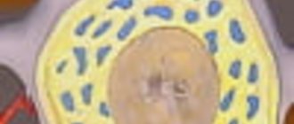
The palatal plates are bilateral rudimentary processes that join along the midline of the oral cavity in the eighth week of fetal development to form the hard palate. They also fuse with the nasal septum, resulting in complete separation of the oral and nasal cavities. In this case, the connective epithelial lining between the plates is destroyed under the action of enzymes, providing the possibility of fusion of the connective tissue. Neonatal palatal cysts, or Epstein's pearls, form from epithelial inclusions along the fusion line of the palatine plates. This disorder is characterized by high prevalence and is observed in 65%-85% of newborns. Cysts are small (1-3 mm) yellow-white bumps along the palatal suture, especially often located at the junction of the hard and soft palate. Histological examination reveals that these cysts are filled with keratin. No special treatment is required, since the cysts atrophy and disappear soon after their contents are removed.
Gingival cysts of newborns
Gingival cysts develop from the dental lamina (ectodermal ligament), which serves as the basis for the formation of primary and permanent teeth. Its remains can proliferate to form small cysts and subsequently cause the development of various odontogenic tumors and cysts. Depending on the location of formation, cysts that appear on the gums of newborns are called Bohn's nodes (present on the buccal and lingual surfaces of the alveolar ridges) or gingival cysts (formed on the process of the alveolar ridge).
Neonatal gingival cysts have a high prevalence: for example, Taiwanese infants screened within three days of birth had a 79 percent prevalence of the disorder.
Typically cysts look like small whitish lesions of constant size. Those that form on the anterior ridge of the lower jaw can be mistaken for congenital teeth. No separate treatment is required as cysts often rupture due to secondary trauma or friction.
Ankyloglossia
The term “ankyloglossia” (tongue-tied) describes the clinical situations of fusion of the tongue with the floor of the oral cavity or insufficient length of the frenulum of the tongue, limiting its mobility. Ankyloglossia can occur in representatives of various age groups, but is most often observed in newborns. According to research, the frequency of this disorder in newborns ranges from 1.7% to 10.7%, in adults – from 0.1% to 2.1%. Based on this, it can be assumed that some milder forms of ankyloglossia resolve with age.
Ankyloglossia of an infant can cause difficulty in breastfeeding and even cause pain in the nipple area for its mother or wet nurse. The preferred treatment for this disorder in newborns is simple frenectomy, where the frenulum is cut off at its thinnest point with small scissors. The procedure can be performed under superficial anesthesia, which ensures minimal discomfort and reduces the likelihood of bleeding. But bleeding is not necessary. Thus, according to the results of a study involving 215 newborns who underwent frenectomy without anesthesia, 38% of children had no bleeding, and 52% had only a few drops of blood. In 80% of cases, nutrition improved within 24 hours after the start of the procedure.
Congenital epulis of the newborn
This disease is a rare tumor of unknown histogenesis. As a rule, the lesion forms on the alveolar ridge of newborns. The course of the disease is as follows: the tumor does not increase in size from the moment of birth, sometimes it can decrease over time, which indicates a reactive rather than a neoplastic etiology. Most often, this tumor is found in the frontal part of the alveolar ridge of the upper jaw and has the appearance of a round attached formation, usually less than 2 cm in diameter (but sometimes larger ones are found), with a smooth lobulated surface. These types of tumors are more common in girls, which may indicate the influence of hormones, although estrogen and progesterone receptors have not been identified. In 10% of cases, multiple lesions may occur, confirming the need for a thorough oral examination.
As a result of histological studies of congenital epulis, large granular cells with small nuclei were identified. Unlike granular cell tumors, staining with the S100 protein antigen in congenital epulis gives a negative result. Other markers of neurogenic origin also showed negative results, confirming a nonspecific mesenchymal origin of the tumor. Surgical removal is recommended for the treatment of congenital epulis, especially if there is difficulty breathing or feeding problems, or if there is a need for histological confirmation of the diagnosis. For smaller tumors, a wait-and-see approach is acceptable, since cases of spontaneous regression of the tumor are known. There were no cases of relapse, even with incomplete removal of the tumor, or malignant degeneration.
Authors:
Van Heerden, Van Zyl
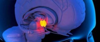
Localization in the brain and symptoms
A subependymal cyst is localized at the site of impaired blood supply to brain tissue, most often in the ventricular region. Symptoms of the disease depend on the location and size of the cyst. The presence of a pathogenic focus in a certain area of the brain leads to disruption of the body functions for which it is responsible.
A cyst in the occipital region affects the functioning of the visual system, visual acuity decreases, double vision, etc. A cyst in the temporal lobe leads to hearing impairment, in the cerebellum – gait and coordination of movements are impaired, in the pituitary gland – hormonal imbalances.
The small size of the cyst may not manifest itself in any way on the child’s behavior. But larger ones lead to convulsions and strokes, which can result in paralysis of the body.
The main symptoms observed in the presence of a cerebral cyst in the head of a newborn child:
- sleep disturbance;
- anxious behavior;
- breast refusal;
- poor weight gain or loss;
- long, causeless crying;
- hyper- or hypotonicity of muscles;
- tremor of the chin, twitching of the limbs;
- hearing impairment;
- blurred vision, sometimes complete loss;
- regurgitation “fountain”;
- loss of consciousness, coma;
- pulsation and swelling of the fontanelle;
- convulsions.
The consequences of growth and lack of treatment of the cyst lead to mental and physical retardation in the development of the child.
Clinic
Subependymal cysts of the brain in newborns can be asymptomatic and resolve within the first year of life, even without treatment. Especially if it does not exceed 5 mm in diameter. But often a cyst, not detected during the first month, after about 6 months, and more often in the second year of life, gives a certain clinical picture.
Symptoms and dynamics of the disease in infants depend on the type, size, and location of the cyst.
A small intracerebral cyst is not dangerous. A growing benign tumor can cause compression and displacement of surrounding brain tissue. A clinical picture similar to brain cancer arises, so an increase in size always alarms specialists.
The localization of intracerebral cysts is determined clinically by focal signs.
The symptom of pressure of a large intracerebral cyst on the occipital lobe is visual impairment. The child has poor vision and at a conscious age complains of blurred vision and double contours of objects.
When the intracerebral cyst is localized on the left, compression of the pyramidal tracts is possible. In this case, on the right there will be an increase in muscle tone and reflexes, the development of paresis or paralysis.
An intracerebral cyst of the left hemisphere, affecting the cerebellar tracts, causes muscle hypotonia, hearing loss and coordination, more pronounced on the left.
Any mass formation gives cerebral and focal symptoms.
General cerebral signs include hypertensive-hydrocephalic syndrome. It is caused by the development of intracranial hypertension and cerebral edema, manifested by a number of cerebral symptoms:
- headache (monotonous crying, causeless anxiety);
- vomiting (constant regurgitation);
- expansion of superficial veins on the head;
- increase in head size;
- bulging and pulsation of the fontanel;
- seizures;
- dizziness, loss of coordination;
- mental changes (mental retardation);
- forced head position;
- hearing impairment.
Complications of cyst development: formation of a brain abscess, rupture of the cyst membrane, transformation into a cancerous tumor. The consequence of a rapid increase in size of an intracerebral cyst with compression of vital centers is coma with possible death.
Diagnostics
In the presence of intrauterine hypoxia, birth trauma, birth by cesarean section, the baby must undergo neurosonography (ultrasound through the fontanel).
The following examinations are also required:
- General and biochemical blood test.
- Magnetic resonance imaging of the brain.
- Dopplerography of cerebral vessels.
- Examination by an ophthalmologist.
- X-ray of the skull in two projections.
- Spinal tap.
Diagnostic criteria
Using neurosonography (ultrasound) in newborns, it is easy to find cysts and other abnormalities in the brain. The fontanel, as a rule, does not close until a year, so this procedure is the fastest, easiest and most reliable.
Neurosonography must be done for premature babies, during severe pregnancy and protracted labor. If the doctor discovers a small subependymal cyst, then the brain tissue returns to normal on its own; you just need to be under the supervision of a pediatric neurologist.
If the cyst exceeds the minimum values, then additional diagnostic measures, MRI and CT are required. Diagnosis using these methods should be carried out twice a year.
Repeated examination of the cyst and recording its growth can lead to serious disturbances in the development of the child and a threat to his life.
Diagnosing a multi-chamber subependymal cyst is very problematic, since the symptoms can be easily confused with other disorders.
MRI of a subependymal cyst in a newborn child
Qualitative approach to therapy
Treatment of a cerebral cyst is aimed at preventing its growth. To do this, you need to find and eliminate the main cause that provokes oxygen starvation. During certain life stages of a child's growth, medical care will vary.
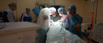
First aid
After birth, a neonatologist performs resuscitation care.
Article on the topic: The benefits and harms of oatmeal with milk and water: should you eat it in the morning?
Removes fluid from the respiratory tract, stimulates artificial respiration; in severe cases, an oxygen mask is used to supply pure oxygen.
The first three days after birth...
A neurologist conducts a daily examination and prescribes a restorative course of medication and rehabilitation therapy. Massage, physical therapy and sedatives are prescribed.
In childhood, the doctor observes the dynamics of the child’s development rate.
Medicines are used to stimulate speech functions and normalize the psycho-emotional state. At this stage, you may need the help of specialists such as a speech therapist and a psychologist.
...and beyond
When a child reaches adolescence, vitamin medications are prescribed to stimulate brain function and normalize metabolism. It is also necessary to take hormone replacement medications.
All physiotherapeutic and medicinal methods are prescribed only after the results of the examination and identification of deviations in the child’s development.
Treatment methods for pseudocysts
Are there ways to get rid of a pseudocyst in the head through therapeutic treatment without surgery? Yes, if this is truly a false formation, then it will resolve on its own. But medications will help improve brain function, strengthen the immune system, relieve increased excitability or stimulate the activity of the nervous system.
Conservative therapy
If a neurologist, based on the diagnostic results, has determined the presence of a pseudocyst in the newborn’s head, the doctor prescribes medications that eliminate the causes that led to the appearance of the anomaly. Similar conservative treatment is prescribed if a true cyst is suspected in the initial stage of growth.
Many of the pharmaceuticals, especially in tablets and injections, are contraindicated or limited for use in infancy, so it is unacceptable to prescribe them to a child on your own. Only a pediatric neurologist will determine whether the baby needs this or that drug and will calculate the safe dose and frequency of use, taking into account his age, the severity of neurogenic manifestations and concomitant diseases.
If a specialist notices that a child has certain signs of neurogenic damage, he can prescribe nootropic medications that improve cerebral circulation and drugs that eliminate the consequences of oxygen starvation (hypoxia) experienced by the child. But only if they are really indicated for a small patient, since many effective medications have serious side effects, cause acute allergic reactions or give complications to the nervous system and hematopoiesis.
Consequences and prognosis of the disease
The formation of small cysts does not have a serious impact on the quality of life, psychological and mental development of the child. Problems appear if it increases in volume. This may be affected by the presence of infections and hypoxia.
As the cyst enlarges, symptoms such as irritability, bronchopulmonary diseases, heart defects, developmental delays, and anemia begin to appear.
The first year of a child’s development does not show any serious deviations. Parents begin to notice violations at 2-3 years of age. The child has hyperexcitability, delayed speech development, and poor coordination. More serious manifestations include coma and death.
Preventive measures
In order to prevent the appearance of a cyst in the head of a newborn, it is necessary to establish the cause of its appearance. During pregnancy this is quite problematic.
It is necessary to do a genetic analysis, including amniotic fluid, but this analysis is done only in case of serious abnormalities in the development of the fetus.
Therefore, a woman needs to lead a healthy lifestyle, both before conception and during pregnancy, monitor her diet, and protect herself from intoxication and inflammatory processes.
Surgical treatment is performed very rarely, so the diagnosis of a subependymal cyst is not as scary as many people think. All that is necessary is observation by a neurologist for timely treatment of the disease and prevention of serious deviations in the development of the child.
Pseudocysts in adults
A pseudocyst in an adult patient is most often diagnosed in the pancreas, causing severe pain. Such a node threatens the life of the patient, as it is prone to suppuration, perforation, malignant degeneration, and the formation of fistulas. With this disease, competent surgical treatment is necessary.
Pseudocysts in the adrenal glands do not initially cause symptoms, but when they enlarge, they cause various manifestations due to compression of adjacent organs. Such neoplasms provoke an abnormal release of adrenaline and are complicated by acute bleeding into the lumen of the capsule. Urinary pseudocyst or urinoma can occur in retroperitoneal or perinephric tissue. When they grow, such structures require surgical removal.
The concept of pseudocyst also appears in the diagnoses of men and women with lesions of toxoplasma and cryptococcal infections, when massive intracellular foci with a concentration of parasitic microorganisms form in the brain and other organs. But most often in such cases the term “pseudocyst” is used.