The vast majority of more than 120 types of primary brain tumors are benign tumors. Their main common differences from malignant ones are slow growth, absence of metastases and relapses. Moreover, they are formed in different places from different types of cells and manifest themselves differently. Accordingly, treatment approaches can also vary significantly.
Benign brain tumors include pituitary adenomas, well-differentiated ependomas, chondromas, schwannomas, meningiomas, choroid plexus papillomas, cysts, lipomas.
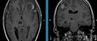
Pituitary adenomas
They make up about 10% of primary brain tumors and are most often found in the anterior third of the gland. They can appear at any age, but usually occur in older people. Women of childbearing age develop more often than men of the same age group. There are hormonally active (secreting) and hormonally inactive pituitary adenomas.
Pituitary adenoma
Most formations are hormonally active. In turn, they are classified according to the type of hormone produced, an excess of which in the body leads to the appearance of characteristic symptoms.
Recovery period and life prognosis
The prognosis for life with benign glioma depends on the timeliness of treatment and the degree of malignancy of the tumor. The necessary information about the tumor can only be obtained through a comprehensive and thorough diagnosis.
Low-grade gliomas are attempted to be completely removed surgically. Usually this does not cause any difficulties due to the localized nature of the pathological focus. Surgical treatment is the gold standard of neurosurgery.
It is impossible to indicate the exact life expectancy after surgery for brain glioma. If the patient seeks help on time, and the neurosurgeon manages to completely remove the tumor, the prognosis for glioma will be favorable. Life expectancy can be increased with subsequent radiation therapy. It allows you to increase the five-year survival rate after surgery to 60%.
It is also important to follow all doctor’s recommendations during the rehabilitation period. During recovery after removal of a brain glioma, the patient is under the supervision of specialists of a narrow and broad profile. They monitor important indicators of the body’s functioning, help normalize overall well-being and restore impaired functions of the nervous system.
Meningiomas
They grow not from brain tissue, but from the soft membranes covering it (meninx), which is why they got their name. They make up about a third of all primary neoplasms. Most often observed in women, starting from middle age (2 times more often than in men). The incidence rate increases after 60 years. They are rare in children. The typical location is in the upper regions, but can also form at the base of the skull. They usually grow inward, causing compression of adjacent parts of the brain. Sometimes they grow outward, which is accompanied by thickening of the cranial bones in the problem area. May contain calcifications, fluid-filled cavities (cysts), and vascular nodes.
Symptoms
Clinical manifestations are divided into focal (primary) and cerebral (associated with hemodynamic disturbances and increased intracranial pressure). The quantity and quality of manifestations depend on the location of the tumor, as well as its size and the degree of tissue damage and compression.
Primary manifestations . Among the focal symptoms, it is customary to distinguish the following disorders:
- memory, attention (with damage to the cerebral cortex) - various psychomotor and cognitive impairments, a slight decrease in the results of intellectual activity may be observed;
- affective (emotional sphere) – anxiety, irritability, unmotivated aggression, psychosis may occur;
- sensitivity - pain, tactile, thermal;
- musculoskeletal system - poor coordination in space, muscle hypotonicity;
- distribution of excitation - if, due to a tumor, foci of stagnant excitation are formed in the brain tissue, then convulsions and epileptic seizures may occur;
- hearing and speech recognition (if the auditory nerve is affected);
- vision and recognition of text and objects (if areas of the cerebral cortex, optic nerve or quadrigeminal region are affected), hallucinations may occur;
- written or oral speech (if the corresponding areas of the brain are damaged);
- autonomic disorders - weakness, fatigue, periodic dizziness, fluctuations in pulse, blood pressure (a tumor can disrupt the innervation of blood vessels and the functioning of the vagus nerve);
- hormonal disorders - with tumors of the pituitary gland, hypothalamus, and sometimes the pineal gland.
General cerebral symptoms . Most intracranial neoplasms can be indicated by such manifestations as:
- headache of a constant nature of high intensity, which is poorly relieved by non-steroidal anti-inflammatory drugs and other conventional non-narcotic analgesics (indicates an increase in intracranial pressure);
- periodic dizziness (can also be attributed to focal symptoms with damage to the cerebellum or indicate tumor growth and deterioration of blood supply to the brain);
- nausea and vomiting not associated with food intake.
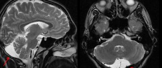
Schwannomas
Develop from Schwann cells of the auditory nerve, also called the 8th cranial, acoustic or vestibulocochlear nerve.
Places of formation of schwannomas
Usually located between the cerebellum and the pons, in the region of the posterior fossa. They grow very slowly, are quite rare (no more than 8%), and develop in middle age. The risk of the disease is twice as high in women.
Localization of neoplasms
Extracerebral formation of the frontal region of the volumetric type almost always appears with virtually no signs. At the same time, the pathology can lead to a significant deterioration in the patient’s health after an increase in size. If it is located on the left side of the frontal area, patients will most often experience problems on the right side of the body. In this case, headaches, atony and muscle seizures may appear. If the brain formation is located on the right side, the symptom complex will be located on the left.
Meningiomas most often occur in women against the background of hormonal changes in the body. Pregnancy often leads to this. Also, they can be triggered by malignant degeneration of the mammary glands with subsequent metastasis, including to the brain.
Craniocerebral injuries can lead to the appearance of various pathologies of a three-dimensional nature. They can develop both after direct damage and after a certain period of time. Among the risk group are patients who have been exposed to radioactive radiation and are also in contact with various toxic substances
It is possible to detect the localization of the disease only by conducting a comprehensive diagnosis:
MRI will detect extracerebral space-occupying formations (temporal region, falx, parietal and other areas). Can be supplemented with angiography and contrast methods. The technique is effective even for diagnosing small formations.
CT scans are excellent for finding meningiomas located in different parts of the brain.
Angiography can be used to assess the flow and flow of blood in the new body, as well as estimate its volume. The treatment and surgical procedure is impossible without assessing the trophism of the extracerebral formation.
Extracerebral space-occupying formations of the posterior cranial fossa, including those arising from the cells of the arachnoid membrane, can reach relatively large sizes. Often the patient is diagnosed with mental disorders that affect behavior, performance and ability to socially adapt.
The extracerebral formation of the middle cranial fossa has approximately the same properties as a tumor of the posterior fossa. The area of occurrence directly affects the symptomatic complex.
The extracerebral formation of the falx is usually located in the area of the falx. The pathology is characterized by epileptic seizures and severe convulsions. In case of complications, paralysis of the legs may appear, as well as problems with the functions of the pelvic organs.
An extracerebral space-occupying lesion in the parietal region is characterized by compression of the orbital pair of cranial nerves. Symptoms may include blurred vision, double vision, and more.
Choroid plexus papillomas
They arise from mutated cells of the choroid plexuses, which are part of the structure of the membranes lining the ventricles of the brain and producing cerebrospinal fluid (CSF). Typically diagnosed in children and young people.
Traditional sites of formation of choroid plexus papillomas
In children with brain tumors under the age of one year, choroid plexus papillomas range from 10 to 20%, from 1 year to 1 year – 2-4%. At the same time, depending on age, the localization of the lesion also differs: the older the patient, the lower the tumor is located.
What it is
A benign brain tumor is an intracranial neoplasm that begins to form as a result of the fact that the cells of one or another brain tissue (or any other tissue located in the skull) lose the ability to control their own division. The ability to differentiate is partially or completely preserved, so the resulting cells resemble in their structure exactly the tissue from which they originated.
Structural origin. What cells can participate in the formation of brain tumors (both benign and malignant):
- glial – auxiliary cells of nervous tissue, their totality is called “glia” or “neuroglia”;
- neurons are structural and functional units of the nervous system involved in conducting, processing and storing information in the form of electrical and chemical signals;
- astrocytes are stellate glial cells with a specific set of functions, their totality is called “astroglia”;
- oligodendrocytes are a separate type of neuroglia that are designed specifically to isolate axons belonging to neurons in the central nervous system;
- ependymal (or ependymal) epithelium - a thin epithelial membrane lining the walls of the ventricles of the brain and the central canal of the spinal cord, can also be called simply “endyma”.
Origin from other fabrics. All the cellular structures listed in this list belong to different areas of the brain. In addition to them, the following cells can participate in the formation of tumors:
- lymphatic tissue;
- blood vessels of the brain;
- cranial nerves;
- meninges of the skull;
- glandular formations - pituitary gland and epiphysis.
Cysts
Although they are considered benign, they can cause serious problems when located in structures that control vital body functions.
Cysts can appear in a variety of areas of the brain
They are varied, the most common are arachnoid (filled with fluid), colloid (filled with a thick iron-like substance), dermoid and epidermoid (filled with soft tissue) cysts.
Causes of benign neoplasms, their symptoms and types
Despite the fact that scientists from different countries have conducted many studies, no clear causes of benign brain tumors have yet been identified. One of the factors influencing its appearance and development is radiation. Moreover, this disease can affect not only those people who were in the zone of radioactive radiation for a certain time, but also their descendants, sometimes even after several generations.
Some doctors believe that the cause of tumors is the negative impact of cellular communications, high-voltage power lines and harmful chemicals used in various industries, for example, in the manufacture of plastic products. Constant and long-term use of a sugar substitute (aspartame) also, according to scientists, leads to the development of the disease. But genetics cannot be ruled out, because this area has also not yet been sufficiently studied, and illnesses of parents and other relatives can provoke the appearance of the disease in future offspring.
In the earliest stages, the disease may not manifest itself in any way. Or have vague and ambiguous signs, which many attribute to simple malaise. It is important to pay attention to them and consult a doctor in time for diagnostic measures. The main symptoms of a benign brain tumor include:
- disturbances in the functioning of the organs of vision, hearing and smell that occur for no apparent reason;
- deviations in the activity of the musculoskeletal system, coordination of movements and balance;
- unreasonable problems with speech, memory, logic and concentration;
- possible convulsions and uncontrolled muscle twitching;
- frequent vomiting and nausea that occur in the absence of food poisoning, changes in blood or blood pressure and other compelling reasons;
- numbness of hands and feet;
- constant pain in the head, most often of a localized nature and indicating the location of the tumor (temporal, occipital, brainstem, and so on);
- partial paralysis of the facial muscles.
Attention! If you experience one or more of the following symptoms for a week or more, you should immediately contact a specialist. It must be remembered that timely diagnosis of the disease is a chance for recovery and complete remission.
To date, a sufficiently large number of types of benign neoplasms have been identified that affect the tissue of nerve endings, veins and arteries. One of the most common types of this disease is meningioma, which occurs in the membranes of the brain. Quite often, acoustic schwannomas are diagnosed, affecting the structures of nerve endings and tissues. A pituitary tumor occurs in fifteen percent of cases. It causes disruptions in the activity of the pituitary gland, like craniopharynoma, but this pathology is more common in children than in adults.
Symptoms of a benign brain tumor
It is necessary to immediately make a reservation: it is impossible to determine by any external signs that a benign and not a malignant neoplasm has appeared in the brain. Moreover, certain disturbances in well-being can only cause the doctor to suspect a tumor process. Only diagnostics can confirm or refute these suspicions. At the same time, based on the patient’s complaints, the neurologist makes preliminary conclusions and draws up an examination plan.
“Even a benign tumor that grows in the head is potentially dangerous,” says Robert Fenstermaker, MD, chairman of the department of neurosurgery at Roswell Park Cancer Center (USA). “The space inside the skull is not unlimited, and even if a brain tumor is benign and grows slowly, it will eventually begin to compress the brain, symptoms will develop and this can be life-threatening.”
Benign brain tumors manifest themselves in a variety of symptoms, the nature of which is primarily determined depending on the location of the lesion and its size.
- An extremely slowly growing chondroma may not show itself in any way for a long time. When the tumor grows in size, it begins to compress nearby structures, which most often leads to headaches, hearing impairment, and visual hallucinations.
- With choroid plexus papillomas, general cerebral symptoms are observed, including characteristic headaches and other signs of increased intracranial pressure.
- It is impossible to identify any general symptoms for cysts due to the wide variety of neoplasms of this type and the possibility of their appearance in a variety of places.
- Ependiomas are characterized by irritability, vomiting, insomnia, and headache. In children under one year of age, one of the first symptoms is often an abnormal increase in head size.
- The symptoms of benign adenoma and malignant adenocarcinoma of the pituitary gland are identical, therefore, when making a diagnosis, other features are taken into account, including the absence of metastases and growth into neighboring sections, scan data, etc. Among the most common manifestations are headache, behavioral disturbances and visual disturbances. Signs of this type of hormone-producing tumors depend on the type of hormone produced.
- Meningiomas grow slowly and can become large before they begin to interfere with normal brain function. Symptoms depend on the location of the tumor. Most often, patients complain of headache, weakness in the arm or leg. Seizures, personality changes, and vision problems may also occur.
- In the vast majority of cases, lipomas do not cause any health problems.
- Schwannomas are characterized by unilateral hearing loss and noise or ringing in the ear. Less often, patients complain of dizziness. If the process affects the 7th cranial nerve, its paralysis may develop. Sometimes there is difficulty swallowing, changes in taste, unsteady gait, and disturbances in eye movements.
Causes of brain tumors in adults and children
According to official statistics, about 6% of all neoplasms are tumors in the skull. In childhood, they are less common; in older people, the risk of developing benign or malignant tumors is higher, and treatment is more difficult. Although there is still no exact data on why cells change structure and begin to grow uncontrollably. A relationship has been identified between the development of tumor processes and the following factors:
- some genetic diseases, oncology in relatives;
- exposure to toxins, carcinogens, ionizing radiation;
- immunodeficiency states;
- traumatic brain injuries;
- intrauterine disorders of the development of fetal brain tissue.
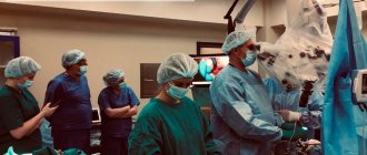
Treatment of benign brain tumor: basic principles and choice of method
In some cases, the doctor may choose active surveillance. This applies to patients with arachnoid cysts and meningiomas, as well as lipomas, which do not cause significant health problems and, as a rule, are detected by chance or during preventive cancer screening.
If a patient is diagnosed with a benign brain tumor that is “unfortunately” located, interferes with the quality of life for other reasons, or has a high risk of malignancy, treatment is aimed at removing the tumor (surgery) or destroying it using modern radiosurgical methods.
For example, some meningiomas can be removed by traditional surgery, while others are inoperable. In the latter case, the tumor can be destroyed using CyberKnife, Gamma Knife, TrueBeam installations. Depending on the characteristics of the tumor, tomotherapy or proton therapy may also be the optimal treatment method.
Operable colloid, epidermoid and dermoid cysts are most often removed in the traditional way or using endoscopic or microsurgical technologies. If indicated, additional antibacterial drug therapy may be prescribed. Sometimes cyst removal is complicated, impossible, or only partially possible.
Radical removal of chondroma, choroidal papilloma, ependyoma, pituitary adenoma can be carried out surgically or radiosurgically.
When treating patients with schwannomas, microsurgical methods or stereotactic radiosurgery are usually used. According to indications, medication, hormonal and radiation therapy are additionally prescribed.
Leading neurosurgeons have high hopes for intraoperative fluorescent control for the purpose of total resection of tumors, in particular pituitary adenomas, since incomplete removal of neoplasm leads to relapses in 20% of patients. But although pituitary adenomas are rarely malignant, they can cause serious problems by compressing nearby areas of the brain, which can lead to Cushing's disease, gigantism, blindness and death.
At the medical school. Perelman University of Pennsylvania developed the first organ-specific marker OTL38, which consists of two parts: folate and an infrared-luminescent dye. Since folic acid is actively used for the development and growth of neoplasms, they are characterized by a high content of folate receptors on the cell surface (which is sometimes more than 20 times higher than the normal level). By binding to receptors, OTL38 causes luminescence of the affected tissues, making it possible to accurately identify the boundaries of the tumor and completely remove it.
As noted by Yu.K. Lee, MD, PhD, professor in the Department of Neurosurgery at the University of Pennsylvania, said, “This study heralds a new era in personalized tumor surgery. Surgeons can now see the molecular characteristics of a patient’s tumors, not just light absorption or reflectivity.”
If you need a second opinion to clarify your diagnosis or treatment plan, send us an application and documents for consultation, or schedule an in-person consultation by phone.
+7 499 490-24-13
Expert opinion
Symptoms and signs of brain cancer
Many other diseases have similar symptoms to the signs listed above. The initial stage of development of brain cancer in a patient is indicated by the combination of these signs. It is at this stage that the disease is easiest to overcome. But most do not pay attention to the primary symptoms, and the disease begins to progress.
The patient's intracranial pressure gradually increases, and the growth of pathological tissue increases the impact on the lining of the brain. The second stage of cancer development begins. Changes in tissues have already occurred, but the pathology can still be treated quite successfully. During the second stage of the disease, the patient may experience muscle weakness on one side of the body, sudden dizziness, severe fatigue, loss of sensation in the limbs, and blurred vision (double vision).
However, all these symptoms are similar to those of epilepsy, hypotension and neuropathy. Only by conducting appropriate examinations can an accurate diagnosis and the reason provoking the patient’s state of health be established.
Since each area of the brain is responsible for its own functions, the symptoms of the disease can be focal. Thus, it is possible to determine the location of the pathological neoplasm already at the initial stages:
| Damage area | Focal symptoms |
| Occipital lobe | Visual impairment in one eye, blindness, blurred vision or double vision. |
| Frontal and temporal lobes | Sleep is disturbed, anxiety, depression, aggression occurs, memory deteriorates, consciousness is confused, hallucinations, antisocial behavior, euphoria, and sudden loss of consciousness may appear. |
| Cerebellum | Constant feeling of nausea, vomiting, loss of ability to control the movement of certain parts of the body, muscle weakness. |
| Parietal lobe | Sensory disorders appear. The patient loses the ability to understand speech, is unable to pronounce words clearly, and coordination of movements is impaired. The skills of habitual actions are lost, their sequence is confused. |
| Brain stem | Paresis of the facial nerve occurs, which manifests itself in facial asymmetry, strabismus, tremors in the limbs, difficulty swallowing, hearing loss, staggering when walking, and inability to coordinate movements. |