Methods of functional diagnostics in neurology. Part 1
What methods of functional diagnostics exist, what do they allow you to find out, what are the advantages of these methods, what is an EEG, when is it worth doing an electroencephalogram, says Dmitry Aleksandrovich Ovchinnikov, neurologist, functional diagnostics doctor.
Ovchinnikov Dmitry Alexandrovich Doctor of functional diagnostics.
Hello. My name is Dmitry Aleksandrovich Ovchinnikov. I am a neurologist, functional diagnostics doctor at the Scandinavia clinic. Today we will talk about functional diagnostic methods in neurology. Neurology is a branch of medicine that deals with the diagnosis and treatment of disorders of the nervous system. Functional diagnostics is a very diverse group of methods that are united by one principle. Assessment of not only and not so much the static state of the system, but the functioning and functioning of organs. In this way, functional diagnostic methods compare favorably with other groups of techniques, such as radiation diagnostic methods, where the structure is assessed primarily at each specific point in time, or laboratory diagnostic methods, which evaluate the composition of tissues and fluids, but only at one specific point in time. It becomes clear why functional diagnostics is so important in neurology, because the nervous system is an extremely dynamically changing structure, the main task of which is to respond to external and internal stimuli and change its functioning over time.
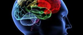
Let's briefly look at the functional diagnostic tools available to neurologists. A large group of functional diagnostic methods are united by their electrophysiological principle. Nerve cells in the course of their work are capable of being excited, changing the polarity of their membranes and generating an action potential. Fixation and registration of this action potential allows a number of functional diagnostic methods to study nerve tissue and draw certain conclusions about the functioning of the nervous system. These methods include electroencephalography, electromyography, evoked potential methods, and a number of other electrophysiological techniques.
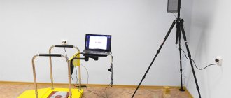
One of the electrophysiological methods of functional diagnostics in neurology is electroencephalography, or EEG for short. Electroencephalography is a technique for assessing the bioelectrical activity of the brain. During the study, electrodes are placed on the patient's head to record brain activity in various areas of the brain. Also during the research, functional tests are carried out, such as photostimulation or tests with changes in the gas composition of the blood, followed by stimulation of the brain with hyperoxygenation. The results of measuring this activity are currently processed by a computer and displayed on the monitor in the form of graphs. The doctor is able to perform various types of analysis of this recording so that the conclusions are as comprehensive and profound as possible. The studies are carried out in a shaded room, but do not involve any discomfort. Lasts from 15 to 30 minutes.
There are also methods for long-term recording of electroencephalograms, but they are used less routinely. With some exceptions, an electroencephalogram does not require any preparation, but on the day of the study it is recommended to wash your hair to reduce skin resistance to electric current.
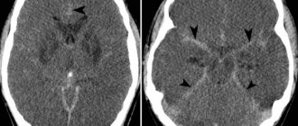
A serious advantage of the electroencephalogram over other research methods is the ability to assess the functioning of the brain over a certain period of time. What no other technique can provide, such as MRI, which allows a very precise and very comprehensive assessment of the structure and anatomy of the brain, but cannot assess its functioning during a specific drive. Or such as computed tomography, which evaluates primarily the bone structure of the skull and the structure of the brain without detailed differentiation. Thus, various types of electroencephalogram are the gold standard in the diagnosis of epilepsy and various epi-syndromes. No other technique currently used in clinical practice is capable of identifying these deviations.
The experience accumulated by the Soviet, Russian and international scientific schools allows the use of EEG not only in the diagnosis of epilepsy. Encelography makes it possible to indirectly judge violations of recoordination, the work of the hypothalamic-pituitary system, the functioning of the brain and cerebral cortex as a whole, and the relationship between the work of the cortex and subcortical structures. Certain changes in the encephalogram can give the doctor the opportunity to judge the functioning of the cognitive component and the exhaustion of the nervous system. Thus, today an electroencephalogram is one of the few methods that allows us to evaluate not only structural changes in the brain, but also changes in its functioning.
Indications for an encephalogram at the moment are loss of consciousness of any origin, functional disorders of the nervous system, such as neurotic, psychological, emotional, psychosomatic complaints of patients. Assessment of the consequences of traumatic brain injuries, assessment of the consequences of neuroinfections. Theoretically, we can evaluate the functioning of the hypothalamic-pituitary system and its impact on brain function. Previously, electroencelography was used to assess changes in the structural properties of nervous tissue to search for focal changes, such as tumors and hematomas. However, at the moment, MRI copes much better with these tasks.
In addition to the indications described above, it is advisable to conduct electroencelography when there are changes in brain function caused by vascular disorders, disorders caused by chronic changes in cerebral blood flow.
Publication date: 04/10/17
Classification of central nervous system diseases
Diseases of the central nervous system can be classified as follows:
- Vascular. Chronic brain failure, which often occurs in combination with cardiovascular pathologies and hypertension. This group of central nervous system diseases also includes acute circulatory disorders in the brain (stroke), which occur most often in adulthood and old age.
- Brain diseases. The most common central nervous system diseases affecting the brain include Alzheimer's disease, Norman-Roberts syndrome, sleep paralysis, hypersomnia, insomnia, etc.
- Infectious. They are usually very severe and pose a serious threat to life. Infectious lesions of the central nervous system include meningitis (inflammation of the membranes of the spinal cord and brain), encephalitis (an inflammatory disease of the brain of a viral nature), poliomyelitis (a severe disease characterized by damage to all brain structures), neurosyphilis (develops from infection with Treponema pallidum).
- Demyelinating. One of the most common demyelinating diseases of the central nervous system is multiple sclerosis, which gradually leads to the destruction of the nervous system. This group also includes epilepsy, disseminated encephalomyelitis, myasthenia gravis and polyneuropathy.
The presented classification is not complete, since diseases of the central nervous system also include degenerative, neuromuscular, neuroses, etc.
As a rule, disorders caused by damage to the central nervous system are irreversible, which is why in advanced cases such diseases lead to disability. If we talk about the causes of the development of diseases of the central nervous system, then the main ones are considered to be infections and parasites, traumatic injuries, pathologies of the heart and blood vessels, and heredity.
Respiratory function test
External respiration function
- Cost: 2,200 rub.
- Duration: 10 – 20 minutes
- Hospitalization: Outpatient
More details
In the CELT clinic, to diagnose respiratory diseases, a study of external respiration function is carried out, including an inhalation test. Another name for the method is spirometry. It is quite simple, but at the same time one of the most informative. It is used both to assess the condition of healthy people and in patients with diseases of the respiratory system. Recommended for risk groups: allergy sufferers, people with a predisposition to asthma, and smokers. Spirometry is often prescribed before surgery under endotracheal anesthesia.
In Moscow, a modern department of functional diagnostics operates at the CELT clinic. Expert class equipment, qualified and attentive specialists are the key to an accurate diagnosis and good health.
Differential diagnosis of spinal diseases
Spinal disorders that often cause pain without neurological deficits
| Vascular | — |
| Inflammatory, infectious | Steroid-dependent meningitis-arteritis (dogs), discospodylitis; osteomyelitis (dogs, cats) |
| Traumatic | Spinal fracture or subluxation (dogs, cats) |
| Abnormal | Occipito-atlanto-axial instability (dogs, cats), syringomyelia; hydromyelia (dogs, cats) |
| Metabolic | Hypervitaminosis A (cats) |
| Idiopathic | — |
| Neoplastic | Spinal tumors (eg, osteochondrosarcomas) (dogs, cats) |
| Degenerative | Degenerative disc diseases; (dogs, cats), spondiomyelopathy (wobbler syndrome); (dogs), degenerative lumbosacral stenosis (Degenerative Lumbosacral Stenosis. DLSS) (dogs.) |
Spinal diseases, often causing pain and neurological diseases
| Vascular | Extra-or intradural bleeding (dogs, cats) |
| Inflammatory, infectious | Bacterial meningomyelitis, fungal meningomyelitis; protozoal meningomyelitis (toxoplasmosis, neosporosis); feline infectious peritonitis; granulomatous meningoencephalitis |
| Traumatic | Spinal fracture or subluxation (dogs and cats) |
| Abnormal | Occipito-atlanto-axial instability; spinal dysraphism; congenital underdevelopment of half a vertebra; syringomyelia; hydromyelia (dogs and cats) |
| Metabolic | Hypervitaminosis A (cats) |
| Idiopathic | — |
| Neoplastic | Spinal tumors (eg, osteosarcomas, chondrosarcomas) (dogs and cats) |
| Degenerative | Degenerative disc disease (dogs and cats), spondiemyelopathy (wobbler) (dogs), degenerative lumbosacral stenosis (dogs), Osteochondral exostosis (various locations (dogs), dural ossification (dogs) |
Spinal disorders usually causing neurological deficits without pain
| Vascular | Emboli caused by fibrocartilaginous tissue (dogs and cats) |
| Inflammatory, infectious | |
| Traumatic | — |
| Abnormal | Arachnoid cyst |
| Metabolic | — |
| Idiopathic | — |
| Neoplastic | Intramedullary tumors (dogs and cats) |
| Degenerative | Degenerative myelopathy (dogs), breed-related degenerative spinal cord disease, lower motor neuron disease (dogs and cats) |
Differential diagnosis of forebrain and brainstem diseases
| Vascular | Bleeding, heart attack (cats, dogs); Feline ischemic encephalopathy (cats) |
| Inflammatory, infectious | Plague, rabies (dogs, cats), infectious canine hepatitis, feline infectious peritonitis, canine herpes virus, feline leukemia virus, feline viral immunodeficiency, encephalitis (Togavirus) (dogs); bacterial meningoencephalitis (cats, dogs); fungal infections (cats, dogs); toxoplasmosis (cats, dogs); neosporosis (dogs); migration of parasitic larvae, granulomatous meningoencephalitis (dogs), feline polyencephalomyelitis, pug encephalitis, Yorkshire terrier encephalitis |
| Traumatic | Concussion, bruises, lacerations (cats, dogs) |
| Abnormal | Hydrocephalus, lissencephaly, agenesis (cats, dogs) |
| Metabolic | Hypoglycemia, hepatic encephalopathy, uremic encephalopathy, diabetic coma, heat stroke, hypothyroid coma, hypoxia, thiamine deficiency, hypocalcemia, Ammonshorn necrosis, various intoxications |
| Idiopathic | Idiopathic epilepsy |
| Neoplastic | Primary tumors (meningiomas, gliomas, choroid plexus papillomas, pituitary tumors, skull tumors, nerve sheath tumors, teratomas); secondary tumors (metastatic tumors, tumors in surrounding tissues) |
| Degenerative | Storage diseases and other degenerative diseases |
Differential diagnosis of cerebellar diseases
| Vascular | Heart attacks, bleeding (dogs, cats) |
| Inflammatory, infectious | Panleukopenia (cats), granulomatous meningoencephalitis, canine distemper (dogs), canine herpes virus, feline infectious peritonitis, Cryptococcus neoformans (dogs, cats), toxoplasma gondii (dogs, cats). Neospora (Neospora) of dogs (dogs) |
| Traumatic | Contusion / rupture / hernia (dogs, cats) |
| Abnormal | Agenesis, hypoplasia, dysplasia (setter, fox terrier, chow chow, miniature poodle, beagle, bulldog, Samoyed dog breeds, Labrador retriever, Australian kelpie, domestic cats), Dandy-Walker syndrome (cats and dogs), Arnold-Chiari syndrome (dogs ) |
| Metabolic | Metronidasole (dogs), lead intoxication, organophosphorus compounds, hexachlorophene |
| Idiopathic | Idiopathic cerebellitis, “White Shaker Syndrome” (dogs) |
| Neoplastic | Primary tumors (cats, dogs), medulloblastomas, secondary tumors (dogs, cats) |
| Degenerative | Abiotrophy (Gordon Setter, Collie, Airedale Terrier, Terrier, Finnish Harrier, Bernese Mountain Dog, Kerry Blue Terrier, English Spaniel, Border Collie, Beagle, Samoyed, Fox Terrier, Labrador and Golden Retriever, Great Dane, Chow Chow, Rhodesian Ridgeback, domestic cats)GM1-gangliosidosis (Beagle, mixed breeds, English springer spaniel, Portuguese water dog, Siamese, domestic cats).GM-gangliosidosis (German shorthaired pointer, Japanese spaniel, domestic cat).Globoid cell leukodystrophy (West Highland white -terrier, cairn terrier, spitz, basset, poodle, domestic cat);Sphingomyelinosis (Siamese, Balinese and domestic cats)Mannosidosis (Persian and domestic cats) |
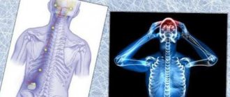
Localization of the lesion in patients with monoparesis (monoparlysis)
Monoparesis of the forelimbs is most often caused by diseases of the peripheral nerves or nerve roots. Diseases of the endplates of vertebral bodies or muscles affecting only one limb are very rare. Diseases of the spinal cord in the cervical region usually cause hemiparesis or tetraparesis. Thus, monoparesis of the forelimbs is usually caused by pathologies of the nerves and nerve roots of the affected limb.
Monoparesis of the pelvic limb is also usually caused by damage to a nerve or nerve root. The damage may be outside the spinal canal (peripheral nerve) or inside the canal (cauda equina nerves). In addition, unilateral spinal cord lesions caudal to Th2 can lead to monoparesis of the pelvic limbs. This is an exception to the general localization pattern. Vascular lesions are the most common cause of such unilateral spinal cord disease (eg, related to fibrocartilaginous embolism (fibrocartilage embolism)).
Differential diagnosis of monoparesis
| Vascular | Fibrous emboli, ischemic encephalopathy (cats, dogs) |
| Inflammatory, infectious | Brachial plexus neuritis (cats, dogs) |
| Traumatic | Trauma (neuropraxia, axonotmesis, neuronotmesis) of the brachial plexus or individual nerves (cats, dogs) |
| Abnormal | — |
| Metabolic | Hypothyroidism (dogs) |
| Idiopathic | — |
| Neoplastic | Malignant tumor of the nerve sheath (cats, dogs). Secondary: lymphoma, skeletal tumors (cats, dogs) |
| Degenerative | Lateralized disc disease, degenerative foraminal stenosis causes compression of the nerve roots (nerve root compression) |
CNS diseases: general symptoms
Diseases of the central nervous system have a wide range of symptomatic manifestations. These include:
- movement disorders (paresis, paralysis, akinesia or chorea, impaired coordination of movements, tremor, etc.);
- tactile sensitivity disorders;
- disturbances of smell, hearing, vision and other types of sensitivity;
- hysterical and epileptic seizures;
- sleep disorders;
- disturbances of consciousness (fainting, coma);
- mental and emotional disorders.