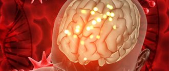
What is cerebral encephalopathy
Encephalopathy is a non-inflammatory disease of the brain.
As a result of various diseases and pathologies, the blood supply to the brain is disrupted, and oxygen starvation of the brain tissue occurs. Due to lack of oxygen, nerve cells die, areas of the brain shut down and encephalopathy occurs. The disease can be congenital or acquired:
- Congenital encephalopathy of cerebral vessels occurs due to intracranial birth trauma, abnormalities in brain development, and infection during pregnancy.
- Acquired encephalopathy occurs in adults as a result of illness and injury.
Hypoxic-ischemic encephalopathy (HIE)
What is perinatal encephalopathy?
Perinatal lesions of the nervous system in newborns are a number of conditions and diseases of the brain, spinal cord and peripheral nerves, combined into a common group according to the time of exposure to damaging factors. The perinatal period includes the antenatal, intranatal and early neonatal periods. The antenatal period begins with the 22nd week of intrauterine development and ends with the beginning of labor. The intrapartum period includes the act of childbirth from the onset of labor to the birth of the child. The neonatal period is divided into early neonatal (corresponding to the first week of a child’s life) and late neonatal (from the 8th to the 28th day of life inclusive) periods.
Why does perinatal encephalopathy occur?
Perinatal encephalopathy is a common complication of the pathology of pregnancy and childbirth. This condition is caused, as a rule, by oxygen starvation of the central nervous system, which occurs with various deviations in the course of the perinatal period, which is difficult for the child. Perinatal brain damage accounts for more than 60% of all pathology of the nervous system in early childhood, and is directly involved in the development of diseases such as cerebral palsy, epilepsy, and minimal brain dysfunction. Depending on the degree of damage to the central nervous system, as well as the availability of correct and timely treatment, different outcomes of perinatal encephalopathy are possible - a third of patients, as a rule, require minimal rehabilitation support for recovery, a third require serious, including drug treatment, and a third, even with competent and long-term medical care, will still have certain health problems in the future.
Among the causes of perinatal brain damage, the leading place is occupied by intrauterine and intrapartum (during childbirth) hypoxia of the fetus, the second most important place belongs to the factor of mechanical trauma to the child during childbirth - usually in combination with one or another severity preceding intrauterine hypoxia. The structure of etiopathogenetic factors of perinatal pathology also includes infectious (including viral) and toxic-metabolic variants of damage to the nervous system. Thus, among the factors causing perinatal damage to the central nervous system, the following are distinguished:
- Intrauterine hypoxia (oxygen starvation) of the fetus
- Fetal hypoxia during labor
- Mechanical trauma during childbirth
- Infectious (viral) factors
- Toxic factors
- Hereditary factors
- Combination of these factors
Among the risk factors for perinatal encephalopathy in children, a large share belongs to maternal morbidity, which, according to numerous studies, not only violates preconception health, that is, the health responsible for the birth of healthy offspring, the parent couple, but also has a direct negative impact on the fetus.
When studying the structure of the general pathology of pregnant women over the past five years, a significant increase in their morbidity is noted, primarily due to an increase in the frequency of diabetes mellitus and thyroid diseases. During pregnancy, these diseases lead to placental insufficiency and impaired absorption and assimilation of nutrients through the placenta, as well as a deficiency in the transport of oxygen and carbon dioxide, which is manifested by fetal growth retardation syndrome, intrauterine hypotrophy, immaturity of the lungs and surfactant. It has been established that a decrease in uteroplacental blood flow serves as an objective indicator of hypoxic brain damage.
How does perinatal encephalopathy manifest?
Clinically, perinatal encephalopathy is manifested by several symptom complexes, the most common of which are: “delayed motor and mental development”, “hypertensive-hydrocephalic syndrome”, “muscular dystonia syndrome”, “convulsive syndrome”, “brain stem lesion syndrome”.
What is psychomotor development delay and how to treat it?
The most difficult period in a child's life is the period of infancy. The rate of growth and development of babies is simply rapid. At the same time, a child is born with far from complete development of all organs and tissues, which after birth are gradually improved and approach the organs of an adult in their structure and functions, therefore a detailed study of the main aspects of the development of a child in the first year of life is an urgent need for pediatric doctors of all specialties. Tracing the development of a baby from the neonatal period to 1 year, experts focus on the child’s anthropometric data, his psychomotor development (motor activity), speech development, and the formation of skills and abilities.
Based on the characteristics of the psychomotor status of a healthy newborn child, carefully monitoring the decay of innate reflexes and the acquisition of motor and social skills over time, one can assess how correctly and harmoniously the baby’s development proceeds in the first year of life.
Delayed psychomotor development and the formation of motor skills is observed in various hereditary diseases, in children with pathologies of the central nervous system, leading to the formation of persistent motor disorders, including cerebral palsy. In this case, a delay is understood as a lag in development of 2 or more months. Particular attention should be paid to children with regression of previously acquired motor and psycho-speech skills, which may indicate degenerative diseases of the nervous system.
A mild (tempo) form of developmental delay is often found in premature infants, with chronic non-neurological diseases, rickets. For moderate to severe delays, special examination and treatment are recommended.
Non-drug correction of psychomotor delays in children of the first year of life includes:
Physical rehabilitation: a variety of therapeutic massage, therapeutic exercises, “position” treatment (styling, splints, “collars”, etc.), Vojta therapy; exercises in water and hydromassage; dry immersion (imitation of weightlessness); physiotherapy (alternating magnetic field, sinusoidal modulated currents, electrophoresis, paraffin therapy, laser therapy, light and color therapy)
Psychological and pedagogical correction: correctional (conductive) pedagogy; psychotherapeutic correction in the mother-child dyad (skin-to-skin contact, kangaroo care); music therapy, aesthetic therapy; tactile-kinesthetic stimulation. For premature babies, a combination of two or three “soft” methods of physical influence with psychoemotional and psychosensory correction is especially recommended, which helps simulate the effect of the so-called “sensory rooms” used in the rehabilitation of older patients.
Drug therapy , which is used by neurologists to correct motor and mental development in the first year of life, is mainly represented by peptide drugs that trigger endogenous compensation mechanisms and return the child to normal development.
What is muscular dystonia syndrome and how is it treated?
Tonic disturbances in an infant can be local (associated with one muscle group) or widespread (associated with several muscle groups). Based on the nature of changes in muscle tone, they are divided into hypotonia (decreased tone), hypertonicity (increased tone) and dystonia (incorrect combination of increased and decreased tone).
The typical and most common local tone disorder can be considered infantile torticollis.
Torticollis, as a rule, is caused by mild underdevelopment or traumatic damage to the sternocleidomastoid muscle and roots emerging from the C5-C6 segments of the spinal cord or accessory nerve; by the age of 6-9 months, torticollis usually disappears. However, mild symptoms of torticollis can also be detected at an older age. Resistance is felt when turning in the direction opposite to the affected muscle.
As treatment, your child may be prescribed applications, electrophoresis or traumeel injections, Vibrukol rectal suppositories, and placing the head in the correct position using a steering wheel pillow.
Muscular hypotonia ("floppy child" syndrome) is manifested mainly by a decrease in resistance to passive movements and an increase in their volume (the appearance of looseness in the joints). In severe cases, hypotension affects the child's posture: the extreme expression of diffuse muscular hypotonia is the “frog posture.” “Floppy baby” syndrome can occur in a wide range of diseases: severe non-neurological diseases, rickets, severe perinatal hypoxia (oxygen deficiency) and birth trauma, leading to the formation of cerebral palsy, metabolic disorders, neuromuscular diseases, developmental abnormalities and degenerative diseases of the nervous system, chromosomal syndromes, some forms of endocrine pathology.
Diffuse muscle hypotonia often occurs with hereditary pathology. In this case, insufficient weight and height gain, epileptic seizures, and episodes of acute cerebrovascular accident may occur. A pronounced decrease in muscle tone is often found in premature infants, and by 3-6 months they may transform hypotension into spastic syndrome.
In case of severe persistent muscle hypotonia, especially in combination with delayed psychomotor development, it is necessary to conduct regular rehabilitation courses (massage, exercise therapy, physiotherapy, hydro and balneotherapy, acupuncture, speech therapy classes, drug treatment).
Muscle hypertonicity can manifest itself from minor (not impeding movement and development) to severe up to spastic syndrome (an extreme form of hypertonicity). An increase in muscle tone can be combined with activation of tonic reflexes and a delay in the extinction of unconditioned reflexes.
Tonic reflexes in combination with increased muscle tone have a pathological effect on the child’s posture. This picture is often observed in premature and immature children, as well as in the formation of cerebral palsy. In the latter case, unconditioned reflexes may even intensify.
Dystonia and hyperkinesis (irregular, unnecessary, pretentious and excessive movements) - these clinical manifestations often appear delayed, after 3-6 months of life, but can also be observed from birth.
Hyperkinesis, as a rule, characterizes damage to the subcortical nuclei of the brain, which are often combined with other neurological symptoms (for example, hearing loss). Both hypertonicity and dystonia, forming long-term incorrect postural attitudes in a child, over time lead to deformations of the baby’s skeleton. Therefore, if there are motor disorders, in addition to a neurologist, such a child must be observed by an orthopedist.
From the moment of diagnosis, it is recommended to begin a course of rehabilitation treatment. Restorative therapy (in the absence of contraindications from the cardiovascular, respiratory systems, absence of epileptic paroxysms, etc.) includes massage, physical therapy, orthopedic styling, physiotherapeutic treatment, hydro and balneotherapy, acupuncture, speech therapy classes, classes in sensory rooms.
Early development methods and drug therapy are chosen individually, depending on the severity of the syndrome and its combination with developmental delay, hyperkinetic and dystonic syndromes.
Drug therapy prescribed to correct muscle tone in children in the first year of life is divided into drugs that reduce muscle tone (antispastic drugs) and drugs that correct muscle hypotension.
What is hypertensive-hydrocephalic syndrome and how to treat it? Normally, the increase in head circumference at 1 year of life is 11-12 cm:
- During the first trimester of life, head circumference increases by 4 cm (1.5 cm/month)
- During the second trimester of life - by 3 cm (1 cm/month)
- During the second half of life, head circumference increases by 3-4 cm (0.5 cm/month)
A pathological increase in head circumference as a symptom of hydrocephalus develops as a result of blockage of the cerebrospinal fluid ducts at various levels and when the relationship between the processes of production and absorption of cerebrospinal fluid is disrupted.
Hydrocephalus:
Congenital hydrocephalus is a disease that develops in the fetus during pregnancy and the child is born sick. The main causes of congenital hydrocephalus are developmental defects, less often intrauterine infection, much less often the cause of hydrocephalus is hemorrhage into the ventricles of the brain in the fetus.
Acquired hydrocephalus - the disease develops after the birth of a child, sometimes in the earliest stages of life. The causes of acquired hydrocephalus are intraventricular hemorrhages, infections affecting the central nervous system - meningitis, encephalitis, traumatic brain injury, brain tumors.
Clinical symptoms of intracranial hypertension:
- Changes in the child's behavior: restlessness, frequent and monotonous crying, throwing back the head, frequent regurgitation
- Delayed mental, motor and psycho-speech development
- Opening of the sagittal suture more than 0.5 cm, bulging, tension of the large fontanelle
- Changes in the shape of the skull with a high forehead (tower skull) or with a sharply protruding occipital protuberance in combination with excessive growth of head circumference, predominance of head circumference over chest circumference
- Graefe's sign, “stagnant” changes in the fundus
- Increased muscle tone, mainly in the hands and feet
- Tremor (shaking) of the hands with a tendency to open them
In addition to hydrocephalus, children with the following conditions may have a “hydrocephalic shape” skull with a large head circumference:
- Rickets
- Constitutionally large-headed children
- Infants with syndromic conditions and hereditary diseases
To adequately assess the situation, it is always necessary to compare the head circumference with the chest circumference, assess the size of the head of the child’s parents, and focus on the results of additional research methods (fundus examination, NSG, CT, MRI).
Drug therapy for the correction of hydrocephalic syndrome in the first year of life is mainly represented by drugs with diuretic and vascular effects.
Hydrocephalus is a disease that is treated not by a neurologist, but by a neurosurgeon. Without consulting a neurosurgeon, a neurologist cannot make a decision on the treatment of hydrocephalus. If for some reason surgical treatment of hydrocephalus is not indicated for a child, a neurologist and a neurosurgeon observe such a patient together.
Insufficient growth of head circumference is observed in progressive hereditary degenerative diseases, in severe organic lesions (secondary microcephaly - when the brain does not grow and, as a result, skull growth slows down), in craniostenosis (pathology of the skull bones, requiring surgical treatment by a neurosurgeon).
It should be noted that the rate of overgrowth of the large fontanel in each child is strictly individual, and is more likely related to the characteristics of mineral metabolism than to the pathology of the nervous system. The preservation of an open large fontanel by the end of the second half of the baby’s life in the absence of neurological complaints is not a cause for great concern, just as the accelerated closure of a large fontanel is in no case a reason to stop the planned prevention of rickets in an infant.
What is seizure syndrome and how to treat it? In the practice of a pediatric neurologist, there are many conditions that occur paroxysmally, i.e. arising suddenly, short-lived and abruptly ending. An example of paroxysms are epileptic seizures, usually accompanied by loss of consciousness. However, not all paroxysms occur with impaired consciousness; some of them are characterized by very unusual symptoms, which can cause diagnostic errors.
It is particularly difficult to identify and differentiate various paroxysmal conditions in infancy. To choose the right treatment tactics, it is necessary to distinguish between paroxysmal states of epileptic and non-epileptic nature; however, only an experienced neurologist-epileptologist can clearly distinguish between the cause of paroxysms and their prognosis.
Affective-respiratory paroxysm is a short-term cessation of breathing at the height of crying with pale or blue discoloration of the skin. It is necessary to differentiate affective-respiratory paroxysms from apnea syndrome (cessation of breathing), which often occurs in premature and immature infants, as well as in children with pathologies of the cardiovascular and respiratory systems, and the brain stem. For clear differentiation, a polysomnography study is carried out, which allows, using simultaneous recording of an EEG, electrocardiogram and spirogram (breathing graph), to determine what is the root cause of breathing problems in the baby. By themselves, affective-respiratory paroxysms are not epileptic, but the frequency of their further transformation into epilepsy is quite high.
Paroxysmal sleep disorders represent a fairly large group of conditions that need to be differentiated from both non-neurological diseases and epileptic paroxysms. The most typical representatives of paroxysmal sleep disorders are nightmares, during which the baby sharply and piercingly begins to scream and cry, without fully waking up and reacting little to the environment. The presence of frequently recurring nightmares may indicate problems in the child’s mental sphere.
Febrile seizures are the most common type of paroxysmal conditions in childhood. Febrile seizures (FS) are paroxysms of varying duration, occurring primarily in the form of convulsive seizures in infants and young children at a body temperature of at least 37.8-38.5°C. FS have an increased likelihood of transformation into epilepsy.
There are typical and atypical FS. The first ones have a short duration (up to 15 minutes), a generalized nature (all limbs are involved, consciousness is briefly lost); indicators of the child’s psychomotor development usually correspond to age; there are no typical changes on the EEG. In atypical FS, the duration of the attack is more than 15 minutes (up to several hours), there is a predominant involvement of the limbs on one side; sometimes after an attack Todd's paresis occurs - transient muscle weakness in the limbs affected by the spasm (in 0.4% of cases); epileptic focal changes are not uncommon on the EEG.
Neonatal seizures (NS) occur during the first 4 weeks of life of a full-term newborn (from the 1st to the 28th day) and somewhat later in premature infants. Neonatal seizures are quite difficult to distinguish by clinical manifestations from benign movements of the child - their presence is established using an EEG with video recording. Myoclonic seizures have the most severe prognosis and may indicate the onset of early severe epilepsy.
Epilepsy is a chronic brain disease characterized by repeated unprovoked seizures that cause impairment of motor, sensory, autonomic, mental or mental functions resulting from excessive neural discharges in the cerebral cortex. The prevalence of epilepsy among the child population is about 10 cases per 1000 children.
Epilepsy is a very unpleasant but very common neurological disease, especially in childhood. In addition to creating life-threatening conditions for the infant during prolonged and complicated convulsive seizures, epilepsy leads to long-term negative consequences, disrupting the cognitive, psycho-social and motor development of the child. Modern anticonvulsants provide control over most forms of epilepsy, but parents are afraid to give them to children because of the poor reputation of older anticonvulsants for fear of their side effects. All your concerns should be discussed with a specialist. Don't deprive your child of a healthy future because of your prejudices.
What are the symptoms of brain stem damage and how are they treated?
The brain stem, or brain stem, is a traditionally distinguished part of the brain, which is an extended formation that continues the spinal cord.
The brain stem contains vital centers responsible for breathing, swallowing, cardiac activity, coordinated eye movements, etc. In newborns with neurological problems, the activity of these centers may be insufficiently developed or damaged. Underestimation of symptoms of brain stem damage can lead to critical consequences.
Bulbar and pseudobulbar disorders in children with damage to the nervous system are noted from birth. They manifest themselves as sluggish sucking, then they begin to choke on saliva and food, choke, food pours out through the nose, the tone of the tongue changes, and speech formation is disrupted.
Oculomotor disorders are very typical for infants with central nervous system pathology. The range of possible disorders includes nystagmus (trembling of the eyeballs), various types of strabismus. It is extremely important to isolate patients from this group who are visually impaired, since oculomotor disorders may be the first symptoms of impaired visual function (for example, the so-called “floating nystagmus of the blind”). Patients require joint supervision by a neurologist and an ophthalmologist.
Respiratory and heart rhythm disturbances are also typical for patients with impaired function of stem structures. Infants born premature and low birth weight are especially susceptible to breathing problems (losses) or apnea and arrhythmias. In such a situation, the fundamental task is the differential diagnosis of neurological and non-neurological (cardiological, respiratory) causes of the identified disorders. Such patients require careful diagnosis and observation by a neurologist, pulmonologist, cardiologist and ENT doctor.
Be sure to ask specialists about the presence of brainstem disorders in your baby. Their consequences can be very serious (for example, respiratory disorders such as apnea cause sudden infant death syndrome, and swallowing disorders and chronic “choking” lead to severe aspiration pneumonia), but with proper and careful care (correct position of the child during sleep, feeding with a raised head end and a special diet) troubles can be avoided even without additional medications.
Do all developmental abnormalities in an infant require special treatment?
Variants of the norm in the first year of life are the following events:
- Physiological tremor - rhythmic twitching of the arms or chin during crying or feeding. Normally, it occurs in half of children under 3 months of age. Tremors are often observed in premature newborns up to 6 months of age.
- Physiological astasia-abasia - from 2 to 5 months, the child, when “suspended”, does not rest on the plane with his legs, presses them in, and accordingly there are no stepping movements (due to the fading of the support reflex)
- Yactation - rocking (self-soothing) movements of the body and/or head, which appear in a child mainly before bedtime, are characteristic of children raised without parental care
- The revival complex is a variety of chaotic movements that accompany emotional, usually positive, “outbursts.” Particularly common in children with immobilized limbs (for example, those receiving casting or orthopedic devices)
Thus, all children with perinatal encephalopathy require long-term dynamic observation by specialists and individually selected repeated courses of rehabilitation treatment, with an interval of no more than 3 months and with regular monitoring of its effectiveness, during the first year of life in outpatient rehabilitation centers, hospital departments and district clinic
However, in order for the rehabilitation process to be continuous, healthcare professionals are not enough - it is necessary to involve the child’s family in the rehabilitation process. Professionals interested in the results of their work will be happy to train mothers in the basics of exercise therapy, infant swimming, and methods of early child development. Contrary to popular belief, restorative treatment of children with perinatal pathology of the nervous system should begin immediately after diagnosis, and stop only after the child “catch up” with his peers in all parameters of physical, motor and psycho-speech development.
O.V. Bykova
Doctor of Medical Sciences, Chief Researcher of the Scientific and Practical Center for Pediatric Psychoneurology
Department of Health of Moscow
Symptoms
Symptoms of the disease include sleep disturbances, lethargy, headaches, tinnitus, irritability, absent-mindedness, and coordination problems. A person quickly gets tired, his vision and hearing deteriorate, he may see double, and his mood changes sharply. The patient's mental abilities and memory deteriorate: he forgets where he was going or does not remember events from the past. In some cases, mental disorders are observed: hallucinations, delusional disorders.
With severe brain damage, the following symptoms of encephalopathy are possible: anxiety, headaches (usually in the back of the head), nausea, blurred vision, dizziness, numbness, lethargy. A person becomes depressed, becomes apathetic, has difficulty pronouncing some words, and his circle of interests narrows.
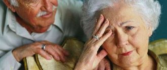
Relatives' eye disease
In a situation where the patient does not agree to see a doctor on his own because he does not see the need for this or cannot describe changes in his condition, the family psychiatrist asks the relatives what worries them about the behavior of their loved one.
It is family members who initiate treatment for encephalopathy in the absence of criticism from the patient.
The severity of symptoms and their combination may vary at different stages of the disorder, but relatives or friends say that a loved one:
- became uncollected, absent-minded;
- gets easily irritated and lashes out at relatives;
- became more demanding, intolerant;
- weak-hearted, easily cries for no apparent reason;
- forgets about important dates, current events and plans;
- has difficulty performing normal tasks;
- withdraws from his routine duties;
- looks depressed, but cannot explain the reason for the bad mood;
- has difficulty remembering any words or names;
- uncritical of ongoing changes;
- could get lost on the street, stopped going to familiar places;
- sleeps poorly at night, sleepy during the day.
Why is encephalopathy dangerous?
Complications of the disease depend on the degree of brain damage. The disease leads to impaired intellectual abilities, problems with movement, muscle tone and speech. In severe cases, cerebrovascular encephalopathy can cause coma and even death.
Untreated hepatic encephalopathy causes swelling of the brain with herniation. Uremic encephalopathy leads to apathy, hallucinations, muscle twitching, and convulsions.
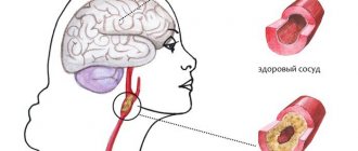
Against the background of discirculatory encephalopathy, a heart attack or hemorrhage in the brain often develops. A serious consequence of the disease is a stroke. It leads to impaired sensitivity, coordination, speech and mental disorders.
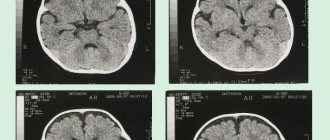
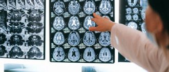
Diagnostics
Early detection of encephalopathy is of great importance in case of vascular and metabolic lesions of the brain. Patients with these types of disorders often seek help when painful symptoms begin to limit normal daily activities. The examination begins with a consultation with a neurologist. If necessary, he refers the patient to a therapist, ophthalmologist, endocrinologist, or narcologist. Instrumental diagnostic methods include:
- MRI, CT;
- EEG;
- Doppler ultrasound of the vessels of the head and neck.
Laboratory methods include: general blood test, coagulogram, determination of sugar and cholesterol fractions.
Classification
Taking into account the prevailing disorders, encephalopathy of vascular origin can be venous, atherosclerotic, hypertensive and mixed. In most cases, the disease progresses slowly; rapidly worsening and remitting forms are less common. According to severity they are distinguished:
- 1st degree. There are no neurological symptoms, and there is a slight decline in cognitive function.
- 2nd degree. Includes obvious motor and cognitive impairments, progressive emotional disturbances.
- 3rd degree. Essentially, it is dementia combined with severe mental and neurological changes.
Treatment of encephalopathy in adults - how to choose a clinic
The issue of choosing a medical institution and specialist worries both patients and relatives. Treatment of encephalopathy in Moscow is possible in three formats:
- managing patients at home;
- outpatient observation;
- hospitalization in a psychiatric hospital.
To decide on the format of medical care, it is important to clearly assess not only the patient’s health, but also his age and social environment. Often relatives are faced with a lack of criticality of the patient towards the condition:
- refuses to see a doctor;
- does not want to undergo examination in a regular clinic;
- has a negative attitude towards hospitalization.
To discuss the specifics of treatment with a specialist, it is enough to contact the clinic without the patient at any free time: doctors will give a detailed and free consultation over the phone. Obtaining reliable information will allow you to make an informed decision. You should not choose the first institution you come across with lower prices. Please note the following conditions:
- Is there a license to carry out medical activities? In boarding houses and private homes for the elderly, treatment is actually not provided;
- availability of own doctors on staff. Visiting specialists are not interested in the outcome of therapy and help only in acute situations;
- availability of valid certificates in psychiatry and psychotherapy among the clinic doctors;
- the ability to examine the patient in a short time. It is necessary to clarify which diagnostic methods are possible - this is of particular importance when working with negatively inclined or immobile patients;
- possibility of providing primary care at home (calling a doctor). This is important in the treatment of acute toxic encephalopathies, when refusing to visit the clinic;
- hospitalization on the day of treatment without paperwork. Some conditions are life-threatening. Treatment of encephalopathy may be required immediately, and getting to a clinic with decent conditions without waiting in line is not easy;
- comfortable conditions in the psychiatric department. Hospitalization is stressful for the patient. An unfavorable climate in the hospital, crowding of patients in the ward, and the inability to receive care from medical personnel reduces confidence in the doctor and worsens the prognosis.
Encephalopathy is a chronic disorder in which damage to brain tissue develops with the occurrence of mental, neurological, and somatic disorders. Competent treatment allows the patient to maintain quality of life, and in case of severe symptoms, reduce the burden on caring relatives.
Forecast
In most cases, the manifestations of initial vascular encephalopathy can be stabilized; patients retain their ability to work and clear thinking for a long time. With a rapidly progressing form, the results of therapy are less noticeable; each stage takes only about 2 years. A relatively unfavorable life prognosis is observed with severe hyperglycemia, stroke, frequent hypertensive crises, and concomitant degenerative changes in cerebral tissue.
Take care of your health in a timely manner. Sign up for a consultation at the Leto clinic by phone. and learn how to treat encephalopathy before it causes irreparable damage to your health.
Symptoms of encephalopathy in the elderly
There are three stages in the development of DEP. And if in the first stage cognitive functions are practically not impaired, the ability to work and everyday independence are preserved, in the third stage the intellectual deficit reaches its peak, and the patient requires constant care.
First stage
Characteristic:
- increased fatigue;
- partial memory loss;
- disturbances in the perception of information;
- general weakness;
- sleep disturbances, nightmares, disturbing dreams;
- decreased performance.
Patients complain of frequent attacks of headaches , and relatives note unusual irritability and anger.
Important ! It is at this stage that drug treatment is most effective. A correctly selected treatment regimen allows you to almost fully compensate for impaired functions.
Second stage
In grade 2 DEP, the clinical picture is dominated by cognitive disorders . Memory impairments progress, the patient finds it difficult to perform any sequential actions (he may forget what he wanted to do in the middle of the “process”). Typically, there is a disregard for generally accepted social norms, inappropriate jokes, and exaggerated foolishness.
often develops . When treatment begins at this stage, the prognosis is no longer so favorable. In many cases, even intensive drug therapy can only slow down the progression of the pathological process.
Third stage
Characterized by the development of dementia with total intellectual deficit. The patient does not recognize loved ones and is completely disoriented. Personality changes are typical, and the development of delusional and manic ideas is possible. An elderly person is convinced that they are persecuting him, they want to “lime” him in order to take possession of his property, they are “poisoning” him with the help of waves, “oscillations,” they are pouring poison, etc.
The ability to work is completely lost, the patient is unable to perform even basic actions, take care of himself in everyday life, practice hygiene, etc. Sometimes a person is subject to the influence of others, especially if the interlocutor agrees with him in everything, and quite often elderly people suffering from senile encephalopathy become victims of scammers.
Severe impairments in coordination and walking functions are also noted, up to the impossibility of independent movement; signs of parkinsonism appear (tremor, stiffness of movements, etc.). Loss of control over urination and bowel movements.