Angioencephalopathy is a vascular pathology in which brain activity is disrupted due to constant poor circulation. The prevalence of this disease among the population is 5%.
Among vascular disorders of the brain, this disease occupies one of the first places. Mostly adults are at risk. Hypertensive angioencephalopathy of the brain is diagnosed mainly in people over forty years of age. The disease is more likely to occur in people with significant mental stress.
Unlike stroke and other related diseases, this pathology does not manifest as an acute condition. The basis for the ontogenesis of encephalopathy is prolonged oxygen starvation of brain tissue.
Who's at risk
The following categories of people are at risk:
- with atherosclerosis of cerebral vessels;
- with unstable blood pressure;
- with diabetes mellitus (treatment prescribed by an endocrinologist);
- alcohol abusers;
- after a brain injury;
- with liver disease (consult a hepatologist);
- working in hazardous industries (fumes of caustic chemicals);
- over 60 years old;
- with a sedentary lifestyle;
- with obesity (consulting a nutritionist will help to cope with the problem).
- smokers;
- suffering from vasculitis, arteritis.
Prevention of vascular diseases of the brain
Vascular diseases of the brain are easier to prevent than to cure. To do this, experts recommend leading a healthy lifestyle: engaging in moderate sports, drinking about 1-1.5 liters of water per day (to avoid dehydration), eating properly and following a diet. The diet involves limiting table salt and foods containing large amounts of animal fats (for example, sour cream and fatty meats). Fresh vegetables and fruits should prevail in the daily diet. A split diet is recommended (4-5 meals per day).
The intake of alcoholic beverages should be minimized (or completely abandoned). The emotional component is the key to good health, so you should not be exposed to stressful situations. Sleep should be complete (at least 7 hours of sleep per day are required). Moderate physical activity should be regular; it significantly improves the condition of the body. These include: swimming, yoga, Pilates, cycling.
Exercising and proper nutrition help prevent the appearance of excess weight, while they train the cardiovascular system. At least once a year it is required to undergo a study of the state of cerebral circulation (especially for those who are at risk). This will allow timely detection of pathology.
In order to prevent the progression of multi-infarction conditions, patients are prescribed combination therapy (antiplatelet and anticoagulant). The most suitable anticoagulants are selected depending on blood clotting parameters. If any signs of bleeding appear, it is important to contact a specialist promptly.
Vascular diseases of the brain are often accompanied by dizziness. To prevent them, doctors prescribe medications that affect the autonomic nervous system. To prevent cognitive impairment (memory deterioration, increased inattention), drugs that improve metabolism are prescribed. In the presence of movement disorders, therapeutic exercises, physiotherapy, massage and other methods of rehabilitation therapy are useful.
Types of encephalopathy
Hypertensive
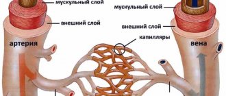
With increased pressure, damage to the vascular bed of the brain occurs. It has been proven that with long-term hypertension, a change in the walls of the arteries occurs: the replacement of muscle cells with connective tissue (sclerosis), which leads to a decrease in vascular permeability.
In addition, small arteries lengthen and acquire pathological tortuosity, which also disrupts the metabolic processes of the brain.
For the vessels of the brain, pressure changes are much more dangerous than simply increased pressure. Often, degenerative processes develop in the first and second degrees of hypertension, when there are no “prohibitively high” blood pressure values.
Diabetic
In diabetes mellitus, pathological processes occur in the inner lining of blood vessels (endothelium), which leads to disruption of the nutrition of brain tissue.
An increased level of glucose in the blood leads to the glycosylation reaction of protein molecules. This is a “springboard” for the development of vascular pathology. Diabetes mellitus is characterized by damage to small blood vessels (arterioles, capillaries).
Discirculatory
It includes both pathological changes in blood vessels due to atherosclerosis and the presence of lesions in the brain tissue. Atherosclerotic plaques cause vascular obstruction, which leads to a decrease in blood flow to the brain tissue.
In this case, the following changes occur in the nervous tissue: cell atrophy, as well as the breakdown of the myelin (outer) sheath of nerve fibers, which is responsible for conducting nerve impulses.
Alcoholic
It is damage to blood vessels and brain tissue as a result of abuse of alcoholic beverages or their surrogates.
Toxic
Severe poisoning from poisons and chemicals leads to brain damage.
Post-traumatic
It is usually a consequence of trauma and cerebral edema.
Encephalopathy in children
It occurs as a result of a viral infection or against the background of oncological processes.
Hepatic encephalopathy
Develops in severe liver diseases (hepatitis, hepatosis, cirrhosis), when the detoxification function of the liver is impaired. In this case, brain damage occurs precisely as a result of the presence of toxic metabolites in the human blood.
Causes and pathogenesis
The occurrence of this disorder is due to the presence of vascular diseases. The following factors provoke the occurrence of angioecephalopathy are noted:
- atherosclerosis;
- hormonal disorders;
- vegetative-vascular dystonia;
- low blood pressure;
- the presence of thrombosis with inflamed vein walls;
- increased arterial blood viscosity;
- inherited damage to blood vessels;
- manifestation of systemic vasculitis;
- impaired heart rhythm;
- congenital defects of the vertebral arteries;
- presence of decompensated diabetes mellitus;
- malformations in the development of the cervical vertebra;
- previous injuries;
- presence of hypertension;
- violation of the stability of the cervical vertebrae;
- presence of kidney disease.
The basic causative factors for the occurrence of pathology are arterial hypertension and atherosclerosis. An equally significant role is played by defects in the aorta, shoulder girdle, vessels of the neck and brain. Inferiority of cerebral hemodynamics is also associated with venous pathologies. The initial morphogenesis of chronic cerebral ischemia is caused by compression of venous and arterial vessels.
Low blood pressure has an adverse effect on cerebral blood flow.
Very often, circulatory pathology occurs against the background of the development of diabetes mellitus. Other pathological processes also lead to cerebral vascular deficiency: blood diseases, specific and nonspecific vasculitis, rheumatism.
All of the above diseases and conditions cause permanent brain hypoperfusion, in which the brain constantly lacks the necessary metabolic elements (glucose and oxygen).
Progressive damage to small arteries contributes to bilateral ischemic injury, which in turn leads to abnormal brain function and cerebrovascular encephalopathy.
The structure of plaques influences hemodynamic disorder in the brain. Fragile plaques cause arterial blockages and acute cerebrovascular accidents.
When hemorrhage occurs in such a plaque, it rapidly increases in volume, with a further increase in all signs of cerebral circulation deficiency.
Stages of the disease
The first , in which initial symptoms appear: absent-mindedness, forgetfulness, decreased performance, headache, tinnitus. Patients usually associate this condition with fatigue and rarely consult a doctor at this stage.
Doctor's advice
It has been noted that encephalopathy develops less frequently and much later in people who are engaged in mental work or who like to read frequently and have a need to obtain new information. There are 90-year-old professors and teachers who continue to engage in scientific work, and at the same time have excellent memory and intelligence. For those who lead an associative lifestyle and have an initial level of education, the diagnosis of “Encephalopathy” may be relevant at 30 years of age. To prevent the disease, you should solve crossword puzzles every day, read classical literature, periodically memorize poetry, retell the contents of books you read, movies you watched.
Victoria Druzhikina Neurologist, Therapist
Therapeutic effects at this stage are the most effective and can almost completely eliminate the symptoms of the disease.
Secondly , the patient’s performance is significantly reduced, he becomes slow, his speech is “sticky”. Minor speech impediments may occur. A characteristic feature of this stage is a change in the patient’s personality; he becomes irritable, angry, and touchy.
The third is characterized by gross disturbances and manifests itself with significant brain damage. Characterized by changes in speech (gross defects), a shaky, shuffling gait, trembling hands, extreme slowness, and serious memory impairment. The patient's personality may change beyond recognition.
Read also
Atherosclerosis
What is atherosclerosis Atherosclerosis is a narrowing of the arteries caused by the formation of plaque.
As a person gets older, fat and cholesterol can accumulate in the arteries and form plaque. Accumulation… Read more
Alzheimer's disease
47,000,000 people suffer from dementia in the world. Doctors call Alzheimer's disease the most common cause of dementia. Alzheimer's disease is a progressive neurodegenerative disease accompanied by...
More details
Vegetative-vascular dystonia
Vegetative-vascular dystonia is a dysregulation of the autonomic nervous system, which manifests itself in the form of various clinical symptoms. This disease is diagnosed at different times...
More details
Chronic cerebral ischemia
One of the most common diagnoses at an appointment with a neurologist for patients in the older age group is chronic cerebral circulatory failure, discirculatory encephalopathy. According to…
More details
Transient ischemic attack (TIA)
What it is? Why is this happening? Is this condition dangerous? What to do if doctors make such a diagnosis? These questions are always asked by patients who come to see a neurologist. According to classification...
More details
Symptoms
The disease is characterized by the following symptoms:
- forgetfulness;
- attention disorders;
- memory impairment;
- slowness;
- "viscosity of speech";
- speech defects (omission of letters, replacement of words, difficulty finding a synonym, lack of long sentences);
- gait disturbances (unsteadiness, difficulty maintaining balance, tendency to fall, clumsiness when turning);
- headache, dizziness;
- noise in ears;
- drowsiness, fatigue, weakness;
- sleep disorders (difficulty falling asleep, shallow sleep, frequent awakenings at night);
- decreased performance, inability to do more than one thing at a time;
- dysfunction of the pelvic organs: urinary incontinence, up to complete loss of control over urination, in advanced cases - fecal incontinence;
- “poverty” of emotions (loss of interest in others, apathy, withdrawal into oneself, a feeling of detachment, uselessness, lack of expression of emotions);
- decreased intellectual potential (in the severe stage, the patient sometimes cannot count to 10).
Diagnostics
In the initial stages, the manifestations of dyscirculatory vascular encephalopathy are easily corrected with adequate treatment of the underlying disease, therefore patients with vascular pathology are recommended to undergo regular examinations to identify the first symptoms and prevent progression. The examination program includes:
- Neurological examination. At stage 1, the neurological status is unchanged. Subsequently, tremor, limb weakness, and focal symptoms are determined.
- Assessment of mental and intellectual status. This is done through conversation and special tests.
- Consultation with an ophthalmologist. Allows you to detect changes in the fundus. Includes determination of the visual field, ophthalmoscopy.
- Electrophysiological methods. Electroencephalography and rheoencephalography are recommended.
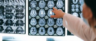
- Visualization of blood vessels. When confirming the probable vascular origin of the disease, MRA, duplex scanning and Doppler sonography are informative.
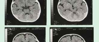
- Magnetic resonance imaging. Performed as part of differential diagnosis with other cerebral pathologies.
To clarify the diagnosis of the causative disease, the patient may be referred to a cardiologist, nephrologist or endocrinologist, and prescribed an ECG, coagulogram, and tests to determine the level of sugar, lipoproteins and cholesterol.
Treatment
Considering the variety of factors causing encephalopathy, treatment should be carried out in the following areas:
- Eliminating the cause of the disease.
- Improving blood supply to the brain.
- Prescription of drugs that protect nerve cells from damage.
For encephalopathy, the following groups of drugs are prescribed:
- Neuroprotective drugs. These drugs are highly effective in treating encephalopathy disorders. They improve trophic processes in nerve cells, provide protection and restoration of cell membranes from damage. The most commonly prescribed drugs are: Cerebrolysin, Nootropil, Encephabol, Cortexin.
- Improving blood microcirculation in the vessels of the brain is achieved by prescribing drugs such as Actovegin and Kurantil.
- Hypotensive (blowing blood pressure). Preference is given to drugs with a long action (about 24 hours) in order to avoid hypertensive crises. If one drug is insufficient, a combination of drugs is prescribed. The most commonly used are ACE inhibitors (Enalapril, Lisinopril, Fosinopril), beta blockers (Nebilet, Metoprolol, Bisoprolol), diuretics (Hypothiazide), Indapamide, Spironolactone. .
- Statins are drugs that lower blood lipid levels. Typically prescribed are Atorvastatin (Atoris), Simvastatin (Simvacard), and Rosuvastatin (Rozart). In addition, to correct lipid levels, you must adhere to a low-calorie diet.
- Antioxidants - these drugs protect the cell from pathological oxidation processes and the effects of toxic free radicals. The use of the drug Mexicor for encephalopathy is effective.
- Antiplatelet agents - by reducing platelet aggregation, blood circulation in small vessels improves. Usually medications such as Aspirin and Clopidogrel are prescribed.
- Monitoring glucose levels is mandatory for diabetes mellitus.
- Detoxification - promotes the removal of toxic metabolites from the body; this therapy is especially effective for alcoholic and hepatic encephalopathy. In cases of severe intoxication, plasmapheresis (hardware purification of blood plasma from toxins) may be required.
After drug treatment, special attention should be paid to the patient’s rehabilitation:
- Compliance with the work and rest regime.
- Normalization of sleep.
- Maintaining a diet if necessary (for obesity, diabetes).
- Dosed physical activity.
- Spa treatment.
Encephalopathy is very insidious, since the initial manifestations of the disease are very nonspecific and can be regarded by a person as chronic fatigue. In addition, it is sometimes very difficult to determine which factor leads to brain damage.
Encephalopathy often develops as a result of several causes affecting the body at once. This means that the boundaries of each type of encephalopathy are very blurred. That is why in the treatment of this condition it is justified to prescribe several groups of drugs with different mechanisms of action.
This article has been verified by a current qualified physician, Victoria Druzhikina, and can be considered a reliable source of information for site users.
Bibliography
1. https://mir.ismu.baikal.ru/src/downloads/2581419b_uchebnoe_posobie_distsirkulyatornaya_entsefalopatiya.pdf 2. https://dep_ninh.pnzgu.ru/files/dep_ninh.pnzgu.ru/hronicheskaya_nedostatochnost_mozgovogo_krovoobrascheniya.pdf
Rate, How helpful was the article?
4.4 21 people voted, average rating 4.4
Did you like the article? Save it to your wall so you don’t lose it!
Intracerebral tumors (a type of brain tumor)
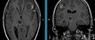
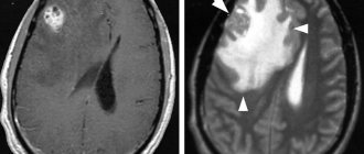
Intracerebral tumors are a large group of primary brain tumors. As the name suggests, these tumors grow directly in the brain tissue. The vast majority of intracerebral tumors develop from glial cells and that is why they are called glial. Given the growth pattern of primary intracerebral tumors, surgical removal is associated with a number of difficulties. These tumors do NOT have clear visual boundaries with the brain substance and radical removal of the tumors is often not possible. The prognosis for glial tumors primarily depends on the histological variant. In the modern WHO classification (WHO), there are four grades, depending on the degree of malignancy.
Piloid astrocytoma (WHO Grade I) is a benign, slow-growing tumor. More common in children and young patients. That is why another name for this type of tumor is juvenile, “childhood” astrocytoma. The presence of cysts is characteristic. Piloid astrocytomas are most common in the cerebellum. Localization in the cerebral hemispheres is more typical for patients in the older age group. If located in the area of the optic nerve, the term chiasmal glioma is used. Piloid astrocytomas can also occur in the spinal cord. Despite their benign nature, surgical treatment of piloidal astrocytomas can be difficult when located in vital structures such as the brainstem. In such cases, after confirmation of the histological diagnosis, radiation therapy may be prescribed. If radical tumor removal is possible, no additional therapy is required. It should be noted that in some cases, “aggressive” - rapidly progressing forms of piloid astrocytomas occur.
Pleomorphic xanthoastrocytoma (WHO Grade II) is a rare form of benign glioma. Most often it is localized as close as possible to the surface of the cerebral hemispheres. With radical removal, the prognosis is favorable. However, if there are signs of malignancy based on the results of an immunohistochemical study, it is advisable to carry out radiation therapy with subsequent dynamic observation.
Subependymal giant cell astrocytoma (WHO Grade I) is a rare benign tumor that is most often one of the manifestations of Bourneville disease. The favorite location of this tumor is the ventricular system. That is why one of the common manifestations of the disease are symptoms of hydrocephalus, due to disruption of normal liquor circulation. If radical removal of the tumor is possible, no additional treatment is required.
Diffuse astrocytoma – (WHO Grade II). These astrocytomas are characterized by infiltrative growth, that is, the absence of boundaries between the tumor and the healthy brain. However, the surgeon should always strive to remove as much tumor tissue as possible. Most often, these tumors occur in young patients and are located in the cerebral hemispheres. Despite their conditionally benign nature, these tumors can become malignant over time, that is, become malignant.
Oligodendroglioma (WHO Grade II) is a diffuse primary brain tumor. The favorite localization is the frontal lobes. The most common, first symptom of the disease is seizures. After surgery, chemotherapy is mandatory.
Oligoastrocytoma (WHO Grade II) is a mixed intracerebral tumor that contains two different types of tumor cells (astrocytes and oligodendroglia) and after removal requires additional treatment - chemotherapy. Just like astrocytoma, oligoastrocytoma can be anaplastic, that is, malignant. In this case, it is Grade III and may be called glioblastoma with an oligodendroglial component.
Anaplastic astrocytoma (WHO Grade III), despite its similar name to other forms of astrocytomas, is a more malignant tumor and, accordingly, with a less favorable prognosis for the patient. In the postoperative period, radiation and chemotherapy are necessary.
Anaplastic oligodendroglioma (WHO Grade III) is a malignant tumor in nature. In the postoperative period, radiation and subsequent chemotherapy are necessary.
Glioblastoma (WHO Grade III) is the most malignant and, unfortunately, common primary brain tumor. Despite the maximum possible removal of the tumor volume (if possible!), radiation and chemotherapy, the relapse-free period in patients with glioblastoma is very short.
Neurocytoma is a rare type of benign neuronal tumor that occurs only within the ventricular system, most often in the lateral ventricles. Due to the peculiarity of localization and characteristic slow growth, tumors can grow to impressive sizes and not cause any complaints. Due to the fact that tumors almost always block normal CSF flow in the ventricular system, the most common manifestations of the disease are symptoms of hydrocephalus. In case of radical removal of the tumor, the prognosis is favorable. Recurrence is possible if there is residual tumor. In this case, radiation therapy may be prescribed.
Chiasmal glioma is a tumor formation that originates from glial cells located in the area of the optic chiasm. Chiasmal glioma is manifested by decreased visual acuity, narrowing or loss of part of the visual fields, symptoms of hydrocephalus and neuroendocrine disorders. The complex of diagnostic examinations for chiasmal glioma includes viziometry, ophthalmoscopy, perimetry, study of visual EP, MRI and CT of the brain, and stereotactic biopsy. Chiasmal glioma is treated depending on its characteristics, location and age of the patient. This may be surgical intervention (removal or partial resection of glioma, restoration of liquor circulation), chemotherapy or radiotherapy.
Brainstem glioma is a tumor formation that is characterized by infiltrative growth, that is, the ability of neoplasm cells to grow into surrounding healthy tissue and replace them. Because of this, it is quite difficult for doctors to establish the boundary between pathological and healthy brain cells and, accordingly, completely remove the tumor. The building material for glioma are glial cells, whose function is to protect and support neurons. Tumors differ in the degree of malignancy, the ability to invade, and according to histological characteristics. Neoplasms that affect glial cells occur in people of various age groups, but most often gliomas are found in children. The peak incidence occurs between the ages of 2 and 8 years.
The symptoms that appear at the beginning of the disease are similar to those that occur with neurological disorders, so the wrong treatment is often carried out at the initial stage. In addition, at an early stage, signs of a tumor usually appear mildly and develop at a slow pace.
The following symptoms should alert the patient, as they may indicate an early stage of brainstem glioma:
- Frequent headaches that are not relieved by medications.
- Nausea.
- Speech dysfunction.
- Epilepsy attacks.
- Impaired vision, memory, associative thinking.
As the glioma grows (grade 2), the patient may experience changes in behavior:
- aggression;
- irritability;
- psycho-emotional instability.
In the absence of treatment, the symptoms worsen and the following signs join the clinical features of the disease:
- intracranial pressure;
- auditory, visual, taste hallucinations;
- vomit;
- dizziness, convulsions, loss of coordination.
Sometimes alarming signs, on the contrary, arise abruptly, this is an unfavorable sign, because indicates the rapid growth of glioma and its high degree of malignancy.
Stage 3, 4 brainstem glioma poses a serious danger to human life. Tumor cells quickly grow and reproduce, and more pronounced symptoms are added to the primary symptoms. In adults and children, the clinical picture may be slightly different.
In adults, the following are added to the primary symptoms:
- vestibular ataxia;
- intracranial hypertension;
- horizontal nystagmus;
- numbness, paresis or paralysis of limbs, body parts, face;
- changes in thinking, behavioral disorders.
Children may develop:
- strabismus;
- facial asymmetry;
- decreased sensation in the arms or legs;
- hearing loss;
- decreased intellectual abilities;
- muscle weakness;
- apathy;
- weight loss;
If the disease lasts for a long time, the child may develop hand tremors.
The most informative way to diagnose brainstem glioma is MRI. This method allows you to identify pathological formations of even the smallest sizes. MRI provides visualization of the brain from various angles. A clear three-dimensional image allows you to accurately determine the location and size of the tumor.
It is possible to completely remove a glioma only at the first stage of malignancy. Glioma of a more severe stage of development infiltrates into surrounding tissues, so it is quite difficult to separate the neoplasm from healthy cells.
The following methods are currently used to treat brainstem glioma:
Operation
Usually, due to the complexity of the area where the tumor is located, surgical intervention may be impossible, so surgical removal is used extremely rarely. Surgery for gliomas is extremely dangerous for the life and health of the patient, therefore it is performed by highly qualified specialists in the best clinics.
Radiation therapy
The brain stem is a thin area and controls various vital functions of the body, so radiation therapy is often the main treatment method. X-ray radiation affects the tumor from different positions, which allows minimizing the effect on healthy cells. This method significantly slows down the growth of the tumor and also helps reduce the symptoms of the disease.
Chemotherapy
This method has a detrimental effect on cells with rapid growth and increased metabolism.
Symptomatic treatment
Used to reduce painful symptoms.
An integrated approach is more effective in treating the disease, but for young patients, doctors try not to use combined methods, since there is a high probability of serious side effects (developmental and growth retardation). When prescribing a treatment regimen, the doctor must discuss with the parents all the nuances, possible risks and choose therapy, taking into account the patient’s health characteristics.
