Brain puncture is a multi-purpose medical manipulation, with the help of which one or more therapeutic and diagnostic purposes are pursued. A puncture (injection) is any penetration with a needle or trocar into the cavity of a vein, another vessel, or an organ, to obtain material for research and diagnosis, optimize functions and remove obstacles, and perform operations.
Modern techniques make it possible to combine operational and diagnostic goals, achieving them simultaneously.
Taking fluid for analysis does not exclude the use of other diagnostic methods. Modern technologies allow parallel ultrasound to determine the location of the dislocation, for example, a cyst. This combination can successfully remove the tumor.
There is no need to be afraid of a puncture - it is not only a diagnostic method, but also a treatment method that was used before, but in an indirect form.
What is a cerebral puncture?
Penetration into the cranium at the location of parts of the brain is carried out less frequently than other manipulations in places that are less dangerous and threaten negative consequences.
Although any puncture can cause complications if it is carried out unprofessionally, affects some important segments or becomes a source of infection. Each invasive procedure has features specific to a particular department, developed techniques and precautions. Brain puncture (cerebral puncture) is a collective name for a therapeutic or diagnostic procedure carried out according to indications at a strictly defined destination:
- in the lower parts of the temporal or frontal lobes;
- over the tympanic space or mastoid process;
- ventricular, in the region of the lateral ventricles;
- within the central nervous system, to obtain a sample to study the spinal cord and brain simultaneously.
To carry out the procedure, a special needle and scalpel are used, the trepanation window is cut out with a special cutter, and bone bleeding is stopped by rubbing wax or electrocoagulation. To regulate the flow of cerebrospinal fluid there is a special device - a mandrin. In most cases, the procedure is performed under local anesthesia, in compliance with all necessary conditions of sterility and preparation of a sterile surgical field.
However, just in case, a large operating room is being prepared, which in rare cases can be used to perform open brain surgery. This scenario is possible when surgical complications arise - damage to a vessel, air entering the cavity, or insertion of a needle to an unexpected height.
Although sometimes the reason for further surgical tactics is an insufficiently studied pathology located directly in the brain (abscess, abscess, cyst, neoplasm).
Spinal puncture technique
The procedure does not require patient preparation. It is performed on an outpatient basis and takes only a few minutes. During the procedure, the patient takes a sitting or lying position. The doctor puts on special medical clothing. According to the algorithm for performing a lumbar puncture, he treats his gloved hands and the patient’s back with antiseptics. Then he dries it with a sterile cloth.
The puncture site is selected by palpating the intervertebral spaces, taking into account bony landmarks. The technique of implementation provides that a spinal puncture is performed in adults at the level of 2-3 lumbar vertebrae, in children - between 4 and 5.
An anesthetic (lidocaine or novocaine) is injected into the selected area using a syringe. The needle is removed and waited 2-3 minutes until the analgesic effect occurs. According to the technique of performing a lumbar puncture, the injection is painless, comparable to anesthesia, which is given in a dental office. As the anesthetic takes effect, bloating or numbness is felt.
According to the lumbar puncture technique, subsequent needle insertion is painless; slight pressure may be felt in the back. Then the mandrin is removed and the pressure of the cerebrospinal fluid is measured. The doctor then collects the cerebrospinal fluid using a laboratory tube. After this, the procedure algorithm involves re-measuring the pressure and removing the needle. The lumbar puncture site is closed and the patient is in a horizontal position.
A person needs absolute rest for 24 hours. The patient should lie still all day, even without raising his head, and consume sufficient fluids.
What is it done for - diagnostic and therapeutic purposes
Obtaining cerebrospinal fluid to determine treatment tactics, analysis and diagnostic prognosis is carried out with the goal of achieving a certain result, and before prescribing a puncture, the tasks are strictly delimited. However, there are situations when a cerebral puncture for the purpose of research, collection of cerebrospinal fluid, as material for diagnostics, turns into shunting or removal of excess fluid to reduce pressure inside the skull.
Ventricular puncture (penetration into the lateral ventricles of the brain) helps doctors achieve several goals:
- performing diagnostics by obtaining important biological fluid for research;
- measuring intracranial pressure or conducting studies with a radiopaque substance;
- operations performed using a special device - a ventriculoscope, or shunting in the cerebrospinal fluid system;
- reducing intracranial pressure by removing spinal fluid if the natural outflow system does not work.
Well-established techniques and precautions allow operations to be performed as needed, using only local anesthesia. The methods and routes of penetration have been developed over many years of practice, and the data obtained in most cases helps to carry out more effective treatment based on objective information.
Bone marrow analysis / Bone marrow biopsy
Bone marrow analysis - the collection of bone marrow using a trephine biopsy from the ilium or sternum. Red bone marrow is located in flat bones. The function of bone marrow is the production of new blood cells: red blood cells, white blood cells, platelets. Trepanobiopsy is the procedure for obtaining bone marrow for laboratory testing. The main indications for trepanobiopsy and bone marrow examination are diseases of the blood system and suspicions of them.
Read more about diagnostic studies of the bone marrow and blood system below.
A bone marrow test can be performed on an outpatient basis or in a hospital setting. When the bone marrow analysis is ready, you and your doctor will receive a myelogram - a table with the qualitative and quantitative composition of bone marrow cells. The result of the study allows us to track the process of formation of new blood cells and its disturbances.
What can be checked with trepanobiopsy? Bone marrow analysis is a specialized test used in hematology, oncology and immunology. for diagnosing the causes of anemia and blood cancer (leukemia, etc.). To assess the condition of the bone marrow, the number of cells in it is determined: myelokaryocytes, megakaryocytes, and the percentage of bone marrow elements is calculated. The result of the bone marrow analysis is compared with the results of the general blood test. This study is prescribed and performed only by a doctor strictly for medical reasons.
Possible abnormalities in bone marrow analysis. A decrease in the number of myelokaryocytes is possible with hypoplastic anemia (severe depression or inadequate functioning of the bone marrow), when the human body is exposed to ionizing radiation (radiation) and certain medications (some antibiotics, gold preparations). A large number of myelokaryocytes is most characteristic of leukemia, B12 deficiency anemia, and a condition after large blood loss. A decrease in the content of megakaryocytes and megakaryoblasts is caused by autoimmune processes (systemic lupus erythematosus), leukemia, and radiation sickness. An increase in the number of megakaryocytes and megakaryoblasts usually indicates the presence of myeloproliferative processes and metastases to the bone marrow (for example, in gastric cancer).
Preparation for performing bone marrow trepanobiopsy. We recommend that you refrain from eating 12 hours before the test.
Main indications for bone marrow trepanobiopsy:
- Leukemia and suspicion of it
- Leukopenia (decrease in the number of white blood cells in the blood)
- Lymphogranulomatosis and suspicion of it
- Anemia – decrease in hemoglobin and number of red blood cells in the blood
- Gaucher disease
- Tumor metastases
- Evaluation of the effectiveness of therapy
How to take a brain puncture
The operation is carried out under local anesthesia with strict adherence to all the rules of sanitary processing, first the incision, and then cutting the bone with a special tool, after which cerebrospinal fluid begins to flow through the hole, which is taken to alleviate the patient’s condition and conduct tests.
The surgical field is limited to sterile tissue, and the outflow of biological fluid is strictly controlled, as well as possible bleeding when a burr hole appears.
Precautions and Rules
It is necessary to take into account all indications and contraindications, possible obstacles to the operation. Thorough sanitization at each stage, preparation of spare instruments, a large operating room, careful monitoring of the patient’s condition at each stage.
Mandrin and other instruments must be thoroughly disinfected
The manipulation is carried out with the patient positioned on his back, with his head bowed to his chest. The neurosurgeon determines the incision line by touch.
There is a method of penetration through the orbit (the so-called Dogliotti method), and there is another method - according to Germanovich, who developed penetration through the temporal bone from below.
Diagnostic and therapeutic methods in neurosurgery
The following methods are widely used in neurosurgical practice:
- spinal puncture - diagnostic and therapeutic method;
- puncture of the ventricles of the brain (ventricular puncture) - a diagnostic and therapeutic method;
- cerebral angiography is a diagnostic method.
Spinal tap
General information
. A spinal puncture is the insertion of a needle into the subarachnoid space of the spinal cord in order to extract cerebrospinal fluid (CSF) or inject medicinal or radiocontrast agents. Spinal puncture is performed at various levels of the spinal column - in the lumbar region, less often in the thoracic region, in the area of the foramen magnum (suboccipital puncture), but most often it is done in the lumbar region (lumbar puncture, or lumbar puncture).
Spinal puncture provides valuable diagnostic information. The resulting cerebrospinal fluid may be clear or contain an admixture of blood or pus, which suggests subarachnoid hemorrhage or meningitis; CSF can flow from the needle under normal, elevated or reduced pressure, which is determined using a manometer, i.e., a glass tube bent at a right angle with centimeter divisions printed on it.
The cerebrospinal fluid should be sent to the laboratory in two sterile tubes: one for studying the cellular composition and protein of the cerebrospinal fluid, and the other for inoculating the cerebrospinal fluid.
Spinal puncture allows for special diagnostic studies - the introduction of radiopaque substances into the subarachnoid space of the spinal cord and brain, followed by radiography. X-ray examination of the spinal cord with the introduction of a contrast agent (iodolipol, mayodil, etc.) is called myelography. If air or oxygen is used as a contrast agent, this procedure is called pneumomyelography. If a sufficient amount of air is introduced through a spinal puncture and it rises into the subarachnoid space of the brain, then an X-ray examination of the brain is called pneumoencephalography.
Rice. 138. Position of the patient during lumbar puncture. a - lying down; b - sitting
For therapeutic purposes, a spinal puncture is performed to extract a certain amount of cerebrospinal fluid, to administer the sodium salt of penicillin or other medications.
A spinal tap is performed in various rooms: in the dressing room - to take cerebrospinal fluid for analysis or to administer medications; in the operating room - during brain surgery to quickly reduce intracranial pressure; in the X-ray room - for myelography or pyeumoencephalography.
Depending on the patient’s condition and the objectives of the study, lumbar puncture is performed with the patient lying or sitting (Fig. 138). When performing a puncture with the patient lying down, the help of an orderly is required, who grasps the patient by the neck and popliteal fossae and bends his back to make it easier to pass the needle between the spinous processes and vertebral arches. When performing a puncture with the patient in a sitting position, which is usually required for pneumoencephalography, the doctor is assisted by an anesthesiologist or a ward nurse who stands in front of the patient’s face, giving him the correct position with his back bent.
Preparation of instruments and material for lumbar puncture. For a lumbar puncture, the sister prepares: alcohol, iodine, 0.25% novocaine solution, 3 syringes with a capacity of 5 ml with needles, a scalpel, 2-3 needles for a spinal puncture, a glass tube-manometer, 2 sterile tubes, gauze balls and napkins, 2 sterile sheets, cleol. In addition, a vial with penicillin sodium salt is prepared. If the puncture is performed for the purpose of X-ray contrast examination, the nurse also prepares ampoules with a contrast agent - mayodil or iodolipol, or an oxygen cushion for pneumoencephalography.
It is necessary to have a 5 ml syringe with needles and ampoules with ephedrine and caffeine ready for subcutaneous injections in case of a collaptoid state, which can develop when air is introduced into the subarachnoid space.
Participation of the nurse in performing the puncture
. To treat the skin of the lumbar region, the nurse gives the surgeon a forceps with a gauze napkin moistened with alcohol, then a forceps with a napkin moistened with iodine, then again a napkin moistened with alcohol, and finally a dry napkin, with which the surgeon wipes the skin in the area of the intended puncture (between II and III or III and IV lumbar vertebrae). It is necessary to wipe the skin with a dry cloth, since during puncture iodine and alcohol can be introduced into the dura mater with a needle, which causes its inflammatory reaction.
With the patient positioned on his side, the nurse covers the puncture area with two sheets. When the patient is sitting, he uses one sheet to fence off the buttock area.
The nurse gives the surgeon a 5 ml syringe with a 0.5% novocaine solution and a thin needle to anesthetize the skin and underlying tissues in the puncture area, then a scalpel to apply a pinpoint puncture of the skin in the area where the puncture needle is to be inserted. This is necessary in order to protect the needle from dulling and deformation when puncturing the skin. A well-sharpened needle causes less severe damage to the hard shell when puncturing it.
After checking that the mandrel slides freely in the needle and that there are no barbs on the end of the needle, the nurse hands the surgeon a needle for lumbar puncture. When cerebrospinal fluid appears, the nurse connects a pressure gauge to the needle pavilion to measure pressure or passes the pressure gauge to the surgeon.
To take cerebrospinal fluid for analysis, the nurse alternately brings one, then the second sterile tube to the puncture needle pavilion. Having collected the required amount of cerebrospinal fluid (as determined by the surgeon), the nurse closes the test tubes with sterile stoppers and hands the test tubes to the ward nurse present at the puncture (the ward nurse writes a referral to the laboratory indicating the patient’s surname and initials, diagnosis and purpose of the study). After removing the required amount of cerebrospinal fluid, the nurse reconnects the pressure gauge to the needle pavilion to measure pressure.
If, when inserting a needle into the subarachnoid space, the surgeon does not receive cerebrospinal fluid (the so-called dry puncture), the nurse gives him a syringe with a capacity of 5 or 10 ml to carefully suction out the cerebrospinal fluid.
If it is necessary to introduce penicillin into the subarachnoid space, the nurse gives the surgeon a syringe containing 25,000* 50,000, 75,000, or 100,0000 units of penicillin sodium salt diluted with saline.
After the surgeon removes the puncture needle, the nurse gives him a cotton swab soaked in cleol, a sterile gauze ball and a napkin to seal the puncture area.
The patient is transferred to a gurney without a pillow, taken to the ward and placed on a bed without a pillow.
Puncture of the lateral ventricles of the brain (ventriculopuncture)
General information
. Ventriculopuncture is the insertion of a needle into the cavity of the lateral ventricle of the brain in order to obtain cerebrospinal fluid or inject drugs or contrast agents. Ventriculopuncture is performed for diagnostic purposes - for the study of ventricular cerebrospinal fluid, which in certain diseases differs from spinal cerebrospinal fluid, for the administration of a contrast agent followed by radiography (ventriculography), as well as for therapeutic purposes - for immediate urgent unloading of the ventricular system by removing cerebrospinal fluid or for long-term drainage ventricular system.
Depending on the indications, the anterior, posterior or inferior horn of the lateral ventricle is punctured. A burr hole is made in the appropriate area of the skull, the dura mater is incised, a section of the cerebral cortex is coagulated, and a Cushing puncture needle is inserted into the ventricular cavity (Fig. 139).
Ventriculopuncture is performed in the operating room or in a specially equipped x-ray room.
It is very important to remember that ventriculopuncture with the extraction of cerebrospinal fluid or the introduction of air into the ventricular cavity can cause severe swelling of the brain or its displacement, which will require emergency brain surgery. Therefore, before performing ventriculopuncture, the nurse must prepare the operating room for a major operation - craniotomy (see below).
Preparation of tools and material
. For puncture of the lateral ventricle, the nurse prepares: alcohol, iodine, 0.5% novocaine solution, syringes with a capacity of 5, 10 and 20 ml with needles, a scalpel, a raspatory, a Jansen screw retractor, a small (ophthalmic) scalpel, a diathermocoagulator with a needle electrode, a vacuum a suction with 2 tips, a brace with a spear and a large-caliber cutter, a Volkmann bone spoon, a Cushing puncture brain needle with a mandrel. If ventriculography with iodine contrast is to be performed, the nurse prepares ampoules with mayodil, 2 sterile tubes, suture needles, silk, needle holder, clips, 2 sterile sheets, napkins, balls, bandages, cotton wool.
Nurse's participation in ventriculopuncture
. To treat the scalp, the sister gives the surgeon a forceps with a gauze cloth moistened with alcohol, then with iodine. Using two sterile sheets, the nurse fences off the surgical field and hands the surgeon the clips. Then he gives the surgeon a 10 ml syringe with a thin needle and 0.5% novocaine solution for anesthesia of soft tissues at the site of the intended incision, a regular scalpel for cutting the skin in the projection of the corresponding horn of the lateral ventricle, a Jansen screw retractor, which holds the edges of the wound and serves for hemostasis , a brace with a spear for making a hole in the bone. He replaces the spear with a cutter and gives the surgeon a brace with a cutter. When the surgeon places a burr hole, the nurse uses a gauze swab on a forceps to free the burr hole from accumulating bone chips. He delivers a Volkmann bone spoon to remove the remnants of the internal bone plate at the bottom of the burr hole, then an “eye” scalpel to open the dura mater, a needle electrode for coagulation of blood vessels in a small area of the cerebral cortex, a brain cannula with a mandrel in it for brain puncture and insertion needles into the ventricle. When ventricular cerebrospinal fluid enters from the needle, sterile tubes are supplied to the outside for collecting cerebrospinal fluid for analysis.
Rice. 139. Puncture of the anterior and posterior horns of the lateral ventricle
If it is necessary to extract a large amount of cerebrospinal fluid to unload the ventricular system, the nurse provides a syringe with a capacity of 20 ml, with which the surgeon gradually removes the cerebrospinal fluid through a cerebral cannula. If iodide contrast agent needs to be injected into the ventricular cavity, she hands the surgeon a 5 ml syringe containing mayodil. For ventriculography, X-rays of the skull are taken in the operating room using a portable X-ray machine or a stationary machine if the operation takes place in a cath lab.
After removing the needle from the ventricle and removing the retractor, the nurse gives the surgeon a swab with iodine to treat the edges of the wound, then a needle holder with a needle loaded with silk for suturing the skin. After the stitches are placed, she gives the surgeon iodine and applies a bandage.
Cerebral angiography
General information
. Cerebral angiography is the injection of a contrast agent into the patient’s brain vessels in order to make them visible on x-rays. Cerebral angiography should be performed directly in the X-ray room, on the X-ray table (or in the X-ray operating room, if there is one in the given medical institution). This procedure requires the well-coordinated work of an entire team consisting of a surgeon, an operating nurse, a radiologist, an x-ray technician, an anesthesiologist, an anesthesiology nurse, and a nurse. To obtain several angiograms in a short period of time (about 6 s), reflecting the arterial, capillary and venous phases of blood flow, special X-ray attachments are used to quickly change cassettes. Currently, there are special angiographic devices that allow up to 16-18 images to be taken in a short period of time while the contrast agent is circulating in the cerebral vessels.
For cerebral angiography, iodide contrast agents are used - diodone, urografin, hypaque, etc. in a dose of 8-12 m, l of 35-50% solution.
There are various methods of cerebral (brain) angiography. If contrast material is injected into the common carotid artery in the neck, the procedure is called carotid angiography. If contrast material is injected into the vertebral artery in the neck, the test is called vertebral angiography. Carotid and vertebral angiography are methods of direct cerebral angiography, since the contrast agent is injected directly into the great vessels of the brain.
There is a technique of indirect cerebral angiography, when a large vessel, for example the femoral artery, is punctured, and through it a long catheter is passed into the carotid artery or brought to the vertebral ostium. Then a contrast agent is pumped through a catheter into the blood vessels of the brain.
In clinical practice, carotid angiography is most often used. It is usually performed by percutaneous puncture of the common carotid artery in the neck. Only if it is impossible to puncture the artery, they resort to exposure of the carotid artery and its subsequent puncture with the introduction of a contrast agent.
For carotid angiography in a closed manner, a special needle a, an adapter flexible polyethylene hose b and a syringe with a contrast agent c are used (Fig. 140).
The needle for puncture of the carotid artery has a length of about 10 cm, a lumen of 1 - 1.5 mm, a cut of 45°. A mandrel is attached to the needle, which tightly closes its lumen, but is inserted easily, without effort. The end of the mandrin is rounded so as not to injure the inner lining of the carotid artery. Free sliding of the mandrel in the needle is very important for accurate and quick angiography. The angiographic needle pavilion has a “lock” for tightly connecting the adapter hose. The same “lock” is located at the end of the hose, fixed to the syringe. Thanks to the tight fixation of the needle, hose and syringe, these parts do not slip off when the contrast agent is injected into the artery. The transition hose is required so that the movements of the syringe in the surgeon’s hands are not transmitted directly to the needle located in the lumen of the vessel and do not displace it.
Rice. 140. Set for cerebral angiography. a - puncture needles with materials; b — transitional polyethylene hose; c - syringe for contrast agent
When preparing instruments for angiography, the nurse must check that the needle is sharp enough (a blunt needle injures the vessel wall), so that the mandrel slides easily in its lumen and that the hose is tightly attached to the needle and to the syringe pavilion.
Preparation of tools and material. Because carotid angiography is performed in an x-ray room, the nurse should have everything needed for this surgical procedure on the operating table and not send to the operating room for missing instruments and material. It must be borne in mind that percutaneous puncture of the carotid artery may not work for one reason or another, and then the artery will have to be exposed and punctured in an open manner. Therefore, instruments should be prepared to expose the vessel.
For carotid angiography, the nurse prepares: alcohol, iodine, 2 sterile sheets, 2 towels, medium napkins (5), gauze balls (10), robe and gloves for the surgeon (robe and gloves for the assistant in case of exposure of the artery), scalpel, 2- 3 angiographic needles with mandrels, 2 adapter hoses, 2 syringes with a capacity of 20 ml for administering contrast, a syringe with a capacity of 10 ml with a thin needle for anesthesia, 0.5% novocaine solution, 2-3 ampoules with a contrast agent, a jar or mug with a capacity of 0. 5 liters of warm water containing an ampoule with a contrast agent (if the contrast is not warmed, then when the ampoule is opened it crystallizes, which makes it impossible to introduce it into the artery), ampoules with papaverine or a noshpa for introduction into the artery in order to eliminate its spasm. To expose the vessel, in addition to the mentioned instruments, you need a scalpel, Farabeuf hooks, hemostatic clamps (8), anatomical tweezers, surgical tweezers, Cooper scissors, 2-3 rubber elastic tubes for placing under the vessel, needle holders, silk, catgut, suture needles .
Nurse's participation in carotid angiography
. The nurse gives the surgeon tweezers with a ball moistened with alcohol, then with iodine to treat the skin of the neck. Separates the surgical field, the right or left half of the neck with sterile linen, depending on the side of the carotid angiography. Provides the surgeon with a scalpel for making a pinpoint puncture of the skin on the neck at the site of the intended puncture, and an angiographic puncture needle with a mandrel in it (without an adapter hose). Before handing the needle to the surgeon, the nurse once again checks the free sliding of the mandrel in the needle.
After the surgeon has punctured the carotid artery, received a pulsating stream of blood from the needle and closed the lumen of the needle with a mandrel, the nurse gives the command to the nurse or anesthesiological nurse to open the ampoule with a contrast agent, which was in warm water, and pour its contents into a 20 ml syringe which the operating nurse keeps it ready.
The nurse gives the surgeon an adapter hose, which he, having removed the mandrin, puts on the puncture needle. After filling the hose with blood (to avoid air embolism), the nurse gives the surgeon a syringe with a contrast agent. The surgeon attaches the syringe to the adapter hose and begins to inject contrast. Having introduced V3 or V2 of the contents of the syringe into the artery, the surgeon gives the command: “image”; The x-ray technician turns on the x-ray machine and the automatic cassette change button, then the surgeon injects the rest of the contrast into the artery.
The nurse holds the mandrin ready and hands it to the surgeon when he disconnects the hose from the needle. The surgeon closes the needle with a mandrel.
The nurse rinses the syringe and hose with saline solution or novocaine and prepares a new portion of the contrast agent for photographs in the lateral projection.
After completing the X-ray images and removing the needle from the vessel, the nurse gives the surgeon a gauze ball soaked in iodine to treat the puncture site and a middle napkin folded in four, which the surgeon presses for 3 minutes to the puncture site to stop bleeding from the carotid artery. Gives the surgeon iodine and applies a bandage.
If percutaneous puncture of the carotid artery fails, exposure of the carotid artery and open puncture of the vessel should be performed.
At the same time, the nurse gives the surgeon a scalpel for cutting the skin, subcutaneous tissue and fascia of the neck, hemostatic clamps and catgut threads to stop bleeding, Farabeuf hooks, with which the assistant pushes the edges of the wound, gives the surgeon anatomical tweezers and a hemostatic clamp with a blunt nose for bloodless isolation of the carotid artery, two thin elastic rubber tubes-holders for bringing under the artery and fixing it. Then the vessel is punctured and a contrast agent is injected according to the method described above. After stopping the bleeding from the puncture of the artery, the nurse gives the surgeon a needle holder with a needle charged with catgut to apply individual sutures to the fascia, then a needle holder with a needle charged with silk to apply sutures to the skin. Gives the surgeon iodine and applies a bandage.
Doctor's competence
Taking biological fluid for analysis or to alleviate the patient’s condition is considered a surgical intervention of a high degree of complexity. The procedure is the responsibility of the neurosurgeon and anesthesiologist. The first one must certainly have extensive practical experience in order to transfer the process from puncture to open brain surgery in case of complications.
No one is immune from errors or complications during the operation. The result may be bleeding and hematoma, damage to the brain substance itself or its vessels, displacement of brain structures or rapid swelling.
When is a lumbar puncture necessary and why not?
lumbar puncture
Lumbar puncture is performed both for diagnostic purposes and for therapy, but always with the consent of the patient, except in cases where the latter, due to his serious condition, cannot contact the staff.
For diagnosis, a spinal puncture is performed if it is necessary to examine the composition of the cerebrospinal fluid, determine the presence of microorganisms, fluid pressure and patency of the subarachnoid space.
Therapeutic puncture is needed to evacuate excess cerebrospinal fluid or introduce antibiotics and chemotherapy drugs into the intrathecal space in case of neuroinfection or oncopathology.
The reasons for lumbar puncture are mandatory and relative, when the decision is made by the doctor based on a specific clinical situation. Absolute indications include:
- Neuroinfections - meningitis, syphilitic lesions, brucellosis, encephalitis, arachnoiditis;
- Malignant tumors of the brain and its membranes, leukemia, when CT or MRI cannot make an accurate diagnosis;
- The need to clarify the causes of liquorrhea with the introduction of contrast or special dyes;
- Subarachnoid hemorrhage in cases where non-invasive diagnosis is not possible;
- Hydrocephalus and intracranial hypertension - to remove excess fluid;
- Diseases requiring the administration of antibiotics and antitumor agents directly under the membranes of the brain.
Among the relative ones are pathology of the nervous system with demyelination (multiple sclerosis, for example), polyneuropathy, sepsis, unidentified fever in young children, rheumatic and autoimmune diseases (lupus erythematosus), paraneoplastic syndrome. A special place is occupied by lumbar puncture in anesthesiology, where it serves as a method of delivering an anesthetic to the nerve roots to provide fairly deep anesthesia while maintaining the patient’s consciousness.
If there is reason to suspect a neuroinfection, then the cerebrospinal fluid obtained by puncture of the intrathecal space will be examined by bacteriologists, who will establish the nature of the microflora and its sensitivity to antibacterial agents. Targeted treatment significantly increases the patient's chances of recovery.
With hydrocephalus, the only way to remove excess fluid from the subarachnoid spaces and the ventricular system is puncture, and often patients feel relief almost immediately as soon as cerebrospinal fluid begins to flow through the needle.
If tumor cells are detected in the resulting liquid, the doctor has the opportunity to accurately determine the nature of the growing tumor, its sensitivity to cytostatics, and subsequent repeated punctures can become a way to administer drugs directly to the area of tumor growth.
Lumbar puncture may not be performed on all patients. If there is a risk of harm to health or danger to life, then the manipulation will have to be abandoned. Thus, the following are considered contraindications for puncture:
- Cerebral edema with risk or signs of herniation of brain stem structures or cerebellum;
- High intracranial hypertension, when removal of fluid can provoke dislocation and wedging of the brain stem;
- Malignant neoplasms and other space-occupying processes in the cranial cavity, intracerebral abscesses;
- Occlusive hydrocephalus;
- Suspicion of dislocation of stem structures.
The conditions listed above are fraught with the descent of the stem structures to the foramen magnum with their wedging, compression of vital nerve centers, coma and death of the patient. The wider the needle and the more fluid removed, the higher the risk of life-threatening complications. If the puncture cannot be delayed, then the minimum possible volume of cerebrospinal fluid is removed, but if wedging occurs, a certain amount of liquid is reintroduced.
If the patient has suffered a severe traumatic brain injury, massive blood loss, has extensive injuries, or is in a state of shock, it is dangerous to perform a lumbar puncture.
Other obstacles to the procedure may include:
- Inflammatory pustular, eczematous skin changes at the point of the planned puncture;
- Pathology of hemostasis with increased bleeding;
- Taking anticoagulants and antiplatelet agents;
- Aneurysm of cerebral vessels with rupture and bleeding;
- Pregnancy.
These contraindications are considered relative, increasing the risk of complications, but in cases where puncture is vitally necessary, they can be neglected if maximum caution is observed.
Important What is psychosis, signs of its manifestation and how to treat
Causes and features of the development of sinusitis
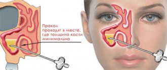
A runny nose is not the only reason for the development of the disease. A number of pathogens that cause sinusitis include:
- injuries to the nasal mucosa;
- staying in drafts or in rooms with dry air for a long time;
- the presence of polyps or adenoids;
- presence of tumors;
- tuberculosis;
- excessive use of vasoconstrictors;
- allergies.
With purulent sinusitis, difficulties arise with the outflow of exudate due to swelling of the mucous membrane, which is why pus is formed. The disease is dangerous with a high risk of developing otitis media, bronchitis and meningitis, vision complications, and infection of the auditory canals. The risk of complications of sinusitis increases in the absence of proper treatment.
Sinusitis can be identified by several symptoms:
- nasal congestion;
- discharge of pus from the nose;
- swelling of the nasal mucosa;
- pain in the forehead and eyes;
- pain when bending down;
- feeling the taste of pus in the mouth;
- bad breath;
- temperature rise to 38 degrees.