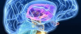
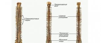
The most common symptoms of damage to the vascular system of the brain and neck are headaches, unsteadiness of gait or weakness in the arm or leg. If symptoms are present, the patient should be examined using MRI of the brain vessels. The purpose of diagnosis is to exclude congenital pathology, namely:
- arterial aneurysm;
- arteriovenous malformation (AVM).
Moreover, symptoms can go undetected for too long, up to 45 years.
The essence of the technique
The study is based on hydrogen atoms, which are affected by a magnetic field, forcing them to be an “exchanger” of energy. This is due to the fact that hydrogen atoms are part of any human organ, including liquids.
Hydrogen atoms rotate around a magnetic axis. When a person is placed in a tomograph, energy is absorbed by hydrogen atoms, and upon completion of magnetic resonance imaging, energy is released. The degree of release by different tissues is somewhat different, since their chemical composition and density play a role. The computer captures this emitted energy, amplifies and re-digitizes it in various degrees of gradation in brightness. Next, the information is displayed on the monitor and recorded on all supported media.
The study is harmless to the patient's health. This is the fastest diagnostic method that painlessly displays all structural and morphological changes in the vascular system of the brain, head and neck.
How is tomography performed?
The tomography procedure is quite simple. It does not require prior preparation of the patient. It can be done at any time of the day or night by appointment. Before the examination, the patient will be asked to come to the MRI center 10 minutes before the scheduled appointment and fill out medical documents. Then I will take him to the examination preparation room. There you will need to leave all items that contain metal and electronic devices - phones, tablets, watches. In the MRI room, the patient will be helped to sit comfortably on the tomography table, and a special signal-amplifying coil in the form of a plastic helmet will be placed on his head. Then slide the table inside the scanning part of the tomograph, and screening will begin.
The patient will understand that the tomograph is taking pictures by a series of noisy signals and tapping sounds, reminiscent of machine gun fire. There is no need to be afraid. If these noises are annoying, you can always ask to wear special noise-canceling headphones.
The average duration of the examination is:
- 15-20 minutes - native version of the examination
- 40-50 minutes - contrast version of the examination.
The damage to diagnostics with contrast tomography is doubled due to the fact that the patient is first given a series of non-contrast images, and then a series of contrast-enhanced images.
Once the scan is completed, the patient has the option of waiting for the results in the clinic or receiving them by email or upon a return visit to the diagnostic center.
Diagnostics MRI of cerebral vessels
MRI of head and neck vessels is an informative neuroimaging method. Give a chance:
- assess the condition of the cerebral hemispheres;
- identify the presence of space-occupying formations;
- determine the presence and development of focal pathological changes;
- assess the condition of the cerebellum and brain stem.
During MRI of the vessels of the head and neck, the entire structure is analyzed with the exception of the skull bones. At the stage of such a procedure, the following occurs:
- assessment of the structure of the cerebral vascular system;
- the presence of an aneurysm in the substance is excluded or confirmed;
- confirmation or refutation of the pathological state of the structure of the vascular system.
What diseases can be detected by MRI of the vascular system?
Angiography is an informative diagnostic method and can identify various pathologies and abnormalities in the circulatory system. MRI can detect:
- Stroke, its previous signs and consequences of a stroke;
- Obstruction of the vascular wall, atherosclerosis;
- Vascular thrombosis, aneurysm;
- Degenerative tissue changes;
- Neoplasms of various etiologies - dimensions and exact location, as well as its circulatory system;
- Assess the extent of damage after brain injuries and concussions;
- Detection of hematomas and contusions.
Possible diagnosable diseases, pathologies
The diagnostic method will help confirm or refute:
- development of deviations due to previous traumatic brain injuries;
- any possible brain infections;
- tumor formations;
- stretching of the walls of the artery with subsequent deformation (most often, immediately after the bed);
- non-standard connection, as well as the location of the capillaries of the vascular system;
- autoimmune diseases;
- carotid artery syndrome;
- artery abnormalities;
- blockage of deep veins.
Such diagnostics can be carried out without the introduction of a contrast agent.
MRI of the vessels of the head and neck is relevant in cases where the patient has been experiencing for a long time:
- headache;
- dizziness;
- attacks of loss of consciousness;
- epilepsy attacks;
- other convulsive conditions.
The patient needs to contact a neurologist with this problem. The latter, using generally accepted medical techniques, will be able to determine how important MRI of the vessels and arteries of the neck is for the patient.
Decoding
It will take about an hour to prepare the tomography results. The patient can spend this time in the waiting room. After processing the results, the person receives pictures, a disc with a video recording of the procedure, and a radiologist’s report.
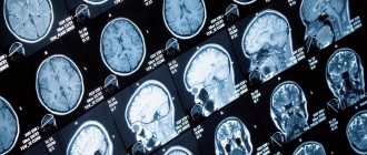
A series of MRIs of the brain, weighted by T2-WI, T1-WI and FLAIR-IP, performed in the axial, coronal and sagittal planes, images of the sub- and supratentorial sections were obtained.
In the left frontal lobe, periventricular to the anterior horn of the left lateral ventricle, an area of altered MR signal measuring 0.7x0.6x0.5 cm is visualized, a hyperintense signal on T2-WI, hypointense with a hyperintense signal along the periphery on FLAIR-IP, hypointense on T1-WI. VI – zone of cystic-glial changes, consequences of a stroke of ischemic type (left MCA basin).
In the white matter of the frontal lobes, a few foci are identified subcortically and periventricularly, with fairly clear, uneven contours, hyperintense on T2-WI and FLAIR-IP, measuring up to 0.2-0.4 cm (most likely foci of vascular origin).
Zones of gliosis are identified periventricular to the anterior horns of the lateral ventricles.
At the level of the semioval centers in the supraventricular white matter and in the projection of the basal ganglia, expanded perivascular spaces are determined along the penetrating vessels.
The grooves of the subarachnoid space along the convexital surface of the frontal and parietal lobes are moderately unevenly expanded.
A cyst of the transparent septum is determined, measuring 5.4x0.7 cm.
The lateral ventricles are asymmetrical (D>S), not dilated - within the age normometry.
III, IV ventricles without features.
The chiasmal-sellar region is not changed.
The cerebellar tonsils are located at the level of the foramen magnum.
Pneumatization of the paranasal sinuses is not impaired.
Conclusion:
MR picture of a previous lacunar stroke of ischemic type in the LSMA basin. MRI signs of a few foci in the white matter of the frontal lobes (most likely foci of vascular origin). MRI signs of expansion of external liquor spaces of a replacement nature.
Author: Shogenov Ramish Kurbanovich
Neurologist with 10 years of experience
MRI of vessels and arteries of the neck and MRI of brain vessels: differences
When performing MRI of cerebral vessels, there is no direct possibility of assessing vascular structures. If a patient has a large aneurysm, most likely this will be visible during a routine native examination of the head.
As for the need to visualize vascular structures, for example, the demanded analysis of blood flow through the vascular system, excluding possible narrowings or other defective variants of the structure of the vascular bed, it is necessary to conduct an examination of the head. Also, if the patient has any ischemic conditions or strokes, with the help of additional brain diagnostics, it is possible to determine at the level of which of the arteries the “catastrophe” occurred.
Indications
The procedure is carried out as prescribed by the doctor. As a rule, the doctor refers the patient to an MRI if the following complaints are observed:
- Frequent headaches that do not go away even after taking painkillers;
- Unreasonable dizziness;
- A sharp deterioration in vision or hearing, incl. episodic;
- Extraneous noise in the ears;
- Suspicion of the presence of aneurysms, atherosclerosis, blood clots;
- Stroke;
- Severe hypertension;
- Suspicions of neoplasms in the brain.
The procedure is also carried out before the upcoming operation and after it - in order to track the dynamics.
The examination reveals the following features:
- Malformations of veins and arteries, occlusions, stenosis;
- Identifying vascular dissections, blockages, or aneurysms;
- Assessment of blood flow to identify possible cerebrovascular accidents.
Decoding of received images
MRI of the head and neck vessels is a complex diagnostic method and requires special skills that only a radiologist and a neurologist can have. A few hours after MRI of the cerebral vessels, you can get a detailed answer from a specialist about the current situation. Also receive recommendations regarding treatment or further hospitalization of the patient.
The examination is carried out using modern equipment, so in addition to the conclusion, as well as photographs, the patient receives a CD with a recording of the examination. It is the use of the latter medium that makes it possible to find abnormal channels of the vascular system of small sizes.
Scan progress
Magnetic resonance scanning of cerebral vessels does not require special preparation, the only exception being the use of a contrast agent in MRI. For a successful and safe examination, the patient must notify the doctor about his or her health status and truthfully answer all questions before the procedure. In most cases, the person being examined is asked to read the list of contraindications and sign as confirmation of consent to the scan.
The examination procedure consists of the following stages:
- The patient is placed on a retractable panel, his limbs are fixed with special belts, his head is tightly held by rollers equipped with wires;
- Next, the panel slides into the device and the scanning process starts;
- During the procedure, the patient hears the voice of a medical professional and can speak to him.
If the patient suffers from a fear of confined spaces or cannot remain without movement for a long time, it is possible to use sedatives; there are also devices that do not have side walls, so the patient does not experience discomfort. It is worth noting that MRI of the brain in Moscow takes place without any pain; the system is designed in such a way that the patient can breathe calmly; for this purpose, an air conditioning system is provided. It is noteworthy that after the scan, the patient can immediately engage in usual activities, for example, going to work. The patient receives the result of the examination in his hands; usually, it is ready 2-3 hours after the examination.
Examination with contrast
An MRI examination of the vessels and arteries of the neck using contrast is carried out exclusively according to the doctor’s indications. As a rule, injection of a dye into a vein is not required. At the time of examination, attention is first paid to the native part of the procedure. If at this stage any pathology or mass formation is detected, the radiologist may decide to carry out such a manipulation.
Despite such a simple procedure, there are a number of diseases that require the introduction of a contrast agent for better diagnosis. These include multiple sclerosis.
What will magnetic resonance imaging of the brain show?
Head tomography data allows for high-quality differential diagnosis of the following pathologies:
- demyelinating diseases, including multiple sclerosis;
- volumetric formations of benign or malignant origin;
- cysts and pseudocysts;
- degenerative changes in brain structures, for example, Alzheimer's disease, Parkinson's disease;
- inflammation of the meninges, including meningitis and encephalitis;
- leukoaraiosis of the brain;
- hydrocephalus;
- epilepsy.
| MRI image of a healthy brain | brain cyst on MRI | brain cancer on MRI |
How often should the examination be carried out?
MRI of the vessels and arteries of the neck is a completely safe diagnostic method and does not leave any traces in the body. It can be carried out countless times, namely as many times as the patient’s condition requires until a complete diagnosis is completed. For some, one study without follow-up is enough. A patient who needs a follow-up examination, for example, after removal of large tumors, undergoes MRI of the vessels and arteries of the neck after 6-12 months.
What symptoms require an MRI of the head?
MR images of the brain in the axial (left) and sagittal (right) planes
MRI is prescribed in the following cases:
- regularly recurring or acute headaches accompanied by nausea, vomiting and weakness;
- increased temperature without signs of viral and colds, especially with symptoms of irritation of the meninges (meningism);
- periodic loss of orientation;
- inhibition of reactions;
- hearing loss, noise in the head;
- frequent nosebleeds;
- blood pressure surges;
- regular dizziness and fainting;
- disturbance or sudden loss of vision;
- speech disorders;
- decreased mental activity;
- high intracranial pressure;
- memory impairment or loss;
- convulsive seizures.
Contraindications
MRI of the head and neck vessels has virtually no contraindications. The only exception is the presence of metals of a certain group in the body. These include products such as pacemakers or other electronics in a metal case. A complete exception may be the presence of titanium structures after osteosynthesis, fractures or joint surgeries. Each situation is considered individually. As a result, each patient can learn about the presence of contraindications.
List of contraindications
Restrictions may apply to patients with pacemakers, fixed metal dentures, pins inside the body and other elements that cannot be removed independently during the examination. Relative contraindications include the first trimester of pregnancy, however, for serious indications that exceed the potential harm, an examination can be performed. Patients weighing more than 250 kg and those with mental disabilities that do not allow them to lie still during the examination are also not allowed to undergo MRI.
What is more informative: MRI of head and neck vessels or computed tomography
CT angiography with the introduction of a contrast agent still remains a more informative procedure. This level of detail also has one significant drawback - the absorption of high load rays by the body. The latter can aggravate the development of cancer and completely excludes dynamic monitoring of certain pathological conditions.
The iodine-containing contrast agent administered during CT angiography is quite allergenic. Therefore, not everyone can conduct computer research. The examination itself is variable. At the time of assessing the clinical picture, the neurologist already understands which study will be more relevant for the patient: MRI of the vessels and arteries of the neck or CT angiography.
Potential Risks
Carrying out magnetic resonance imaging does not involve any risks for the patient. Due to the fact that the subject’s body is securely fixed with special straps, throughout the entire scanning period the likelihood of any injury is reduced to zero.
However, the possibility of unpleasant sensations due to being inside the tomograph cannot be completely excluded. The patient may experience increased heart rate, dizziness, and a feeling of tightness in the sternum, which has a psychosomatic cause. Therefore, you should not ignore the doctor’s suggestion to take sedatives before starting the examination.
Choosing a clinic, specialist and price of service
The mrt-mozga service is a full-fledged resource with a database of diagnostic clinics in Moscow (about 300 centers). The resource’s simple and intuitive interface will suggest the best offer. The patient can sort clinics according to various parameters: geolocation of the clinic, price, type of tomograph used, availability of contrast agent, operating hours, promotional offers. The information on the site is constantly updated and expanded.
You can make an appointment by calling 8 (495) 032-07-59 from 09:00 to 00:00. By taking advantage of the site's offer, you can not only make an appointment for free, but also get the best price offer.
Advantages of MRI over X-rays
MRI, as a method of diagnostic examination, is compared with radiographic examination, let’s figure out what the main differences are. The fact is that during an x-ray, the human body is exposed to radioactive rays, as a result of which different tissues of the human body have different colors in the image, thus, bone tissue in the x-ray has a bright white color. As for diagnosing the brain, X-rays are used only in cases of injuries and fractures of the skull bones; in the study of the vascular system, it is absolutely useless.
In addition, it must be said about safety; active X-rays, despite the minimum radiation dose, cannot be called harmless. For this reason, this examination is not recommended for young children, pregnant women, nursing mothers and people who have received a certain dose of radiation before.
Make an appointment now!
When is contrast needed during MRI of the brain?
A contrast agent is used for magnetic resonance imaging to improve visualization of soft tissues. The contrast is unevenly distributed in healthy and diseased tissues, helping to identify abnormalities and determine the boundaries of the development of the pathological process.
A contrast agent is used when it is necessary to diagnose brain pathologies such as:
- neoplasms;
Tumors have a developed vascular network. Distributing throughout the tissues, the contrast agent brightly colors the pathologically changed areas. Using contrast, you can analyze the size of the tumor and its exact location. Contrast also helps determine the malignancy of the tumor. Benign tumors have clear boundaries and regular shapes, while malignant tumors grow into healthy tissue and have uneven boundaries and shapes. These features are clearly visible on MRI scans obtained using contrast.
- demyelinating diseases.
Diseases such as Alzheimer's and Parkinson's disease manifest themselves in changes and degeneration of the nerve substance. Contrast is used to diagnose the extent of the disease.
In both cases, the use of a contrast agent makes it possible to clearly differentiate healthy tissues from altered ones, and to see the extent and extent of the pathological process.
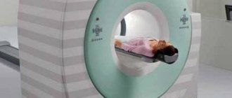
What does the diagnostic equipment look like?
A standard MRI machine is a large cylindrical tube surrounded by a magnet. The patient sits on a movable examination table that slides inside a magnet. Some CT scanners (called short-tunnel systems) are designed so that the magnet does not completely surround the patient's table.
Some devices are open on the sides. Such tomographs are especially suitable for examining obese patients and people who suffer from a fear of closed spaces. Modern open MRI scanners make it possible to obtain very high-quality images for various examinations. However, if an open type machine uses an old magnet, the image quality may be reduced. Some studies cannot be performed on an open tomograph. For more information, consult a specialist.
The computer working system that processes the images is located in the room next to the scanner.
Up
How to prepare before MRI of neck vessels?
Many patients who are faced with such a study are wondering about preliminary measures to prepare for the scanning session. However, there is no preparation for performing MRI of the brain arteries. A person can continue to maintain his daily lifestyle, eat any food and take various medications. The only point that doctors highlight is the patient’s morale. It will be best if you come to your appointment in a calm and balanced state.
If it is necessary to perform an assessment with a contrast agent, the patient is tested in advance for the presence of an allergy to the active component. Also on the day of the session, you must tell the technologist about:
- Presence of pregnancy.
- Claustrophobia or difficulty maintaining one position for a long time.
- Unstable emotional state.
- Provide the doctor with an identification document, past scan results and medical record for review.
In addition, you need to dress in comfortable clothes without metal objects. Before you begin, you should remove all jewelry and other accessories. Also, doctors advise girls not to use cosmetics. In some situations, you can take a sedative.