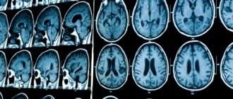
Ultrasound of cerebral vessels is an absolutely painless technique that helps to identify possible pathological processes. It has been scientifically proven that ultrasonic waves do not have a negative effect on organs and tissues, therefore such manipulation can be carried out not only for diagnosis in adults, but also in children. The number of procedures and frequency of examinations are not limited - ultrasound can be performed as often as necessary. Brain examination using ultrasound is carried out both according to the doctor’s indications and at the request of the patient. Let's look at how an ultrasound is performed and how to properly prepare for it.
In what cases is ultrasound of the brain prescribed?
Of course, you can undergo the examination without any complaints. But there are a number of symptoms that indicate that the procedure should not be postponed.
Be sure to consult your doctor if any of the following concerns you:
- Constant feeling of drowsiness and weakness;
- Dizziness, darkening of the eyes with a sharp rise;
- Noise in ears;
- Regular headaches or headaches that occur in the morning;
- Flashing “flies” before the eyes;
- Difficulty in perceiving information (reading or listening);
- Problems paying attention or remembering;
- Blood pressure surges;
- Episodes of incoordination;
- Apathy or low mood;
- Unreasonable nausea and vomiting;
- Deterioration of vision or hearing.
For preventive purposes, ultrasound is also indicated for those people who have high cholesterol or blood sugar levels, as well as people suffering from obesity, the elderly and those who have previously suffered a heart attack or stroke.
Sometimes ultrasound of cerebral vessels is prescribed before an upcoming operation, for example, if tumors in the head area are to be removed.
Indications for use of the procedure
Doppler ultrasound is prescribed to patients for a number of indications.
Causeless migraine and dizziness
If a patient suffers from migraines quite often, but does not associate it with fatigue, stress, or physical exertion, he should undergo an ultrasound scan.
to identify the true causes of pathology. The same can be said about causeless dizziness. Perhaps this is caused by impaired cerebral circulation.
Noise in the ears and head
Noise in the ears and head can be a symptom of atherosclerosis, vegetative-vascular dystonia, hypertension and other diseases. Doppler ultrasound will allow you to accurately make a diagnosis.
Poor health, which is accompanied by attacks of weakness and a feeling of shortness of breath
The feeling of weakness and lack of air cannot always be associated with poor physical fitness of the patient or problems with the breathing apparatus. Perhaps the reason lies in impaired blood supply to the brain, anemia or other pathologies that ultrasound scanning will help identify.
VSD (vegetative-vascular dysfunction)
VSD is manifested by paroxysmal or constant palpitations, increased sweating, headache, tingling in the heart area, redness or paleness of the face, chilliness, and fainting. This pathology is not considered as an independent disease - it always accompanies some organic pathology, which may be associated with impaired blood supply to the brain.
Hypertension
Hypertension, or a sustained increase in blood pressure, can cause many diseases, including those associated with poor circulation in the vessels of the head and neck. Doppler ultrasound of the vessels of the head and neck is the best way to confirm or refute the presence of just such pathologies.
Types of ultrasound of cerebral vessels
Various techniques can be used to study cerebral vessels:
Doppler ultrasound
- used to assess blood flow in vessels, allowing to identify its nature and speed.
Duplex ultrasound.
This technique is a variation of the technique described above. Duplex examination allows you to visualize blood vessels, assess their condition, identify blood clots, plaques, aneurysms and other pathological processes. During this technique, the direction and speed of blood flow are also determined.
Triplex ultrasound.
With its help, an image of the arteries is obtained, colored in different shades, depending on the direction and speed of blood movement. The ophthalmologist can also set other parameters for staining - this opportunity is provided by a computer program. Ultrasound of cerebral vessels using triplex scanning allows you to obtain maximum information about the state of the circulatory system.
Each method has its own advantages and disadvantages. Which diagnostic technology will be chosen is determined by the specialist, depending on what parameters are important to him for diagnosis.
Ultrasound of head and neck vessels
Ultrasound of the vessels of the neck and head is one of the most accessible and effective methods for diagnosing diseases such as atherosclerosis and thrombosis. The examination allows you to assess the condition of the arterial and venous branches and lymph nodes, measure the speed of blood flow and other parameters. Medical invites patients to undergo ultrasound diagnostics using new generation devices. An informative and safe examination will allow timely detection of vascular pathologies and initiation of treatment.
Indications for ultrasound examination of the vessels of the neck and head
An examination is prescribed if the patient notes the following symptoms:
- recurrent headaches, migraines;
- fainting conditions;
- dizziness not associated with hunger or fatigue;
- feeling of heaviness in the head in the morning after waking up;
- sleep disorders, insomnia;
- noise or congestion in the ears;
- sudden deterioration of vision;
- decreased concentration, memory problems;
- impaired coordination of movements;
- chronic fatigue, etc.
In addition, ultrasound examination of the vessels of the neck and head is carried out for such diagnoses and conditions as:
- traumatic brain injuries;
- stroke or heart attack;
- ischemic disease;
- hypertension;
- arrhythmia, angina pectoris;
- thrombosis of veins or arteries.
An ultrasound of the vessels of the neck and head will also be required if changes appear in blood tests, for example, if cholesterol levels increase. An annual examination is recommended for patients over 45 years of age. People who smoke a lot, lead a sedentary lifestyle, or work under constant stress are also at risk.
Contraindications
The procedure is quick and painless. Ultrasonic waves do not have a negative effect on the body and do not cause side effects. Thanks to these qualities, examination can be prescribed even to pregnant women and newborn children. The only limitation for diagnostics may be severe damage to the skin in the area of contact with the ultrasound equipment sensor.
Preparation for ultrasound of the vessels of the head and neck
Before undergoing the examination, the patient is recommended to consult with the attending physician. The specialist will most likely stop taking certain medications that may affect the functioning of blood vessels and distort the picture of the disease.
If the patient is involved in professional sports or heavy physical work, then on the eve of the examination the load should be minimized.
Preparing for ultrasound diagnostics also includes giving up coffee, strong tea, energy drinks, alcohol and smoking. It is not recommended to eat before the test.
How is ultrasound of the vessels of the brain and neck performed?
The main type of instrumental diagnosis of diseases of the cervical spine is a complex of duplex and triplex scanning. It gives an idea of the characteristics of blood flow in the vessels of the neck and head (including intracranial). During diagnostics, a complete picture of soft tissues is displayed on the equipment monitor, against which veins and arteries are clearly visible. The method allows you to record the speed of blood flow in a certain area. This parameter is coded in different colors: shades of red indicate the movement of blood towards the sensor, shades of blue indicate the movement of blood away from the sensor. The study makes it possible to identify such disorders as inhibition of blood flow due to atherosclerotic plaques, thinning of vessel walls, expansion or narrowing of the lumen, inflammatory processes, vascular damage due to diabetes or hypertension, congenital anatomical abnormalities in the structure of veins and arteries, and others.
The procedure takes from 30 to 60 minutes depending on the type of ultrasound and takes place in several stages:
1
The patient takes off his outer clothing, frees his neck and collarbones from jewelry, and then lies down on the couch.
2
The doctor sets up the equipment and treats the sensor with a colorless hypoallergenic gel, which can additionally be applied to the skin in the area of study. Acoustic gel improves the conductivity of the ultrasound signal and the gliding of the scanner.
At the end of the examination, the results are deciphered and given to the patient in writing.
What does an ultrasound scan show?
During the examination, the specialist examines the main arteries that provide nutrition to the brain: common, external and internal carotid, as well as middle, anterior, posterior vertebral and basilar.
The ultrasound protocol contains a detailed description of the anatomical features of the vessels of the patient’s neck and head, data on the movement of blood flow, obstruction or deformation of veins and arteries. The examination allows you to detect the following abnormalities and pathologies:
- Atherosclerotic plaques. At the initial stage of blockage formation, a thickening of the vessel wall of up to 1.5 mm is noted. A reading greater than this value may indicate the presence of plaque.
- Thrombosis. When the disease occurs, the protein fibrin and blood cells, platelets, accumulate in certain areas of the vessels. A thrombus can partially or completely block the lumen of a vein or artery, which leads to impaired circulation, hypoxia and other dangerous consequences.
- Cerebrovascular accident. Pathology can occur as a result of thrombosis, narrowing of the diameter of arteries and veins, changes in blood flow speed, development of osteochondrosis, etc. Identifying hypoxia at an early stage will prevent the occurrence of concomitant diseases, such as stroke.
Many vascular pathologies are irreversible, so patients at risk are required to undergo an annual ultrasound of the vessels of the head and neck - you can do this at our medical center in Moscow.
Advantages of the Miracle Doctor clinic
Experienced doctors
Ultrasound diagnostics of blood vessels is carried out by specialists with extensive practical experience. The total work experience of some doctors exceeds 30 years.
Modern equipment
The clinic has new generation ultrasound machines. The equipment allows you to obtain detailed images of blood vessels without interference.
All types of ultrasound at low prices
By contacting us, you can undergo Doppler ultrasound, duplex and triplex scanning of blood vessels. Ultrasound diagnostics are performed by appointment. To get tested, make an appointment by phone. A description of the examination results is performed in front of the patient.
VIP service
You can undergo a vascular examination any day of the week without a queue. If necessary, we will schedule you for a consultation with a phlebologist or neurologist.
To find out the cost of ultrasound of the vessels of the head and neck, make an appointment and ask other questions you are interested in, call our clinic in Moscow or fill out the feedback form.
Possible contraindications
There are no contraindications to ultrasound examination of cerebral vessels. The exception is severe damage to the skin in the area being examined. In this case, the procedure can be postponed until complete healing.
Another nuance may be an allergic reaction to silicone, which is part of the contact gel. If you have already had cases of allergic reactions to silicone, be sure to notify the doctor who ordered the test and the doctor who will conduct it. A specialist may recommend taking antihistamines to reduce allergies, or purchasing another gel without silicone.
Our advantages
By contacting the Alfa Health Center clinic for ultrasound diagnostics, patients can appreciate the following benefits:
- Quick examination. Patients undergo ultrasound diagnostics at the appointed time. You will not have to wait long for your turn and return to the clinic again to pick up the examination results. You will receive a transcript of the ultrasound 15 minutes after the procedure.
- Availability of ultrasound diagnostics. Ultrasound is one of the most common and accessible examination methods. Patients receive professional medical services on favorable terms.
- High information content. Specialists use expert-class ultrasound equipment, which makes it possible to obtain maximum reliable information about the speed of blood flow and detect indirect signs of its disturbance. This allows the doctor to suspect the development of a dangerous condition, prescribe advanced diagnostics and, if necessary, adjust treatment tactics.
- No restrictions on the number of procedures. Ultrasound is used as often as the clinical situation requires. Ultrasound is allowed to be used during pregnancy, to examine children from the first days of life.
Advantages and disadvantages
The main advantage of the diagnostic procedure is its high information content and 100% safety. Since the manipulation does not imply invasiveness, it has no contraindications. The study has other benefits:
- It doesn't hurt at all;
- Ultrasound does not cause discomfort;
- The ability to study both the physical state of blood vessels and their functionality.
One of the disadvantages is the low accessibility of blood vessels in certain areas of the brain for diagnostics using ultrasound.
Advantages of diagnostics at the Innovative Vascular Center
Our medical center specializes in vascular and endovascular surgery, so it is important for us to obtain detailed information about the condition of the carotid arteries, because ischemic stroke is one of the most dangerous complications in vascular surgery. We perform ultrasound of the carotid arteries on all patients in our hospital and have developed an accurate ultrasound diagnostic algorithm. Prices for research in our center in Moscow are affordable to everyone. An appointment with a vascular surgeon is always accompanied by ultrasound diagnostics.
The advantage of ultrasound of the carotid arteries in our clinic is expert-level ultrasound scanners, evaluation of the results obtained by an ultrasound physician together with the operating vascular surgeon. The accuracy of the results of this study by our specialists is 96%, versus 80% in many other general practice diagnostic centers.
Preparing for the study
If you want to get the most accurate results, you need to properly prepare for the upcoming test. Let's consider the most important stages of preparation:
- Since we are talking about checking blood vessels, it is important three days before the upcoming procedure to give up alcohol, energy drinks and additives that can affect the functionality of blood vessels and the speed of blood flow;
- Try to completely relax and breathe freely at the time of diagnosis;
- Do not engage in sports or physical activity for at least 2 hours before the ultrasound.
- If you are constantly taking any medications, be sure to discuss this with your doctor. If it turns out that the pills affect blood pressure and blood vessels, the doctor may stop them for a day, or postpone taking the drug to a later time;
- 3 hours before ultrasound diagnostics you need to refrain from smoking;
- Before the procedure, long hair must be collected so that nothing interferes with the examination process.
If the examination is being carried out on a child, you should prepare him mentally. Explain to your child that this procedure is completely painless and does not cause discomfort, persuade him to lie still - movements during the diagnostic process can distort the results of the study.
Ultrasound ultrasound: progress of the procedure
An ultrasound scan of the vessels of the head and neck lasts, as practice shows, approximately 45-50 minutes.
The patient lies on his back and throws his head back. To facilitate the movement of the sensor, the doctor applies a special gel to the skin. During the examination, the sensor is constantly moved to assess blood flow in different parts of the head and neck. During the procedure , ultrasound examination of the vessels of the head and neck
It may be necessary to perform some functional tests. To do this, the doctor asks the patient to breathe deeply, and he can press the vessels with his fingers or a transducer.
How to do an ultrasound of the brain
The person lies down on the couch, his head resting on a low pillow. The desired area of the skin is lubricated with silicone gel. In order to determine the nature and speed of blood flow, several sensors are attached to the temples.
If it is necessary to study the condition of the veins and arteries of the brain, the specialist suggests conducting Doppler sonography, during which changes in the displayed sound waves from moving blood cells are recorded. To carry out the procedure, the sensor can be localized in the forehead, back of the head or temples.
In total, the manipulation lasts no more than 40 minutes. Next, the patient receives the test results in hand and goes to the attending physician to decipher the results.
What body functions does ultrasound study?
By performing an ultrasound scan of the vessels of the neck, one can diagnose pathologies of the carotid and vertebral arteries, and scanning the vessels of the head allows one to assess the condition of the carotid, subclavian, vertebral arteries and the main arteries of the brain. Such examinations are usually carried out simultaneously. This allows you to obtain the most complete and reliable information about the state of the vessels supplying the brain. There are several parameters by which the condition of the vessels of the neck and head, as well as the blood flow in them, is assessed.
How elastic are vascular walls?
A decrease in the elasticity of vascular walls can lead to strokes, heart attacks, and varicose veins. The reasons for decreased elasticity are: poor diet, high blood cholesterol, bad habits, age factor. Timely detection of the fact of decreased elasticity of the walls of blood vessels will allow the doctor to prescribe therapy in a timely manner and prevent serious problems with the patient’s health.
What condition is the inner surface of the vessel in?
The inner surface of the vessel is in direct contact with the blood and releases special substances that prevent the clotting process. In other words, the main function of the inner surface is to prevent the formation of blood clots. Violation of the structure of the inner layer of the vessel and its integrity can lead to thrombus formation.
The presence or absence of changes in the integrity of the vessel walls
It is logical that a violation of the integrity of the walls of blood vessels leads to a disruption of blood flow, a change in its movement, and therefore a disruption of the blood supply to the brain. The doctor’s task when conducting an examination using ultrasound dopplerography ( ultrasound Dopplerography of the vessels of the neck and head
) identify ruptures in the vascular walls, assess the complexity of the situation.
Intraluminal formations of arteries or veins
Formations in the lumens of veins or arteries are blood clots that impede blood flow. Accordingly, the blood supply to the brain becomes more difficult, which can result from a stroke, cerebral infarction and other pathological conditions.
Decoding the results
A neurologist interprets the results of an ultrasound examination of cerebral vessels. In order to establish an accurate diagnosis, the specialist takes into account the characteristics of the cerebral vessels and the patient’s medical history, because some changes may be an anatomical feature unrelated to pathology.
If everything is normal, the results should look like this:
- the lumen of the vessels is completely free, various neoplasms are absent;
- turbulent blood flow is not present in the branching of vessels;
- the thickness of the vascular wall is less than 0.9 mm (sometimes 0.9-1.1 mm is also a normal option);
- absence of foreign inclusions, cysts and various neoplasms.
Please note that a diagnosis cannot be made based on ultrasound alone. The disease is determined by a combination of pathological factors, which are indicated by various studies.
What does the study clarify?
After ultrasound with Doppler, the specialist is able to determine a number of factors.
Vessel patency
It is important to assess how freely blood flows through the vessel, whether there are any blood clots or blood clots in the lumens of the veins and arteries.
To what extent does the course of the vessel correspond to the normal trajectory?
The existing direction of blood flow during scanning is compared with the norm along the trajectory of the anatomical course of the vessel. An increase in the tortuosity of blood vessels leads to disruption and slowing of blood flow.
Diameter and location of the vessel lumen
Vasomotor function is a change in the diameter of the lumen of a vessel under the influence of certain factors. This function is necessary to regulate blood pressure in blood vessels, heat exchange, and metabolism. Pathological narrowing of the lumen of the vessel leads to increased pressure in the spasmodic area, and local blood supply is disrupted.
Length of visibility of the changed lumen
When identifying pathological changes in the lumen of the vessels of the neck and head, it is important to identify the area of localization of the pathology, as well as the length of visibility of the changed lumen. This will allow you to accurately diagnose and prescribe treatment.
What can she show?
Duplex scanning allows you to determine areas of narrowing of the lumen of blood vessels, the direction of blood flow, its speed, as well as the condition of the vascular walls. This study can show the location of cholesterol plaques and the presence of blood clots, which is very important when diagnosing atherosclerosis.
A decrease in the elasticity of the walls of the arteries, as well as their thickening, indicate that the patient has hypertension (high blood pressure). If a change in blood flow is diagnosed, this indicates the presence of obstacles. For example, an aneurysm is a sac-like protrusion of a vessel.
Normal head according to ultrasound
Normal indicators, if no abnormalities are detected during the diagnosis, look like this. The structures of the brain are symmetrical, the ventricles of the brain are symmetrical, have smooth boundaries, are anechoic, the subcortical nuclei have average echogenicity, there is no shift in the M-echo.
There should be no foreign fluid in the interhemispheric space or inclusions in the brain structures. There should be no tumors or other formations, as well as aneurysms of large cerebral vessels. Vascular walls must have smooth contours and structure. The patency of blood vessels should be within the age norm; there should be no compression of the arteries.
If during the diagnosis any violations were identified, do not give up. If there are pathologies, the doctor will refer you to additional diagnostic procedures, after which you will be prescribed effective treatment.
How is the head ultrasound procedure performed?
Another advantage of ultrasonography is that it does not require any special training. The results of a brain ultrasound do not depend on the time of the study, food intake, or other factors. But the doctor asks the patient to tell him what medications he took over the past 24 hours.
At the appointed time, the client is invited to the ultrasound diagnostic room, asked to undress to the waist (this is necessary for scanning the vessels of the neck) and lie down on the couch. Then a special gel is applied to the skin, which facilitates the sliding of the sensor and improves the conduction of ultrasonic waves.
Ultrasound of the head is performed in duplex scanning mode, which allows one to assess blood circulation in the vessels of the neck and brain and identify possible pathological changes in the vascular endothelium.
Are there any contraindications?
Ultrasound waves are absolutely safe for humans, so there are no special contraindications to this type of ultrasound. The only exception is a violation of the integrity of the skin. The procedure involves contact of sensors with the head or neck, so the examination is postponed until the wound heals.
However, there are a number of special cases that make duplex examination of blood vessels difficult:
- cardiac arrhythmia, which changes the speed of blood circulation through healthy arteries and veins;
- the area under study is hidden under bone tissue;
- slow blood flow in the patient.
These are not contraindications, but in this case changes in the location of the sensors are required. It will be necessary to navigate non-standard conditions, which requires professionalism on the part of the doctor.