Among neuroimaging diagnostic methods, electromagnetic screening has the highest level of information content and reliability. MRI of the brain is a medical method that helps doctors carefully examine all the structures of the organ, the ventricular section, the substance present, the cavities where there is cerebrospinal fluid. You can find a clinic where you can undergo an MRI of the brain with a contrast reagent or in the usual mode at mrt-v-spb.ru. Call the hotline for a free consultation and get a discount on examination up to 1,000 rubles.
Brief information about the central nervous system
The central nervous system is responsible for the stable functioning of the entire body. When any disorder develops in it, a person experiences painful symptoms, leading to harmful consequences. Factors indicating the progression of abnormalities affecting the performance of the brain are considered: painful sensations, neurological signs (visual, motor, auditory disturbances), fainting, etc.
Non-invasive techniques are used to diagnose lesions in this organ. MRI of the brain produces clear, three-dimensional images that can be zoomed in and out to closely examine the structure of the region. The screening programs installed in the device allow you to visualize cerebral vessels. The results of tomography allow the doctor to determine the cause of the disease, its location, as well as establish a diagnosis and create an acceptable treatment regimen.
How is a tomogram read?
To make the study as accurate as possible, contrast is used. It will help highlight the brain tissue as clearly as possible. If the tissues have undergone pathological changes, they appear darker during a contrast study. The contrast will help you find out the state of the blood supply to the brain structures.
When analyzing MRI results, the doctor evaluates the following parameters:
- Localization . If a pathological process is detected, the doctor evaluates its location. Localization will help to understand which structures are affected and what this entails. For example, if we compare tumors located in the cerebral cortex with tumors at its base, the latter are much more dangerous.
- Size and shape of the found source . If the tumor has smooth edges, this is most often a sign of its benignity and the presence of a capsule. If there are many foci, this is a sign of the appearance of metastases.
- Hue . An experienced doctor, armed with knowledge of pathological anatomy, can draw a conclusion from the shade of the lesion and its structure which pathological process is running. A changed gray tint, for example, may indicate new growth or softening of the tissue.
There are also indirect indicators that help the doctor make the correct diagnosis. For example, a tomogram of the vessels of the neck and head will help determine the cause of stroke or ischemia.
If the outflow of blood is disrupted, neurological symptoms may also appear, for example, increased intracranial pressure. This leads to compression syndrome.
Differential diagnosis of brain lesions on MRI using algorithms - interpretation of images with explanations:
What is an MRI of the brain?
During tomographic screening, doctors can install different diagnostic programs. This helps in targeted testing of different areas of the brain. The main protocols are:
- MRI of the brain - the purpose of the procedure is to evaluate the white and gray matter, as well as the ventricles along with the membranes.
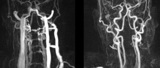
- MR angiography - visualizes the vessels of the organ.
- MRI of the nasal sinuses - the procedure shows the anatomical structure and pathologies in the nose.
- MRI of the eye - the method is aimed at studying the condition of the eyeball along with the optic nerve and orbit.
- MRI of the pituitary gland - diagnosis of the sella turcica area.
The choice of protocol depends on the symptoms, objectives of the study and the goals of the doctor.
Features of head tomography of a healthy person
The radiologist describes a healthy brain in the study protocol as follows: All brain structures must be developed correctly, the intensity of the magnetic resonance signal is normal. The ventricular system is developed correctly, of normal size, neither dilated nor reduced. The perivascular and subarachnoid space are normal. The sulci and gyri are normal. The brain structures are of normal size and not displaced. Sella turcica, pituitary gland without pathology. The eye sockets, nasal sinuses, and ear canals are of normal size and shape, without signs of pathology. Brain tissues do not have focal or diffuse changes. If contrast was used, the brain vessels are developed correctly and are evenly filled with contrast agent.
What are positive MRI results:
- Normal arrangement and “consistency” of tissues;
- Absence of abnormal formations (“plus-shadow”);
- There are no bleedings, purulent accumulations, blood clots, or arteriovenous malformations.
What is shown on the results of an MRI of the brain?
The head diagnostic program includes monitoring of the following departments:
- ventricles of the brain;
- white matter of the subcortical plan, located in the frontal lobe;
- chiasmal area;
- middle sections;
- cerebellar tonsils.
Additionally, during the procedure, the radiologist evaluates the pituitary gland along with the paranasal sinuses.
What does an MRI of the brain show?
A study using electromagnetic equipment makes it possible to visualize the following abnormal conditions in a patient:
- infectious diseases;
- lesions of the nervous system such as multiple sclerosis;
- failures in the structure of the pituitary gland;
- malformations of the brain;
- bleeding from traumatic head injuries;
- malfunctions of the vascular system;
- dementia of a senile nature;
- sources of malfunction of the visual and auditory organs.
Indications for magnetic resonance imaging of the brain
- Neoplasms or their metastases. Diagnosis is prescribed for persistent migraine-like pain, sudden loss of vision and hearing, auditory, olfactory and visual hallucinations, attacks of confusion, sudden reading and writing disorders, often accompanying cancer pathologies.
- Epilepsy and other diseases manifested by fainting, confusion and convulsions.
- Suspicion of single or multiple cystic cavities filled with fluid, bloody or other contents.
- Possible presence of parasites (cysticercus and echinococci) carried through the vascular bed with the bloodstream into the head.
- Inflammations – meningitis, encephalitis, arachnoiditis, myelitis. Lesions caused by infections - measles, herpes, tuberculosis, toxoplasmosis, tick-borne encephalitis.
- Rehabilitation after stroke, traumatic brain injury and surgery. Using magnetic resonance diagnostics, the doctor evaluates the effectiveness of the treatment and predicts long-term results.
- The likelihood of developing multiple sclerosis, Alzheimer's disease and other degenerative processes.
- Children are examined for congenital pathologies and hydrocephalus.
With all these diseases, life and health directly depend on a timely diagnosis. Therefore, at the slightest suspicion of brain disorders in yourself or your child, you need to come to the clinic and be examined.
When should you get an MRI of the brain?
The main symptoms that become the reason for undergoing research are called:
- headache;
- dizziness;
- double vision;
- epilepsy attacks;
- memory problems;
- high pressure readings.
Other manifestations of unknown origin for which MRI of the brain is prescribed include:
- feeling of weakness in the limbs;
- problems with coordination;
- chaotic thinking;
- neoplasms of various origins;
- pathologies of vascular development;
- vascular lesions such as stroke and their consequences;
- demyelinating disorders; degenerative abnormalities; inflammatory lesions;
- encephalitis, including in patients with HIV status, abscess; congenital developmental anomalies;
- arachnoid type cysts; hydrocephalus; Arnold-Chiari pathology;
- defective development of the corpus callosum; neurocutaneous syndrome; Acute TBI; hematomas.
The doctor recommends undergoing an electromagnetic brain procedure to monitor the radicality of the removal of the tumor body and to exclude its further growth in patients after surgery.
When is it prohibited to do an MRI of the brain?
Despite the high level of information content of the procedure, the method is not suitable for everyone. The main contraindications apply to people who suffer from asthma and chronic kidney disease. Pregnant women should be treated with special caution when considering MRI of the brain. Despite the long-term use of tomographic equipment in practice and the absence of adverse effects in pregnant women or newborn children after the examination, patients need to inform the attending physician and radiologist about the current situation.
Relative restriction is called claustrophobia. If the patient is afraid of a closed space (where he will be during the diagnosis), you should choose a clinic with open-type tomographs. Typically, the procedure occurs without consequences for patients with titanium implants.
However, people who have structures made of other types of metal and electronic devices (insulin pump or defibrillator) in their bodies should not undergo MRI of the brain. The presence of such objects in the body can distort the results of the study, disable the device, as well as the devices themselves implanted into the body.
MRI of the brain in an adult and a child
Magnetic resonance imaging is equally safe for all ages and groups. If there are no implants in the subject’s body that react to a magnetic field (pins, endoprostheses, knitting needles, pacemakers, etc.), the procedure cannot harm him in any way. The effect of MRI on the human body can be roughly compared to applying a large magnet to it. There will be no consequences for the body, but special instruments will be able to record minor changes in the nuclei of atoms, on the basis of which a computer program will create a detailed “map” of the area under study. Any myths about the dangers of MRI, its terrible consequences, are nothing more than fairy tales and inventions of uneducated or dishonest people who want to draw attention to their person.
The brain of a child after 1.5 years of life is practically no different from the brain of an adult. The procedure is also the same for both children and adults. The only characteristic of young children is their behavior. Babies are not at all able to lie down quietly for a few minutes, especially in an unknown place, without their mother nearby, and slightly older children may simply be afraid or capricious. Unfortunately, in such situations we have to refuse to perform an MRI on the child or have it done in a hospital under anesthesia.
Do you need to prepare for an MRI of the brain?
No special preparatory actions are required from the patient. There is no need to stop taking previously prescribed medications or change your lifestyle. To undergo an MRI of the brain, you will need to request an appointment in advance. You can do this quickly and without problems at mrt-v-spb.ru, where dozens of medical centers in the city are presented with a brief description, current prices and technical criteria for equipment.
If the person being examined has built-in metal implants in the body, you will need to notify the doctors of the clinic where you are going to be examined in advance. The patient will need to take with him a document for the design or electronic device, which contains a column about compatibility with research equipment.
When planning to undergo an MRI of the brain with contrast, you should have a light snack 40 minutes before the session. This will help reduce or completely eliminate the patient from vegetative processes when the dye is administered. It is better to come to the procedure in loose and loose clothing without metal fittings, in which you will be comfortable lying.
On the day of the MRI, it is best to leave jewelry at home. Women are not recommended to wear makeup as some cosmetics contain traces of metals. Before the diagnosis, the patient will need to leave electronic devices in a safe. It will not be possible to enter the office with a smartphone, watch, player, hearing aid or other devices. If you are concerned about the safety of things, it is better to leave them at home.
Examination of pathologies using MRI involves checking the head. Problems appear in people with metal structures located in the mouth. Braces or dental implants can create artifacts that can distort the image and make it difficult to interpret the data.
Having studied the properties of the material from which the structure was created, the radiologist can change the tomograph settings or recommend an alternative diagnostic method to the patient. If there is an urgent need for an MRI, you will probably need to remove the orthodontic device for a while.
Brain MRI technology
The electromagnetic scanning method is carried out as follows:
- The patient fills out documents at the reception desk at the clinic, where you can make an appointment through. You will need to provide results of past examinations and tests related to your current medical condition, as well as a referral from a doctor (if you have one).
- The subject goes to the diagnostician’s office for consultation, clarification of the absence of restrictions and instructions on the norms of behavior inside the scanner;
- The patient goes into the diagnostic room, lies down on a mobile table, taking a comfortable position.
- The technician may secure the limbs and heads with fasteners to prevent accidental uncontrolled movements during the session.
- During the screening, the entire medical staff leaves the diagnostic room and monitors the process through a glass barrier in the next room.
- When an MRI with contrast is prescribed, an additional enhancer is administered intravenously to the patient before starting the device.
- After completing the examination, the doctor will help the patient get up, take him to the locker room and hand over personal belongings.
Test results can be waited in the break room for 15 to 60 minutes or collected the next day. The patient can also ask the doctor to send the answers by email.
Interpretation of brain MRI results
A radiologist is responsible for compiling a description of the diagnostic procedure performed. After an MRI, a series of informative images are obtained, which helps the physician to carefully examine the anatomical structure of the intracranial areas. During the study, the main criteria and detected signs of anomalies are noted. The radiologist deciphers in detail all existing foci of a pathological nature and indicates their volume. At the conclusion of the diagnosis, a conclusion is drawn up about the structure of the organ.
If necessary, the doctor makes recommendations regarding additional procedures or a follow-up examination after some time. The diagnostician may advise the patient to consult with specialized doctors. MRI information is used to make a correct diagnosis, draw up an adequate treatment regimen, and also when planning surgery.
Tip No. 2: about the dependence of image quality on the type of tomograph
Rule No. 1 also works in the opposite direction: if the power of the tomograph is less than 1.5 Tesla, then no matter how good a specialist the radiologist is, he will not be able to draw reliable conclusions from the patient’s tomograms (images). The fact is that MR tomographs are low-field (up to 0.4 T), mid-field (1 T) and, accordingly, high-field (1.5 T and above). Thus, the quality of images obtained with the above devices increases with the increase in the field of the MRI scanner.
Images on high-field cameras are better in contrast and spatial resolution, which means they are informative and reliable. For this reason, before making an appointment at an MRI center, we recommend that you find out what type of tomograph the clinic uses and undergo the examination only on a high-field machine.
Is it safe to have an MRI of the brain?
An examination based on the electromagnetic method does not harm the patient’s health in any way. It does not involve an ionizing load on the body, which means that the safety of MRI can be compared with ultrasound screening. But the accuracy and information content of tomographic testing is many times higher than that of ultrasound. You are allowed to undergo MRI an unlimited number of times without a long recovery period. The examination can be combined with any other types of hardware or instrumental screening on the same day.
Which is better: CT or MRI of the brain?
The computer monitoring method is based on exposure to radiological radiation. The advantages of the study are the speed of implementation, high levels of information content regarding bones and blood vessels (with contrast). The disadvantages of diagnostics are related only to the radiation exposure to the patient’s body, so there are restrictions on the number of permissible sessions. CT scanning is not performed during pregnancy or children.
It is impossible to determine whether it is better to do an electromagnetic or computer test. CT and MRI of the brain are complementary studies, each of which can be more relevant in a certain clinical situation. Computer scanning is prescribed for:
- damage;
- likelihood of parasitic infection;
- bleeding;
- signs of complications of acute cerebrovascular accidents;
- detection of areas of calcification.
MRI of the head is recommended by doctors to identify ischemic processes, neoplastic lesions and inflammation. The method helps in visualizing vascular diseases that do not cause ruptures in the walls of arteries and veins. For in-depth research, angiographic diagnostic mode or contrast is used.
In some cases, the results of MRI alone are sometimes not enough to make a diagnosis. Therefore, doctors prescribe directions to the patient for two procedures at once in order to collect as much information as possible about the lesion and reduce the chance of an erroneous medical conclusion to a minimum.
Where can I get an MRI of the brain?
Using our search service you can select top-rated and reliable clinics in the city. You will find verified information about each organization: a brief description, current prices, promotions, information about the medical team and technical indicators of tomographs, work schedule, as well as reviews from visitors.
Users are provided with a clear search system for medical institutions for MRI, which can be found near the metro or in a specific area. Additionally, there is an online map with verified addresses and a panel for sorting clinics by price and rating. Registration for diagnostics is carried out remotely.
To make an application to see a doctor at a convenient time and date, call the hotline. Operators provide free consultations and record daily from 8 am to 12 midnight. Call now to get a discount of up to 1,000 rubles on an MRI of the brain.
List of sources used:
- Kornienko V.N., Pronin I.N., Golanov A.V. and others. Diffusion-weighted images in the diagnosis of brain gliomas // Medical visualization. - 2000. - No. 1.
- Konovalov A.N., Kornienko V.N., Pronin I.N. Magnetic resonance imaging in neurosurgery. - M.: Vidar, 1997.
- Antonova O.G. Brain stem strokes as an object of magnetic resonance imaging / Materials of the All-Russian Scientific Forum “Achievements and Prospects of Modern Radiation Diagnostics” M. 2004.
- Warach S, Gaa J, Siewert B. et al, Acute human stroke studied by whole brain echo planar diffusion-weighted magnetic resonance imaging // Ann Neurol, - 1995 - Vol 37.
- Bigler ED, Kerr B, Victoroff J. el al. White matter lesions, quantitative magnetic resonance imaging, and dementia H Alzheimer, Dis, Assoc. Disord, 2002, No. 3 (16).
- Dolgushin, M.B. 3D diffusion-weighted magnetic resonance imaging of the whole body in patients with metastatic brain lesions / M.B. Dolgushin, L.M. Fadeeva, A.Yu. Zaitseva // Neurovisualization: selected articles / ed. V.N. Kornienko, I.N. Pronina. - M.: TM Andreeva, 2008.
- Belichenko O.I., Dadvani S.A., Abramova N.N., Ternovoy S.K. Magnetic resonance imaging in the diagnosis of cerebrovascular diseases. M.: Vidar, 1998.
- Barber PA Identification of Major Ischemic Change: Diffusion-Weighted Imaging Versus Computed Tomography. // Stroke 1999; 30:2059–2065.
- Kornienko V.N. Magnetic resonance imaging in the diagnosis of intracranial hematomas: Abstract. report All-Union Symposium on Computational Tomography / V.N. Kornienko, A.M. Turkin. Tashkent, 1989.
- Clinical application of new MRI techniques / Yu.N. Belenkov, O.I. Belichenko, V.E. Sinitsyn and others. Medical radiology. - 1990. - No. 3.
Contraindications
- Installed pacemakers and other electronic devices that are disrupted by the surrounding electromagnetic field.
- Fixed dentures with metal elements present in the mouth, crowns containing metal, braces and other orthodontic structures. The metal they contain is heated by a magnet and deteriorates, damaging surrounding tissue.
- Tattoos applied to the skin, made with metal-containing ink. Due to the heat caused by the electromagnets, burns may occur in these areas. In the absence of information about the pigment. used when applying a tattoo, it is better not to risk it and do a CT scan, ultrasound or x-ray. Examination is also prohibited for any metal piercing that cannot be removed.
- MRI examinations with contrast are not performed during pregnancy or if contrast agents are intolerant. Such an examination is not prescribed for severe kidney pathologies that make it difficult to eliminate gadolinium.
Magnetic resonance imaging is a safe and highly informative procedure that detects pathologies in the early stages. Therefore, in case of migraine-like phenomena, coordination problems, a sharp decrease in hearing and vision, fainting, or progressive memory deterioration, you must definitely go to the clinic and be examined. The price of MRI diagnostics is low and quite affordable for Muscovites and residents of Moscow Region.
Get an MRI of the brain >>>