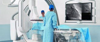
Contrast computed tomography angiography is a very informative method for diagnosing vascular pathology. CT scans use a combination of a high-tech X-ray scanner and powerful computer processing to provide detailed, 3D images of the blood vessels in the brain and neck. CT angiography provides the doctor with comprehensive information about the structure of blood vessels, the presence of narrowings or aneurysms, and the characteristics of blood circulation in the brain.
Since the Innovative Vascular Center deals with high-tech treatment of diseases of the carotid and vertebral arteries, we need an accurate diagnosis of vascular pathology of the cerebral arteries.
We conduct studies of the main arteries of the neck and head at the stationary base of our clinic in Klin, or at a partner organization in Moscow. Vascular surgeons at our clinic perfectly interpret MSCT angiography data and, based on these data, plan surgical intervention. In the hospital, we use a modern 64-slice computed tomography scanner with excellent image characteristics.
Why is MR angiography needed?
The study of the circulatory system is used in surgery, oncology, neurology and other areas of medicine. MRI of a given area with MR angiography allows you to assess the functionality, fullness, lumen, and condition of the walls of veins and arteries.
Based on the scan results, the nature of the blood supply to the area under consideration is determined, and foci of ischemia and necrosis are identified. In case of injuries, tomograms visualize hematomas, a violation of the integrity of the vascular wall.
Indications for prescribing MRI scanning in angiography mode are:
- dizziness, fainting;
- lack of coordination;
- severe headaches (cephalgia) that cannot be relieved with medication;
- decreased visual acuity, flickering of spots before the eyes;
- hearing loss, tinnitus;
- paresis and paralysis of the facial muscles;
- numbness, weakness of the upper and lower extremities;
- speech disorder;
- memory impairment;
- “shaky” gait;
- convulsions;
- leg pain;
- swelling of the lower extremities;
- dryness, peeling, ulceration of the skin of the legs, etc.
Decreased tone, decreased lumen, and ruptures of vessel walls lead to the development of pathological changes in surrounding tissues and organs. Impaired patency of the coronary arteries causes destructive, dystrophic processes in the heart muscle, which can cause a stroke or heart attack.
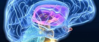
MRI angiography of cerebral vessels (sagittal section)
Pathologies of the aorta, superior and inferior vena cava are accompanied by dysfunction of the abdominal and pelvic organs and contribute to the development of diseases of the lower extremities.
In the diagnosis of cerebral vascular lesions, MRI using the MR angiography mode visualizes the slightest changes in the functioning of the circulatory system.
The procedure allows you to diagnose:
- stenosis, occlusion - narrowing and blockage of the vascular bed;
- acute disturbance of blood circulation in tissues;
- aneurysm - protrusion of the arterial wall;
- thrombosis - the formation of a clot in the lumen of a vein;
- vascular wall ruptures;
- atherosclerosis - cholesterol deposits in the lumen of the artery;
- vasculitis - inflammatory changes, thickening and stratification of the walls;
- embolism - blocking the lumen of the artery with foreign particles (gas, parasitic, etc.);
- extravasal compression syndrome - external compression of the vascular bed;
- arteriovenous malformations - pathological anastomoses (connections) of blood channels;
- congenital anomalies of the vascular system - kinks, bifurcations, etc.;
- dissection (dissection) of the aorta;
- thrombophlebitis - inflammation of the walls of the veins with the formation of bloody clots;
- vascular tumors.
The results of magnetic resonance angiography are analyzed by an MRI physician. The study protocol indicates:
- diameter, patency of the vascular bed;
- the condition of the walls, the fullness of the vessel;
- the presence of foreign particles (emboli) in the lumen;
- localization of the affected area;
- condition of surrounding tissues;
- functionality of veins and arteries;
- degree of circulatory disturbance in tissues;
- causes of hemorrhagic and ischemic phenomena.
MRI angiography examination is prescribed before surgery. Based on the scan results, the location and extent of the affected area are determined, the nature of the pathology and the scope of the upcoming operation are clarified.
During the rehabilitation period, recovery processes are monitored using MR angiography; MRI helps to timely diagnose the development of complications.
Scanning allows you to track the course of chronic diseases. In this case, vascular MRI angiography is prescribed at a frequency of 2 times a year. Early detection of relapses facilitates timely correction of treatment and helps achieve stable remission.
There are two types of MR angiography in MRI. The methods are identical, the difference lies in the purpose of the study.
MRI of the neck with MR angiography
Venography is aimed at studying the work of the vessels of the same name. Scanning allows you to diagnose inflammatory, degenerative processes, signs of reverse flow, and determine the causes of pathological phenomena.
Arteriography is indicated in case of symptoms of vascular insufficiency. If necessary, a comprehensive study of the vascular system is performed.
Angiography of the arteries of the neck and brain
Two related vascular studies are related by the fact that the carotid (localized in the neck) and vertebral arteries are the main ones for the blood supply to the brain, so the indications for their examination are similar:
- Headache that persists for a long time;
- Dizziness of unknown etiology;
- Severe causeless drowsiness;
- Frequent or prolonged fainting;
- Traumatic injuries to the neck and skull;
- Presence of vascular pathology in the past.
MR angiography with contrast
Magnetic resonance imaging of the circulatory system involves various scanning modes:
- time-of-flight;
- phase contrast;
- MRI angiography of vessels with an intensifier.
Time-of-flight tomography provides T1-weighted images. The liquid medium under study is brightly colored against the background of the surrounding soft tissue.
Phase contrast MR angiography involves receiving a signal from blood moving through the vessels. The method allows you to determine the characteristics of fast arterial and slow venous flow with equal accuracy. The simultaneous use of time-of-flight and phase-contrast MR angiography helps to differentiate between hematomas and other static areas.
Magnetic resonance imaging with enhancement visualizes the bloodstream in the area of interest. A solution of gadolinium salts is used as a contrast agent. The drug has paramagnetic properties and provides a hyperintense signal.
Contrast for vascular MRI angiography is administered intravenously. The injection is given using a catheter connected to an automatic device. This system allows the “coloring” solution to be supplied at a constant speed.
Layer-by-layer images during MRI angiography of cerebral vessels
Data recording begins after the lumen of the bloodstream is filled with contrast. 1-2 ml of solution is pre-injected, which helps to identify the initial moments of the arterial and venous phases. In this way, the signal registration time during vascular MRI angiography is determined. For further measurements, the peak arterial concentration of the gadolinium solution is used.
Angiograms make it possible to study the nature of the blood supply to a certain area. A broken line with a hyperintense signal indicates stenosis and occlusion of the vascular bed. The consequence of the pathology is ischemia and necrosis of the underlying tissues.
MRI angiography of vessels with contrast does not show calcifications, blood clots, or cholesterol plaques. In case of atherosclerosis, embolism, the images show changes in the lumen, fullness, blood channel, and parietal foci with a hypointense signal.
Contrast MR angiography for MRI diagnostics of neoplasms visualizes arteries and veins in the area of interest, shows the condition of surrounding tissues, and helps determine the nature of the disease.
The hemodynamics of the tumor circulatory network depends on the malignancy of the process. The vessels of a malignant tumor have a large number of anastomoses and bends, so the tumor tissue accumulates and releases the contrast agent more slowly.
Subarachnoid hemorrhage on MRI with MR angiography
The final diagnosis is made by the attending physician. The results of MRI angiography of veins and arteries for vascular neoplasms are supplemented with data from clinical tests, biopsy and other research methods.
Preparation and technology
Best materials of the month
- Coronaviruses: SARS-CoV-2 (COVID-19)
- Antibiotics for the prevention and treatment of COVID-19: how effective are they?
- The most common "office" diseases
- Does vodka kill coronavirus?
- How to stay alive on our roads?
Angiography does not require any significant preparation. The most important thing is to refrain from eating approximately four hours before the scheduled examination time.
You should also take care to choose comfortable clothing for the examination that does not contain any metal components. Any decorations, as well as gadgets, should be left in the locker room so that their impact does not spoil the visualization. Even dentures and hearing aids, if any, will have to be removed for a while.
The procedure lasts about half an hour if the appointment involves the administration of contrast. After the solution is administered via intravenous injection or drip, the patient will be asked to lie down on a medical couch. Since he will have to lie motionless, to make it easier to maintain the position, a cushion will be placed under his head. And to fix the position, auxiliary straps are used.
After activating the equipment, the couch is sent under the scanner arch, which will rotate, taking pictures. Unlike MRI, which takes quite a long time, CT angiography takes much less time, which appeals to people who suffer from claustrophobia.
While the detectors are operating, they can emit sound signals, which do not need to be alarmed, so as not to accidentally flinch.
Only a diagnostician can tell you the exact time of the examination, since it depends on the specific model of equipment. New versions work a little longer, since their layer-by-layer scanning step is only 0.5 mm. Increased clarity comes at the cost of longer examination times.
Typically, the diagnostic results are given to the patient within the next hour, or the next day they are sent directly to the attending physician. The conclusion is not only an image on film and on electronic media, but also an accompanying transcript with a conclusion, revealing the health status of the blood vessels in the patient’s area under study.
There are no contraindications after completion of the procedure. Doctors simply advise drinking more water to speed up the elimination of the iodine-containing contrast drug.
What types are there
The area of interest in MRI determines the type of MR angiography. Main areas:
- head;
- neck;
- lower limbs;
- abdomen;
- pericardium;
- upper limbs;
- small pelvis
MRI angiography of the vessels of the brain, neck, and lower extremities is most often prescribed.
A study of the blood supply system of the central nervous system can prevent the development of organic damage to the cerebral substance. MRI of the brain with MR angiography involves studying the lumen of the arteries and determining the functionality of the veins.
A neck scan is performed to assess the patency and fullness of the great vessels.
MRI of the lower extremities with MR angiography is prescribed for damage to the venous network, inflammatory processes, and thrombosis. Early diagnosis of diseases contributes to the effectiveness of treatment measures.
Angiography of cerebral vessels
Acute and chronic insufficiency of cerebral blood supply is manifested by decreased performance, inhibition of certain functions, and deterioration of well-being. In the absence of treatment, an increase in symptoms is observed:
- cephalgia;
- loss of memory, intelligence;
- paralysis;
- seizures, etc.
Severe disease can lead to disability and death.
MRI angiography of cerebral vessels with contrast is prescribed for diagnostic and preventive purposes. Patients at risk for cerebrovascular pathologies are recommended to undergo regular tomographic examinations.
MRI of the head with vascular angiography allows you to diagnose:
- arteriovenous malformations - there is no intermediate capillary network in the junction zone, the surrounding tissues are atrophic, signs of gliosis are observed;
- carotid-cavernous anastomosis - due to the intensive discharge of blood from the internal carotid artery into the sinus cavity, the pressure in the sinus increases, dilation of the ophthalmic vein occurs on the affected side, and pulsating exophthalmos develops (displacement of the organ of vision forward);
- aneurysms of cerebral vessels - there is a uniform increase in the diameter of the artery in a limited area or protrusion of the wall in the form of a bag (diverticulum);
- intracranial atherosclerosis - against the background of degenerative processes, a narrowing of the lumen of the blood channel occurs with subsequent expansion;
- vasculitis of cerebral vessels - the formation of multiple foci of cerebral infarction is possible;
- stenosis, deformation of the main arteries - pathological tortuosity, coiling (loop formation), kinking (curvature at an acute angle).
Brain MRI with vascular MR angiography is used in the diagnosis of strokes. Magnetic resonance imaging is informative already in the first 6 hours after an attack.
MRI angiography of head vessels
Acute disturbance of cerebral circulation can be caused by ischemic and hemorrhagic phenomena. In the first case, changes in the hemodynamics of the arteries are observed on MRI images of the brain with MR angiography. Intracranial hemorrhages occur when the integrity of the vascular wall is disrupted. Venography images reveal single or multiple hematomas.
Angiography of the lower extremities
The venous system of the legs experiences high loads, which is due to the structural features of the human body. Blood rises through the vessels, overcoming the force of gravity. The veins of the lower extremities have a system of valves that prevent reverse flow.
Vascular diseases of the legs are accompanied by swelling, a feeling of heaviness, and impaired motor function. When blood clots form, there is a risk of the latter breaking off and being transported through the arteriovenous network.
MRI angiography of leg vessels
A serious complication is blockage of the pulmonary circulation by an embolus (high risk of death).
MRI angiography of the veins and arteries of the lower extremities allows you to diagnose:
- thrombosis;
- atherosclerosis;
- thrombophlebitis;
- traumatic vascular lesions;
- stenosis, occlusion of the arteries of the lower extremities;
- diabetic foot syndrome.
Timely detection and treatment of thrombosis during venography reduces the risk of life-threatening complications.
Angiography of the neck
The study of the great vessels makes it possible to assess the blood supply to cerebral structures, soft tissues, the thyroid gland, etc. In the diagnosis of diseases of the central nervous system, complex MRI angiography of the neck with examination of the arteries and veins of the brain is often used.
Scanning reveals:
- thrombosis of intracranial venous sinuses;
- causes of cerebrovascular accident;
- stenosis, occlusion, thromboembolism of the main and brachiocephalic vessels;
- aneurysms;
- vasculitis of neck vessels;
- malformations;
- disruption of venous outflow from the cranial cavity, etc.
MRI of the neck with MR angiography (series of layered photos)
The scan results in detailed images of the vasculature and surrounding tissues. MRI of the brain and neck with MR angiography allows you to assess the patency, fullness of the vertebral arteries, the quality of cerebral blood supply, and the functionality of the internal jugular veins with the main tributaries.
Preparation for vascular angiography
MRI examination of the arteriovenous system with contrast is performed by appointment. You need to bring to the procedure:
- passport;
- Compulsory medical insurance policy, VHI policy;
- results of previous examinations;
- referral from the attending physician.
You should arrive 10-15 minutes before the scan starts.
Before MRI angiography of cerebral vessels, it is recommended to rest well and avoid stressful situations. It is advisable to monitor your blood pressure several days before the procedure.
Alcohol and smoking should be avoided 2-3 days before MRI with MR angiography. Avoid taking tonic substances, including strong tea, coffee, and energy drinks.
Before MRI angiography of veins and arteries, you need to warn your doctor about possible contraindications, diseases, and use of medications.
In case of suspected pregnancy, the woman needs to undergo additional examination. To clarify the condition, an analysis is performed to determine the level of the hCG hormone, and an ultrasound is performed. MRI angiography of blood vessels is a contrast procedure, which is a contraindication for diagnostic purposes.
During lactation, women are advised to prepare milk or formula to feed the baby. After administration of the contrast solution, the breasts must be expressed for 6-12 hours.
Before MRI angiography of veins and arteries, the patient removes jewelry and metal accessories. You can change into a comfortable home set and pajamas.
Who is the examination contraindicated for?
If we put aside the most important prohibition for testing - pregnancy, then all other contraindications can be classified as relative. In practice, this means that the decision to ignore them is made by the doctor individually, based on the preponderance of benefit over possible harm.
Such relative contraindications include:
- renal failure;
- heart failure;
- diabetes;
- problems with the endocrine system;
- an allergic reaction to iodine in its pure form or seafood in particular.
All of the above applies only to situations where, in addition to the traditional part of the examination, the patient was prescribed a contrast stage. But since a thorough study of the condition of the vascular bed is almost never possible without it, contraindications are relevant for almost all patients.
The main danger when examining with contrast is the iodine-containing components of the solution for the procedure. If the patient is allergic to them, then they try to replace the drug with rarer analogues.
If the patient is not aware of whether he has an allergic reaction to iodine, then at the stage of preparation for the study he should undergo an allergy test.
Separately, situations are considered when the patient is breastfeeding at the time of diagnosis. To avoid negative consequences for the child, doctors insist on refusing to feed at least 36 hours after the diagnosis. During this time, most of the harmful components of the contrast solution will be excreted by the kidneys naturally - with urine. To speed up this process, you need to drink more fluids. In this case, it is necessary to express the milk and pour it out.
Also questionable will be the use of CT scans for young children, elderly people, and patients with uncontrolled movements. The latter include a group of people with mental disorders and nervous disorders. The most important condition for obtaining clear visualization is to remain completely still throughout the entire procedure. Otherwise, the photo will have to be redone again.
Another relative contraindication is the short time interval between CT and x-ray examination. Doctors recommend maintaining an interval of about four weeks between repeated tests.
How is MR angiography done?
The procedure takes 40-45 minutes.
Scanning steps:
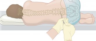
- The patient is placed on the tomograph conveyor, the limbs and body are fixed with special rollers. Random movements during an MRI session lead to distortion of MR angiography results. The device may produce noise when operating; use headphones for protection.
- After briefing, medical personnel are located in an adjacent room. Communication with the patient is maintained using an intercom. For emergency communication, the patient can use the panic button.
MRI of the neck with MR angiography on a closed tomograph
- For more accurate scanning, a gradient coil is placed above the area of interest, creating an alternating electromagnetic pulse. The table with the subject is moved into the tube of the apparatus, where the constant force field generator is located.
- After a series of native images, the patient is injected with a gadolinium solution. Scanning continues after filling the vascular bed with a contrast agent.
- During an MRI session, angiography of veins and arteries may require holding your breath for 10-20 seconds.
The shooting is carried out in axial, sagittal and frontal projections. Based on the results of MR angiography, a three-dimensional image of the arteriovenous network can be reconstructed. The 3D model helps to clarify the localization of the pathologically changed area. The thickness of the scanned section is from 1 mm.
MRI angiography of vessels is performed using open and closed type devices. The first ones do not have a tunnel module, the power of the tomograph is a maximum of 0.8 Tesla. Such devices are recommended for MRI in patients with claustrophobia and overweight patients.
Closed tomographs provide a magnetic field strength of 1.5 Tesla.
MRI of the neck with contrast MR angiography
High power allows you to make high-quality images of the studied area.