MR angiography of cerebral vessels is a modern diagnostic instrumental study aimed at visualizing blood vessels using a magnetic resonance imaging scanner. MR angiography of the brain allows one to evaluate the anatomical and functional features of blood flow and identify existing pathological conditions. The process may use a contrast agent, which is safe for the human body and is eliminated from it within a few hours after the procedure. MR angiography of the cerebral arteries can easily be called the “gold standard”. It allows you to achieve clear visualization of brain vessels and determine even minimal changes in them and adjacent tissues.
MR angiography of cerebral vessels - 4,000 rubles.
2-5 hours
(duration of procedure)
You can make an appointment and undergo MR angiography of the cerebral arteries at the CELT multidisciplinary clinic. We employ leading domestic diagnostic radiologists, in whose hands our modern equipment turns into a highly effective instrument capable of providing almost one hundred percent diagnostic accuracy!
Indications
- Frequently recurring headaches;
- Clinical manifestations in the form of dizziness and noise in the head;
- The need to exclude/confirm vascular pathologies;
- Postoperative control.
Contraindications
Absolute contraindications:
- The patient has a pacemaker;
- The patient has implants made of metal and metal alloys;
- The patient has an electronic or ferromagnetic middle ear implant;
- The patient has hemostatic clips of cerebral vessels.
Relative contraindications:
- The presence of metal implants;
- The patient has prosthetic heart valves;
- Pregnancy.
Indications for examination
Among the indications:
- stenosis (narrowing);
- occlusion (blockage);
- atherosclerosis;
- cardiovascular pathologies;
- aneurysms;
- thrombosis of blood vessels in the legs;
- retinal diseases;
- study of liver function;
- identification of tumors, blood clots, fusions of arteries and veins in the brain;
- surgery on the heart or brain is planned;
- determination of the extent of vascular damage during wounds and traumas.
Traumatic brain injury
If there is a risk of vascular damage due to traumatic brain injury, MR angiography is prescribed. A modern research method makes it possible to diagnose false aneurysms and disorders in patients with gunshot wounds in the first few days after resuscitation. The examination is carried out to exclude dissection and damage to the carotid and vertebral arteries.
Atherosclerosis, inflammation of the walls, pathological vasodilatation
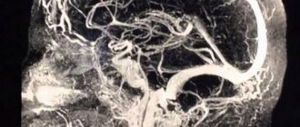
Using this method, you can determine the slightest disturbances in the structure of the walls of blood vessels. A visualization of the arterial circle of the brain is created, obtained by exposing the examination area to radio waves in an external magnetic field. Angiograms clearly show the posterior, anterior, middle and carotid cerebral arteries, as well as the basilar artery. MR angiography of intracranial arteries allows you to quickly see disturbances in vascular architecture, as well as problems with blood flow caused by atherosclerosis, tumors, and aneurysms. At the same time, an examination of the arteries of the neck can be carried out. The procedure is aimed at identifying pathology in the structure of the vascular walls. MR cerebral angiography of cerebral vessels provides a complete picture of blood flow functions.
Aortic dissection
The technique makes it possible to determine the exact location of the dissection and where the blood flow is changed. A contraindication to diagnosis is the presence of a metal valve in the patient.
External vessel compression syndrome
Magnetic resonance, or MRI, angiography is prescribed for external vessel compression syndrome. In a diagnostic center, a contrast agent is injected into the patient's artery and three-dimensional images of the vessels that have undergone deformation are taken.
Congenital heart defect
Angiography is the main method for studying patients with congenital heart defects. During the procedure, the pressure gradient and oxygen saturation of the blood are measured. As a result, flows, resistances and shunts within the heart are calculated, and the function and anatomy of this organ is accurately assessed.
Benefits of diagnostics
The main advantage of magnetic resonance imaging of vessels in the angiography mode is the high detail of the images, which make it possible to diagnose even the slightest deviations. Other advantages include the following:
- Absolute painlessness and absence of discomfort during the examination;
- A minimal list of contraindications and no side effects;
- No negative impact on the body;
- Possibility of receiving results on the day of the study.
Methodology and preliminary preparation
The technique is based on the influence of a magnetic field and electromagnetic waves. As a result of the study, multidimensional images of the vessels of a certain area are obtained. Particularly valuable is the diagnosis of blood vessels in the neck and brain. The duration of MR angiography is 30-40 minutes.
The procedure can be performed with or without the administration of a contrast agent. Before angiography, an allergy test for a contrast agent is performed. It is not recommended to eat food 8 hours before the procedure. The doctor must be warned in advance about taking medications. Before the procedure, you must remove all metal-containing products.
Among the preparatory activities:
- eliminate alcohol 15 days before the procedure;
- the woman needs to make sure that she is not pregnant;
- take blood clotting tests;
- do an ultrasound of the heart;
- do an enema.
The study is carried out with an empty bladder.
MRI or CT scan of cerebral vessels?
MRI and CT have their advantages in different situations. People who have electronic devices (pacemaker) or objects made of magnetic metals in their body are allowed only CT scanning.
An MRI scan takes tens of minutes and requires you to lie still. A CT scan takes less time, but this procedure uses X-rays. CT scans should not be done too often. The procedure is contraindicated for pregnant women at any stage. MRI is performed as many times as necessary; it is contraindicated only in the 1st trimester of pregnancy.
MRI of cerebral vessels is a safe procedure that will provide a detailed picture of the disease. At the medical office, you can undergo an MRI scan by appointment on any convenient day and time.
The patient's condition after the procedure and possible complications
Bed rest is recommended during the day. The attending physician monitors the patient’s general condition (examination of the intervention area, temperature measurement). If the person’s condition is satisfactory, the bandage is removed on the second day and sent home. The risk of complications is minimal. For most people, the study is not dangerous.
Possible problems:
- allergy to contrast agent, anesthesia;
- in the presence of chronic diseases there is a risk of acute renal failure and myocardial infarction;
- bleeding from the place where the puncture was made.
What types of angiography of cerebral veins and arteries exist?
Several research methods are used in medicine:
- Survey - contrast agent is injected into a large artery. The technique allows you to see smaller vessels.
- Selective - contrast is injected into the artery locally to affect a specific area of the body.
Magnetic resonance angiography of the brain is divided into:
- MR venography
(without contrast); - MR arteriography
.
Features of angiography of various organs
The method allows you to study vessels of any size (from the smallest capillaries to the aorta), organs and systems of the body. The procedure is carried out before surgery.
Angiography of cerebral vessels
This is the most common way to diagnose disorders in the blood supply to the brain. Visualization helps to identify pathological processes, cysts, tumors, and micro-stroke.
Cerebral MR angiography of blood vessels is prescribed for:
- dizziness and nausea for unknown reasons;
- prolonged headache that cannot be relieved with medication;
- preparation for neurosurgery on the brain.
Angiography of the heart
Indications for the procedure are:
- angina pectoris in a progressive stage;
- myocardial infarction;
- heart rhythm disturbances.
Angiography of the lower extremities
Diseases of the veins and arteries of the lower extremities are widespread. MR angiography is performed if the following diseases are present or suspected:
- thrombophlebitis;
- thrombosis;
- endarteritis;
- atherosclerosis.
Angiography of the neck
To assess the condition of the vascular stack, localize disorders and clarify the diagnosis, neck angiography is prescribed. The procedure is indicated for bruises and injuries, headaches, dizziness, disturbances of consciousness and sleep. With its help, Takayasu's disease, stenosis and developmental anomalies, vascular neoplasms, compression of blood vessels by scars and tumors are determined.
3. How is the procedure of MR angiography of the cerebral arteries performed?
When going for diagnostics using an MRI scanner, you need to expect that the procedure will take a significant amount of time - from 30 minutes to 1.5 hours.
Special preparation for the study is not required, however, the doctor must warn the patient about the need to remove all metal objects, including dentures, and also asks to inform about the presence of implants in the body, their location, volume, and the material from which they are made.
The patient is placed horizontally on the surface of the tomograph, after which the head is fixed with a special helmet. For the accuracy of the diagnosis, any head movements must be excluded.
If the patient is prone to claustrophobia, he will be asked to take sedatives.
The operation of the tomograph is accompanied by rather sharp sounds, so special protective headphones are put on the ears of the person being examined. The scanning unit makes rotational movements around the patient's head and transmits a three-dimensional image to the monitor. The doctor deciphers the data received and draws conclusions about the state of the patient’s vascular system of the brain.
The results of MRI diagnostics are given to the patient in the form of a report, which may contain a printed scan of the image or a disk with files.
About our clinic Chistye Prudy metro station Medintercom page!
Contraindications for the procedure
- Renal, liver and heart failure during the period of decompensation.
- Poor blood clotting.
- Pregnancy and lactation period.
- A number of mental illnesses.
Important!
MRI technology does not allow simultaneous visualization of arteries and veins. Therefore, conventionally called MR angiography consists of two different methods:
- MR venography;
- MR arteriography.
MR venography
– is a diagnosis of sinuses and intracranial veins. No contrast is applied. Can be performed independently or in combination with MRI of the brain. MR venography allows you to identify aneurysms, vascular malformations, thrombosis, various developmental and localization anomalies, tumor formations growing into blood vessels, etc.
MR arteriography
is a visualization of the arteries, with which you can identify blockages, their location, and the degree of threat to health and life. Allows you to identify hemorrhage, thrombosis, vascular spasms, etc. This technique is especially important because it allows you to control the treatment of those patients who are at very high risk.
Cerebrovascular diseases
Headache is often overlooked. Patients believe that such pain, if it is not too intense, can be ignored. People live with “ordinary” headaches for years, and during this time pathological processes occur in their brain. MRI makes it possible to distinguish migraine from, for example, an aneurysm, and psychosomatic headache from tumors and impaired blood supply to the brain.
What diseases are detected using MRI of cerebral vessels?
Most often, cerebral vessels suffer due to three pathologies: atherosclerosis, high blood pressure, type 2 diabetes, which can lead to the development of serious complications.
Stroke
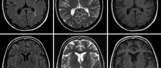
A stroke is an acute circulatory disorder in the brain, a very dangerous condition that can lead to serious health consequences. MRI is often used to diagnose strokes as a high-tech method that allows one to obtain accurate images of the pathology.
Stroke is divided into several types depending on the cause:
- Ischemic. It occurs when an area of the brain stops receiving blood due to blockage of a vessel by a blood clot or cholesterol plaque. The brain matter dies, not receiving oxygen through the bloodstream. Heart failure, diabetes mellitus and atherosclerosis can contribute to the occurrence of the disease.
- Hemorrhagic. Occurs due to hemorrhage into the brain tissue from a damaged vessel. A risk factor for this type of stroke is hypertension.
Timely diagnosis of stroke is very important. Timely measures taken to identify and assess the consequences of a stroke help to begin early rehabilitation and restore damaged functions.
In the event of a stroke, MRI gives a clear and detailed picture of the course of the disease, the location and extent of brain damage.
Atherosclerosis
This disease develops to one degree or another in almost every adult, but the severity of the disease and the speed of its development are strictly individual. When the disease occurs, cholesterol and fats are deposited in the artery wall and a plaque forms. The following options for the development of the disease are possible:
- the plaque narrows the lumen of the vessel, the brain does not have enough blood;
- expansion of the artery walls (aneurysm) is formed;
- the vessel wall ruptures with subsequent bleeding;
- a blood clot forms at the site of the plaque, further interfering with the movement of blood;
- the contents of the plaque and/or the thrombus breaks off and clogs the vessel.
The following diseases contribute to the emergence and rapid progression of atherosclerosis:
- High blood pressure (arterial hypertension, hypertension) damages the vessel wall, which becomes less elastic and cannot cope with the load. Fats are deposited in it more easily and atherosclerosis develops. Due to high pressure, thin or severely damaged vessels, such as vessels with an aneurysm, rupture.
- Diabetes mellitus type 2. Excess glucose also damages the vessel wall. People with type 2 diabetes mellitus develop atherosclerosis and hypertension more quickly. Often one person has a combination of atherosclerosis, hypertension and type 2 diabetes mellitus. They reinforce each other and increase the risk of stroke.
- Heart diseases (heart rhythm disturbances, myocardial infarction, heart defects).
Conditions that are less common: autoimmune diseases (vasculitis, systemic lupus erythematosus, type 1 diabetes, etc.), infectious diseases, sepsis, traumatic brain injuries.
Cerebral small vessel disease
A new diagnostic method sometimes opens up a whole world of diseases about which not much was known before. MRI technology helps study a group of diseases of small vessels in the brain, those with a diameter of less than 2 mm.
Small vessel disease of the brain is dangerous for cognitive decline and/or stroke. About a quarter of ischemic strokes occur due to capillary damage. There are many causes of damage to small vessels and they are still being studied.
Some older people may develop cerebral amyloid angiopathy. It affects approximately 20% of people 61-70 years old; The older the age, the more often it is detected. A special protein called amyloid is deposited in the brain vessels, gradually the blood supply to the brain is disrupted and neurons die.
Aneurysm
An aneurysm is a pathological enlargement of an artery, which occurs on average in 2-3 people out of 100. The disease occurs more often in women, as well as in the age group over 30 years. Aneurysms can be congenital, but over time, the vessel wall is also deformed by atherosclerosis, blood pressure, trauma, and an infectious process, which aggravates the course of the disease.
A vessel affected by an aneurysm may burst. Most often, the gap occurs between the ages of 20-40 years. Before this, a person may suffer from headaches similar to a migraine attack. MRI is indispensable for early diagnosis of the disease. Modern tomographs are capable of visualizing even the slightest pathological lesions in the brain with great accuracy.
Risk factors for the formation of aneurysms include smoking, high blood pressure, the use of hormone replacement therapy during the postmenopausal period, and if there are people in the family with aneurysms and other diseases of the cardiovascular system.
Angioma (vascular malformation)
A benign tumor consisting of overgrown and intertwined veins and arteries of the brain. The formation does not pose a threat to life, but as it grows, it can compress the structures of the brain, resulting in headaches and disrupting the normal functioning of the areas of the brain near which the pathology is located.
Based on size, malformations are divided into:
- Capillary. They consist of tiny vessels and usually do not cause symptoms.
- Venous. Includes larger veins and arteries. May cause specific symptoms.
- Tricky. The vessels intertwine so closely that they form entire cavities in which blood accumulates. Such a tumor can grow quite strongly and put pressure on the brain tissue.
The causes of angiomas have not been fully studied. Often the disease is congenital or occurs in early childhood. A small percentage of angiomas occur after infectious diseases.
MRI of the brain vessels will accurately show not only the location, size and shape of the tumor, but also the degree of compression of the surrounding tissues. Thanks to magnetic resonance imaging, the attending physician will be able to develop an effective plan for subsequent therapy.
Advantages of MRI at the Literary Fund clinic:
- You receive high-quality transcripts of images from leading MRI diagnostic specialists.
- After the MRI, without wasting time, you can get advice from experienced doctors (neurologist, rheumatologist, orthopedist-traumatologist) with detailed treatment recommendations and undergo treatment under the supervision of highly qualified doctors!
- The Literary Fund clinic offers the full range of procedures necessary for the diagnosis and treatment of headaches and cerebrovascular accidents, diseases of the spine and joints. All types of physiotherapy, reflexology, intra-articular injections, hardware spinal traction, manual therapy, therapeutic massage are at your service.