and other chronic diseases, you must notify your doctor.
It is worth knowing that spinal myelography is performed in the presence of doctors who will provide assistance. In some cases, doctors help administer the enema and are also responsible for giving the patient a sedative to suppress the swallowing reflex.
How is the examination carried out?
Before you sign up for myelography of the lumbar spine, it is worth learning about the methodology for performing it. To begin with, the patient is asked to lie down on a special table, after which the skin at the site of the future puncture is numbed with a local anesthetic and disinfected. After this, a puncture is carried out, the needle will move inward under the control of a fluoroscope. A contrast agent is injected, after which the needle is removed and the puncture site is disinfected again. Depending on which part of the spine is being examined, the person may be asked to lie on their stomach, side, or sit up when the needle is inserted.
After the contrast is administered, the patient should lie on his stomach, and the doctor will carefully tilt the table so that the substance is distributed along the subarachnoid canal of the spine. To take a photo, a person may be asked to turn over on their side and lie still, otherwise the result will be blurry.
The procedure will take from half an hour to an hour. After this, the patient will be sent to a ward, where he will be observed for two to four hours. At this time, the person will have to lie on an elevated (about 45 degrees) headboard and drink a lot, which will allow the contrast to be removed from the body more quickly.
Myelography with three-dimensional reconstruction
In this case, contrast myelography will be performed using a magnetic resonance imaging scanner. This involves data processing using special software. As a result, three-dimensional images will be obtained that will detect any abnormalities and pathological processes in the spine and spinal cord.
Contrast myelography
This is done using a computed tomograph. The peculiarity is that the procedure involves examination using x-rays. This is not permissible in all cases, so the decision will be made by the attending physician.
Preparation for MRI of the spine with myelography
You must complete your last meal 8 hours before the test. If the cervical spine is to be examined, you need to take a sedative to suppress the swallowing reflex (prescribed by your doctor). Before MRI of the lumbar region with myelography, it is necessary to give a cleansing enema.
Before the examination, the doctor explains what the procedure is and how it will take place. He clarifies that the patient is not allergic to iodine, asks what medications the person is taking, whether he has circulatory problems or chronic diseases (asthma, epilepsy, diabetes, kidney pathologies).
Decoding the results
Myelography with an iodine-containing drug is interpreted by a doctor with the appropriate qualifications, usually a radiologist. Based on the results of decoding, a conclusion is drawn up, which is given to the patient and with which he can go to the attending physician.
Advantages and disadvantages of the procedure
The advantages of the procedure are that it allows you to quickly identify problems that neither CT nor MRI without contrast would reveal. You can examine not only the spine, but also the spinal cord and nerve roots, which are almost impossible to see with other studies. As a result of the procedure, no traces of radiation remain in the body.
The disadvantages are that the administration of contrast does not give the most pleasant sensations: there is often a salty taste in the mouth, redness of the facial skin, a feeling of heat or burning, dizziness or headaches. Nausea or even vomiting may occur after contrast administration is stopped. Also, the procedure is not possible in all cases, since there are contraindications.
When is myelography required?
The examination of the spine using the method under consideration can be carried out when:
- compression fractures;
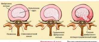
- myelopathy of discogenic type;
- osteochondrosis of the spine in a severe stage;
- any spinal cord injuries;
- arachnoiditis (inflammatory process);
- infectious diseases of the spinal canal or processes.
In addition, as a diagnostic procedure, myelography is performed in case of suspected damage to the nerve roots of the spine, progression of malignant tumors, or problems in the functioning of the spinal vessels.
Contraindications for
It is impossible to undergo myelography in the following cases:
- state of fever;
- diseases of the kidneys, heart, liver in the stage of decompensation;
- severe arthritis;
- pregnancy;
- previous spinal surgeries;
- anatomical features or defects that prevent the introduction of a substance into the subarachnoid cavity;
- infectious processes on the skin in the place where contrast needs to be administered;
- inability to maintain one position during the procedure.
The final decision should be made by the doctor, since sometimes it happens that the possible risk is less than the benefit of the x-ray procedure.
How should you prepare for research?
Detailed instructions for preparing for myelography are provided to the patient by the attending physician.
The physician should be notified of all medications the patient is taking and of any allergies, especially to barium or iodinated contrast materials. It is also important to tell your doctor about recent and any chronic illnesses.
In particular, the physician needs to be aware of: 1) all medications that should be stopped several days before the test; 2) the presence of allergic reactions to the contrast material used during myelography. A couple of days before myelography, you should stop taking certain medications. These include some antipsychotic medications, antidepressants, any blood thinners, and medications used to treat diabetes, especially metformin. The most important thing is to stop taking anticoagulants, which thin the blood. When using such drugs, alternative methods of maintaining blood viscosity during myelography should be discussed with your doctor. For example, after stopping long-acting anticoagulants, temporary intravenous administration of other drugs, such as heparin, may be possible.
Before the study, it is necessary to stop taking certain anticonvulsants. In addition, medical personnel should be warned in advance about the presence of diseases accompanied by seizures, which will allow them to develop the most adequate plan for temporarily stopping the use of anticonvulsants several days before myelography. Although reactions to iodinated contrast materials are extremely rare, it is important to notify your doctor if you have had previous allergic reactions to contrast or other medications. In addition, your doctor should be informed if you have bronchial asthma or allergies to any other substances. In such situations, when administering contrast material, medical personnel monitor the patient's condition especially carefully. Allergic reactions to any iodine-containing substances deserve special attention. As a rule, the day before myelography, patients are advised to expand their drinking regime, since sufficient hydration of the body at this time is extremely important. A few hours before the test, you should stop eating solid food, but liquids are not limited.
During the examination, you will need to remove some or all of your clothing and wear a special hospital gown. In addition, you should remove all jewelry, glasses, dentures, and any metal or clothing that could interfere with the x-ray image.
Women should inform their doctor and radiologist of any possibility of pregnancy. As a rule, X-ray examinations are not performed during pregnancy to avoid exposure of the fetus to radiation. If x-rays are necessary, special precautions should be taken to protect the developing child.
After the myelogram is completed, the patient usually remains under medical supervision for several hours before being allowed to go home. Arrangements should be made in advance to have someone help the patient get home unless he is staying overnight in the hospital.
Up
Where can I do it?
Myelography of the spine is a complex procedure that is not performed in all clinics. You can sign up for the study at the following medical institutions.
| Name of institution | Clinic address | Reception phone number |
| OAO "Medicine" | Moscow, st. Mayakovsky, per. 2 Tverskoy-Yamskaya 10 | +7 |
| Medical | Moscow, st. Alley Zhemchugova 5/4 | +7 |
| Scientific clinical | Moscow, Volokolamskoe highway, 84 | +7 |
| St. Petersburg, st. Marata, 6a | +7 | |
| Neurosurgical Institute | St. Petersburg, st. Mayakovsky, 12 | +7 |
Indications for scanning are:
- pain in the back, in the lower extremities, of varying severity, unilateral or bilateral
- sensory disturbances (paresthesia, numbness) or motor disturbances in the legs (up to paresis/paralysis)
- previous spinal injury
- search for metastases or primary cancerous node
- upcoming or previous surgery
- presence of signs of neurological impairment of other organs (for example, breathing problems, vision problems, heat intolerance)
What is myelography: MRI contrast of the spinal cord
MR myelography is a research method based on the study of about 14 types of various biochemical information (differences in the structure of water and fat tissue) without the use of radiation exposure, but requiring invasive injection of contrast into the subarachnoid space.
Magnetic tomography visualizes the membranes of the spinal cord and brain well without contrast, but a contrast agent is added to identify the cause of compression. Using magnetic resonance angiography (MRA), it is possible to visualize the state of the blood supply to the veins and arteries.
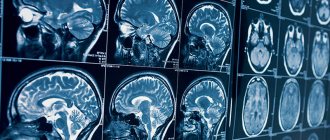
MR myelography allows you to identify malignant and benign brain tumors that may cause compression of brain tissue. The study has broad potential for diagnosing pathology of soft tissue structures.
MR angiography allows one to detect vascular abnormalities with a high degree of certainty. Used to assess the condition of veins and arteries. The study determines the state of blood flow, which changes in the presence of malignant and benign neoplasms.
In the images, areas of demyelination are marked as foci of signal with increased intensity. The shape of such a section is oval or rugged. If 4 similar fragments are detected, verification of the diagnosis is beyond doubt.
Price for MR myelography of the spine
The research results (images) can be obtained:
on disk
on a flash drive
on film
You can find prices for MRI of the coccyx and other studies in the MRI prices section. The cost of an MRI includes a radiologist’s report, as well as recording of images on a CD. If necessary, for an additional fee, the results of the study can be recorded on film or a flash card.
A radiologist is consulted to develop an individual plan for an MRI study if it is necessary to combine several studies with specific parameters.
Side effects
Despite the information content and relative painlessness of the procedure, it has some disadvantages. Myelography of the spine can cause the following side effects:
- when a needle is inserted, soft tissue damage and internal hematomas occur;
- cerebrospinal fluid pressure increases;
- allergic reactions often occur - itching, rash, difficulty breathing;
- If disinfection is not carried out correctly, inflammation may occur at the needle insertion site;
- Harmful X-ray radiation can trigger tumor development, so it is safer to do MR myelography.
Complications
After the procedure, the following side effects are possible:
- headache;
- nausea and vomiting;
- numbness of the limbs;
- limited mobility;
- increased body temperature, chills;
- allergy;
- bleeding in the puncture area.
In this case, you need to go to the hospital.
What the pictures show
The images are interpreted by a radiologist or a functional diagnostics doctor. The 3D images are compared with previous images of the patient's or healthy person's spine. This method reveals pathological changes: osteochondrosis, hernia and others. The stage of development of the disease is determined and treatment methods are selected.
MRI of the spine can be performed an unlimited number of times to clarify the diagnosis. The photographs clearly distinguish bones and soft tissues - dark colors, the spinal cord - light shades. A tomogram helps the doctor:
- determine the degree of damage to the spine, identify developmental pathologies in the early stages;
- identify inflammation and neoplasms in soft tissues;
- detect dysfunction of the spinal membranes, determine the stage of osteochondrosis;
- identify the condition of nerves and blood vessels in the diagnosed area;
- recognize a herniated spine, protrusion of longitudinal ligaments, muscles, intervertebral discs;
- determine the nature and extent of the injury.
Advantages
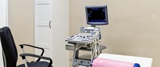
Carrying out such a complex study as spinal myelography requires high-tech equipment. For this purpose, the MART clinic uses a modern high-field tomograph made in Germany by Siemens Espree, an expert-class device.
Its main difference from older models is its comfortable shape: due to the ultra-short length and wide diameter of the magnet, examination can be prescribed for patients suffering from claustrophobia. The weight limit is 160 kg, which is much higher than for devices released several years earlier.
—>
Appointment with a neurologist after MRI RUB 1,500. instead of 2,000 rubles.
Sign up
Rules for the patient
To avoid discomfort during the procedure, as well as complications after it, it is very important to know the features of myelography and the rules for its implementation. Painful sensations rarely occur, but if the patient is afraid of pain, it is better to tell the doctor about it. In this case, he will be given a sedative to help him calm down. During the procedure, you must follow all the doctor’s recommendations and warn him about all side effects and discomfort.
After myelography, the most important thing is to quickly remove the contrast agent from the body. To do this, you need to drink as much fluid as possible, and after 2-3 days start doing certain exercises that would increase blood flow and metabolic processes.
Myelography of the spine is one of the most reliable methods for examining it. With its help, the doctor can examine the condition of all tissues, see pathologies of its structure, disturbances in the functioning of even the smallest vessels and nerves.
Diagnostic equipment
To perform myelography, you need a special table that can change the angle of inclination, an X-ray tube, and a monitor on which the image during fluoroscopy will be received. A special device called a fluoroscope converts X-rays into video images. To improve the picture quality of the monitor, an amplifier is used.
You will also need a puncture needle with a mandrel (a removable rod that clogs the lumen of the needle to prevent premature leakage of cerebrospinal fluid), an antiseptic solution (iodine, alcohol or another), painkiller solutions (1% lidocaine solution), and a contrast agent. As a rule, gas (nitrous oxide or oxygen) or special X-ray contrast agents are used as the latter.
To identify hypersensitivity to a contrast agent, the subject is given an allergy test: 2 ml of this drug is administered intravenously. If symptoms indicating intolerance occur, myelography is not performed or is performed using the contrast that the patient tolerates well.
Hemorrhage without myelography: MRI provides quality and contraindications
During operation, MRI scanners are accompanied by a strong magnetic field, so any metal objects in the human body begin to move under the influence of radiation. Because of these features, some patients are contraindicated for MR myelography or other MRI procedures.
Magnetic resonance imaging cannot be performed on the following categories of patients:
• With the presence of pacemakers, pacemakers; • Electrocochlear devices, neurostimulators; • Postoperative clamps of aneurysms; • Orthopedic joint prostheses; • Sternal suture wire; • Third trimester of pregnancy; • Patients with claustrophobia.
Scanning creates a lot of noise. To suppress it, headphones are recommended for a person. Pleasant music can relax the patient, but in case of neurological disorders or fear of closed spaces, it is advisable to give the person a sedative before the examination.