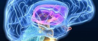
The brain is a complex and poorly understood human organ that is difficult to diagnose. However, the brain is the control center responsible for the operation of all systems in the body. Therefore, any pathological processes in it affect the physical and mental health of a person.
MRI is one of the methods for studying the brain to determine negative conditions in it. Diagnostics is prescribed for adults and children and is a safe examination method.
What it is
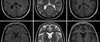
The method refers to a non-invasive examination using radio waves and a magnetic field. During the scanning process, the doctor receives an image showing parts of the brain. Diagnosis is carried out without radiography. The study reveals disturbances in the functioning of the nervous and vascular systems, aneurysms, and tumors.
Also, based on the results of the examination, the level of activity of the cerebral cortex is determined. When using a contrast agent, tissues are better visualized, which helps to more accurately identify pathologies.
Advantages of the method
Advantages of brain tomography:
- the patient does not experience pain during diagnosis;
- During the examination, no foreign substances are introduced into the human body;
- no ionizing radiation;
- clear images help to accurately recognize pathology and prescribe treatment;
- as prescribed by a specialist, an examination of the head and cervical/upper spine is carried out to assess the functional activity of the brain - based on the results, a safe method of surgical intervention is prescribed;
- areas of the brain hidden by the bones of the skull are examined - other methods do not diagnose these areas;
- MRI helps to obtain a complete picture of the brain and vascular system even without the use of a contrast agent;
- tumors and other pathologies are detected in the early stages of development.
Make an appointment now!
Decoding the results
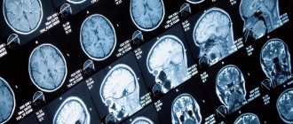
At the final stage of the study, the doctor interprets the obtained images. After studying the substance of the brain, vessels, cavities, membranes and other structures, a description of the study is performed, which reflects all the changes compared to the normal version. When pathological foci are identified, their size, volume, shape, location, and structural features are indicated. At the end of the description, the doctor leaves a conclusion, which must indicate a preliminary, differential or exact diagnosis. If the patient has provided previous results of magnetic resonance imaging of the brain, the doctor will compare them with new images, evaluate the dynamics of changes and reflect this information in the conclusion. Along with the doctor’s conclusion, the patient is given a CD on which the tomograms themselves are recorded. If necessary, the images can be printed on a special film for an additional fee.
Why is an examination prescribed?
MRI diagnostics helps to recognize pathological changes in the soft tissues of the lining of the brain, inflammatory reactions, and disorders of the central nervous system. Also, with the help of an examination, the conditions of the parts of the head are studied: the visual and occipital lobes, the pituitary gland, the cerebellum, areas of thinking and memory, and the ventricles of the brain.
Before the procedure, the patient undergoes tests, the results of which determine the type of diagnosis. For example, tests showed that the patient had an excess level of the hormone prolactin. The doctor writes a referral for examination of the cerebellum.
Examination of the brain in a magnetic field helps determine the presence of:
- Multiple sclerosis - lesions of the myelin sheath of nerve fibers. The diagnosis determines the stage of the disease, the level of damage, after which the doctor prescribes therapy.
- Benign and malignant tumors. Brain scanning is used to identify the affected area, monitor the development of the tumor, as well as the treatment process.
- Disorders in the cerebral cortex - Parkinson's disease, Alzheimer's disease. MRI determines the density of white and gray matter, the presence of pathologies in the subcortex and cortex of the head.
- Ischemic strokes, heart attacks. The images determine the area and density of ischemic lesions, the presence of cortical necrosis, the appearance of edema, and the stage of development of the disease.
- Brain injuries are damage to blood vessels and tissues due to trauma. Diagnostics reveals the first signs of VSD.
- Mental disorders of endogenous and exogenous types. The method helps to identify hereditary pathologies, changes after traumatic brain injury, viral infection, and poisoning with toxic substances. MRI diagnostics determines schizophrenia.
For children, head tomography is prescribed for the following pathologies:
- head injury, concussion after injury, intrauterine infectious processes;
- hypoxia, ischemia and other disruptions in brain development;
- epileptic seizures, cerebral hemorrhage;
- the first stages of multiple sclerosis;
- dangerous diseases such as pituitary adenoma, brain tumors, cysts and suspicions of them;
- a sharp deterioration in vision and hearing, malfunction of the inner ear;
- high intracranial pressure.
Diagnostics helps to study the state of the brain and its parts, identify the causes of headaches in children, and prevent the development of autism.
Differences between MRI and CT scan of the brain
Magnetic resonance imaging of the head differs from other diagnostic methods in the following:
- the examination is performed in 3-4 projections, which provides more information for identifying diseases and choosing treatment methods;
- pathologies are detected in the early stages of development - ischemic changes are detected 2-3 hours after a stroke;
- even minimal changes in the brain that cause multiple sclerosis are determined;
- parts of the brain that are not diagnosed by computed tomography (CT) are studied - the brain stem, the cerebellum.
What will a brain scan show?
The effect of MRI is based on the effect of nuclear magnetic resonance. Electromagnetic waves, passing through the human body, affect hydrogen atoms, changing the frequency of their vibrations. The energy released by the nuclei in this process is captured by a tomograph and then converted into an image on a computer, which allows one to obtain a detailed picture of the anatomical structures of the brain under different planes - coronal, transverse and sagittal. The image of the diagnosed area is formed by combining many layer-by-layer sections with a thickness of 1-3 mm, which makes it possible to detect the slightest deviations and pathologies.
Appearance of the Philips Intera 1.5T tomograph
When performing magnetic resonance imaging of the brain, the slice thickness can be set at values of 1-3 mm, which makes it possible to accurately examine such small structures as the pituitary gland and optic nerves.
Benefits of MRI
The advantages of magnetic resonance imaging include:
- speed of the procedure and obtaining results;
- high accuracy;
- obtaining three-dimensional images of organs;
- safety for the patient;
- possibility of carrying out in late pregnancy and during breastfeeding;
- early diagnosis of diseases.
When performing MRI, unlike radiography, no harmful radiation is used. The magnetic waves created by the tomograph are absolutely safe for humans. MRI diagnostics can be performed as often as necessary.
When is a diagnosis prescribed?
A brain examination is prescribed for the following ailments:
- developmental anomalies and diseases of cerebral vessels;
- head injuries and bruises, cerebral hemorrhages;
- sharp deterioration of vision and hearing;
- tumor development;
- infectious reactions in the central nervous system - meningitis, HIV infections, abscesses;
- pituitary adenoma, epilepsy;
- pathologies of the base of the skull;
- multiple sclerosis, sinusitis, neurodegenerative diseases;
- aneurysms, thrombosis and other anomalies in the vessels of the head.
Diagnosis is also carried out after surgery. Prescribed for frequent migraines and fainting, tinnitus, sudden deterioration of memory and attention, impaired coordination of movements, and mental disorders.
Contraindications
Head tomography is not prescribed if the patient has absolute contraindications, but is allowed at the discretion of the doctor in case of relative contraindications. Diagnostics is prohibited if the following conditions exist:
- the patient has prosthetic heart valves, pacemakers or neurostimulators;
- presence of insulin pumps, inner and middle ear prostheses, cochlear implant;
- the patient has an Ilizarov apparatus;
- the presence of metal implants, ferromagnetic particles, fragments in the body.
Relative indications include:
- tremor, the patient’s inability to hold his breath for a long time during examination;
- presence of dental braces, dentures, stents, vena cava filters;
- heart failure, clip instead of gallbladder;
- pregnancy;
- claustrophobia;
- severe pain while standing still;
- coronary artery bypass grafting.
Preparatory stage
The doctor, based on the test results, determines whether to perform a tomography with or without the introduction of a contrast agent. If the first option is chosen, then the patient does not take food or liquid 5 hours before the diagnosis. Before the procedure, you must remove all jewelry and accessories, watches and objects with metal elements.
During a consultation with a doctor, the patient reports the presence of chronic diseases, allergies to medications, claustrophobia, and pregnancy.
When preparing a child for a head tomography, it is not recommended to give him anything to drink or eat 3 hours before the diagnosis. If an MRI is performed with the administration of contrast, the examination is performed on an empty stomach. Before the procedure, the child should be shown to an allergist to check for an allergic reaction to the injected substance.
Procedure for MRI diagnostics
No special preparation is required for the study. The only limitation is that it is not advisable to perform tomography soon after eating. If you have already had an MRI of your brain, take the results of previous examinations with you. Before starting the procedure, you need to remove all things containing metal.
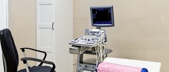
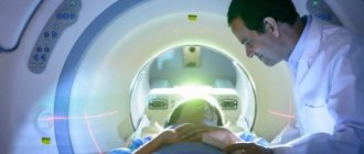
The scan is carried out in the tomograph tunnel, where the patient spends 30–40 minutes in a lying position, remaining motionless. All this time the doctor is nearby. If necessary, you can contact him using a special call button located inside the tomograph.
Get specialist advice or make an appointment
+7 (499) 400-47-33
Diagnostic features
If a brain tomography is performed with the introduction of a contrast agent, the examination takes 1-1.5 hours.
Stages:
- the patient removes objects and clothing with metal elements;
- lies on the tomograph table on his back;
- the doctor administers a contrast agent intravenously;
- if there is tremors or inability to lie still, the patient takes sedatives;
- legs and arms are secured with belts, bolsters are placed under the neck;
- the table is pushed into the tomograph capsule - the doctor monitors the procedure from a special room with equipment;
- during the diagnosis, the patient hears slight noises of the tomograph operating - the procedure is safe and painless, a slight sensation is felt in the area of the injection of the substance;
- head diagnostics lasts 15 minutes - the patient is recommended to remain in a stationary position to obtain accurate scan results.
Expert equipment
The Clinical Hospital on Yauza uses the latest generation 1.5 T Philips Ingenia fully digital magnetic resonance imaging scanner, which provides:
- Reduced scanning time (less time to lie in the magnet)
- Maximum possible tunnel diameter (73 cm)
- Highest quality images
- Full range of studies due to maximum equipment
- Additional information through advanced software
- Environmental control system (music from the patient’s playlist, choice of lighting, personal climate control)
- Automated quality control system for research and descriptions
- IT platform with triple control of research results - with the support of professors, leading specialists from Russia, Europe and Israel (“second opinion”)
Features of examination of children
Brain tomography in children is carried out under the drug n8a8p00ko70z0o-7m9, -6v5 Propofol is administered. Anesthesia helps keep the child still during diagnosis. Children over 5 years old are given a sedative and given psychological adjustment and support before the tomography.
During the examination, the baby is shown cartoons and toys. Some clinics use open tomographs, when only the child's head is placed in the capsule, and the parents stand nearby and hold his hand.
Before diagnosis, it is recommended to take the child to the toilet, remove electronic items and jewelry with metal inserts from clothes and body. Next, the child is dressed in special clothes, “introduced” to the tomograph and allowed to listen to how it works. This helps to calm the baby before the procedure and obtain his consent to the examination.
Side effects
After tomography, the patient may experience nausea, dizziness, vomiting, weakness, and disorientation in space. This is a normal reaction. It occurs in people with increased sensitivity, when there are metal objects on the patient’s body, and when the rules for preparing for an MRI are not followed. Unpleasant sensations go away on their own. However, if side effects do not disappear for a long time, it is recommended to consult a doctor.
If the rules are followed, the procedure takes place without pain or side effects. The images help doctors accurately determine the cause of the disease and prescribe effective treatment.
Indications for MRI of the brain
Brain diagnostics are carried out according to the doctor’s indications. As a rule, MRI is prescribed in the following cases:
- Suspicion of the presence of benign or malignant neoplasms, as well as metastases in the brain;
- Manifestation of epilepsy of various types;
- Previous head injuries, suspected concussion or functional damage;
- MRI of the brain is performed after operations, for example, after removal of tumors, to track dynamics;
- Suspicion of meningitis and other inflammatory diseases located near the brain;
- Symptoms of cerebrovascular accident.
Also, a diagnostic procedure can be carried out if the patient complains of the following symptoms:
- Unreasonable fainting;
- Absent-mindedness, memory lapses;
- Frequent causeless headaches;
- Disorientation in space;
- Impaired sensitivity of the upper or lower extremities, face;
- A sharp decrease in hearing or vision;
- Dizziness accompanied by nausea and vomiting.