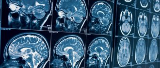
Computed tomography is a method of diagnosing internal organs that allows one to obtain layer-by-layer images to detect serious diseases.
CT is a radiation method, which is based on the different absorption of X-ray radiation by tissues. It is the similarity with radiography that makes many patients doubt whether it is harmful to have a computed tomography scan and how often can this study be done? In addition, they are concerned about the need to use a contrast agent when performing bolus-enhanced CT, which may be accompanied by the development of allergic reactions.
Radiation exposure during computed tomography
Everyone knows the fact that in one year it is permissible to expose the human body to only a certain amount of radiation, which does not exceed normal limits. The permissible annual dose of radiation exposure is 150 m3v. If this standard is observed, radiation does not harm human health.
For example, with regular use for the purpose of preventive fluorography, examination of the mammary glands, and an image of the jaw at the dentist, on average, a person receives at least 15 m3v per year. When performing a computed tomography scan on a standard apparatus for examining the brain, the radiation dose ranges from 1 to 2 m3v, and with a CT scan of the pelvic organs, lungs or abdominal cavity - 6-11 m3v.
According to research, even when undergoing a CT scan several times a year, the dose of radiation received, as a rule, does not exceed the permissible norm.
How is it carried out?
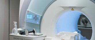
The patient is placed on a special table located near a powerful scanner. Gradually the scanner moves along the table or vice versa. Data about the state of a specific organ or entire system is sent to a computer with a special program. The doctor can visually see them, even in three-dimensional form. On average, diagnosis takes about half an hour, depending on the organ being examined (the abdominal cavity takes the longest to scan and can take up to an hour).
As a result, the doctor will receive detailed images of organs and systems in several planes. This method will help make an accurate diagnosis and identify pathology at an early stage. Scanning provides the most complete information about the organ being examined.
An iron pin or other metal foreign body is a contraindication for tomography.
Is it harmful to have a CT scan with contrast?
Radiation exposure, according to some patients, is not the only danger. To some extent, a radiopaque contrast agent used in some cases for computed tomography can compete with it.
As a rule, it is an inert substance that is not absorbed into surrounding tissues. However, the components included in its composition can cause harm - in some patients they can cause the development of allergic reactions.
This complication may occur in the presence of the following factors:
- hypersensitivity to seafood and iodine;
- renal failure;
- cardiovascular diseases;
- diseases of the gallbladder and liver.
The development of minor side effects is observed in only 1-5% of patients. They experience mild nausea, vomiting, skin reactions, and impaired sense of taste and smell. As a rule, these symptoms do not require special treatment and disappear on their own.
There are isolated cases of the development of side effects of moderate severity: Quincke's edema, acute respiratory failure caused by narrowing of the lumen of the bronchi and sudden involuntary contraction of the muscles of the larynx, shortness of breath. To eliminate such conditions, emergency medical care is required.
In extremely rare cases, severe adverse reactions develop: sudden cardiovascular failure, which can result in loss of consciousness and death. Most often, this harm to CT is caused to allergic patients. In such cases, immediate resuscitation measures are required.
If there is a history of a negative reaction to drugs containing iodine, an antihistamine is administered to the patient before starting a contrast-enhanced computed tomography scan. Some patients require special tests to help identify the allergen.
The development of allergic reactions in patients prone to them occurs in fairly rare cases. Rapid intravenous administration of a contrast agent is accompanied by the occurrence of side effects much less often than slow infusion using a dropper.
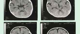
How the brain behaves
Is MRI harmful to human health if it scans the brain? This organ, like any other, should not be irradiated for any reason, although radiation from cell towers and wireless Internet is almost everywhere around us.
MRI brain examinations are carried out only if there are strict indications:
- constant headache;
- regular dizziness;
- suspected development of a tumor in the brain;
- disturbance of brain activity.
So is it harmful to have an MRI of the brain? Can tissues be irradiated? If it is safe, then in what cases is there a risk? Are there any complications? Let's consider the features of the procedure when studying the brain.
The patient is placed on a special platform that moves continuously. The brain is exposed to a strong electromagnetic field. Data is supplied to the equipment - the reaction of hydrogen nuclei after a powerful electromagnetic pulse. As detailed studies have shown, no harm is caused to either the brain or other organs. The benefits of such research are hundreds of times higher than the possible risks. You should not expect any side effects. But the effect is simply colossal. You can see the human body from the inside in detail and in different projections.
Sometimes a special contrast agent may be used. This is what sometimes leads to negative effects. Therefore, before carrying out the procedure, the staff must ask the patient what substances he is allergic to. If there is a negative reaction to the contrast agent that will be administered for the procedure, it is replaced with an analogue.
Contraindications for MRI of the brain may include:
- renal failure;
- presence of pacemakers;
- middle ear implants;
- There are other foreign bodies made of metal in the body (shards, iron crowns, metal knitting needles, etc.). An exception may be implants with titanium elements;
- the patient experiences severe claustrophobia. This is not an absolute contraindication for the procedure. Only preliminary preparation is required. With proper use of sedatives, such patients can remain in the device for up to 40 minutes;
- mental illness. There are a number of mental illnesses that can be a barrier to research.
There are many studies whose purpose was to determine whether MRI is harmful to the body and, in particular, to the brain. None of them could prove any potential danger of the procedure. Therefore, it is generally accepted that MRI can be performed as often as necessary for diagnosis.
The patient's health will not be harmed in any way. There will be no harmful effects. In this case, patients of any age can be examined, even immediately after surgery.
In a hospital setting, a doctor can prescribe an MRI to a patient quite often - several times a month.
Hospital staff must explain to the examinee the essence of the procedure before it begins: the stages of implementation, duration.
MRI can be replaced by other research methods, but efficiency and accuracy will suffer. The tomograph is highly informative and has the ability to amplify electromagnetic radiation.
Indications and contraindications for CT
Computed tomography allows you to identify the pathological process and clarify the diagnosis in patients with various conditions:
- diagnosed with cancer, metastases, suspected cancer;
- frequent, prolonged headaches without obvious causes;
- cerebrovascular accident and the accompanying consequences of this disorder;
- attacks of seizures, convulsions, loss of consciousness;
- conditions after injuries;
- inflammatory processes localized in various parts of the body.
Computed tomography has undeniable advantages - with the help of this study you can assess the condition of almost any organ. In addition, computed tomography is also used to clarify pathology previously identified during other examinations. This study can only harm patients with the following contraindications:
- syndrome of impairment of all renal functions;
- applied plaster or metal structure in the examined area;
- claustrophobia (fear of closed spaces);
- violent behavior caused by mental disorders.
In addition, the use of CT is contraindicated in patients with excessive body weight exceeding 150 kg, pregnant women (especially in the first three months) and children under 14 years of age (except in cases of extreme necessity).
When is it prescribed?
MRI is prescribed when there are specific indications. The patient’s desire alone is not enough, and this should only be done by the doctor. Indications for tomography may include:
- suspicions that a spinal hernia has appeared;
- suspicions that the patient is developing a tumor, mastopathy, fibroids, etc.
Sometimes a contrast agent may be used to better see blood vessels and organs. When administered correctly and at the correct dosage, it is not dangerous.
Which is less harmful: CT or MRI?
One of the modern informative diagnostic methods, in addition to CT, is magnetic resonance imaging (MRI). CT and MRI are not considered alternative methods. MRI is used to study organs that have a high fluid content, but are reliably protected by the bone skeleton: the brain and spinal cord, intervertebral discs, joints and pelvic organs. And with the help of CT it is preferable to examine the musculoskeletal system and lung tissue.
Both CT and MRI have almost equivalent information content when studying the genitourinary and digestive systems. However, computed tomography, compared to magnetic resonance imaging, requires much less time to perform, so it is preferred in emergency cases.
MRI and X-ray – which is safer?
These hardware diagnostic methods are among the most popular. It should be noted that:
- During X-rays, the patient is exposed to a stream of ions. It has low power, so one x-ray session cannot cause any harm. The radiation dose is too low.
- The specificity of X-rays is that the received radiation doses are added. It cannot be used repeatedly in a short period of time. Doses accumulate, and with repeated examination the negative effect intensifies. It is highly undesirable to repeat x-rays several times in a short period of time. The negative impact from the previous dose simply will not have time to wear off. Over time, tissues are restored, and if the ionizing effect is repeated frequently, cells and tissues can be damaged.
- X-rays are contraindicated in certain pathologies and pregnancy. Radiation can be harmful to the fetus.
- X-rays are also undesirable during breastfeeding. A nursing mother should be examined by other methods.
Some patients equate the magnetic resonance method with x-rays. It is not right. Both methods, although hardware-based, are based on completely different physical processes. With MRI, there is no risk of harm to health even with repeated procedures, and there is absolutely no ionizing radiation.
Principles of protection
Patients who doubt the safety of radiation diagnostic methods should familiarize themselves with some principles of reducing radiation exposure:
- reduced time period: the duration of screening can be reduced by refusing to perform screening simultaneously in the sagittal and transverse projections, reducing the current strength of the X-ray tube, as well as the number of tomography phases;
- conducting computed tomography through bismuth screens: in this way, it is possible to reduce radiation exposure without compromising the quality of the images;
- increasing the distance: reducing the radiation dose can be achieved by increasing the distance between the X-ray tube and the body of the subject. You can protect other parts of your body that may be exposed to radiation by using lead shielding.
In cases where CT is used in pediatric patients, the use of sedatives is recommended, since immobility of the subject is important to obtain good quality images. For this purpose, you can also use special belts and pillows to ensure the child’s immobility during the examination.
Computed tomography is often the only possible method for diagnosing certain pathologies, for which there is no high-quality alternative, so the question of whether CT scanning is harmful is often inappropriate. This examination is used to confirm complex diagnoses and immediately begin treatment, especially when it comes to preserving the patient’s quality of life. If all recommendations are followed, the patient should not worry that a CT scan will cause irreparable harm to their health.
CT scans of any organs can be done at affordable prices at the Yusupov Hospital in Moscow. The clinic is equipped with a modern tomograph of the latest generation, thanks to which the experienced, highly qualified doctors of the Yusupov Hospital receive high-quality images. Based on the results of computed tomography, doctors create the most effective treatment tactics, individually for each patient.
Features of MRI of the spine
Research using a tomograph does not require any diet or other type of preliminary preparation from the patient. However, it is important to know the body's reaction to iodine, since this element is part of the contrast agent often used in certain situations. There are other contrasts that are injected into the vertebral area. Therefore, in each specific case, it is important to know whether there is an allergy to the components included in the composition.
In addition, to maintain the accuracy of the images, it is important to lie still inside the device. If a person suffers from claustrophobia or other disorders associated with a fear of narrow and confined spaces, you should tell your doctor about this. The slightest movement will make the examination uninformative.
Why is an MRI prescribed?
In addition to the fact that MTP is prescribed to obtain detailed information about the condition of bones and tissues, bypassing surgical intervention, this study is often used to monitor various neoplasms.
For example, when a cyst or other growth, the nature of which is unknown, is discovered inside bone tissue or the spinal cord, the attending physician first prescribes regular examinations with a certain frequency in order to determine the state of the formation over time: is it growing or not, and at what speed. This allows you to understand the etiology of the growth and determine the correct tactics of medical behavior.