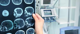
The cranium performs a vital function in the human body. This bone structure is the protective shell of the brain, and therefore has a certain strength. However, there are situations when the integrity of the skull, and, accordingly, the safety of brain tissue, may be at risk. Injuries, diseases and abnormalities in the development of the cranium can directly threaten not only human health, but also human life. Considering the structural features of the skull, as well as the density of its structure, the value of non-invasive methods for examining this bone structure cannot be overestimated. One of the most common and accessible diagnostic methods is skull radiography. It is this that doctors often prescribe as the first stage of patient examination, preceding the more complex and expensive one - computed tomography and magnetic resonance imaging.
How does the skull work and what functions does it perform?
The cranium is part of the human skeleton. Essentially, it forms the bone frame of the head.
Content:
- How does the skull work and what functions does it perform?
- What does a skull x-ray show and why is it prescribed?
- Indications and contraindications for x-rays of the skull
- Preparation requirements, procedure for performing skull x-rays
- Types of skull radiography
- Features of radiography of the skull in children
- How are skull x-rays interpreted?
This part of the skeleton has its own characteristics, for example, the growth and development of the bones of the skull occurs before a person reaches the age of 30-32 years. In addition, as a person grows older, the proportions of the relationship between the brain and facial parts change, the cartilage located between the bones of the base of the skull disappears, and the fontanelles (non-ossified areas of the cranial vault connecting its parts) become overgrown.
The anatomical structure of the skull includes 23 bones, two sections - the brain and the face, while the first is significantly larger in volume than the second.
In the facial part of the skull there are paired and unpaired bones: the vomer, the ethmoid and hyoid bones, the lower jaw, the inferior nasal concha, the upper jaw, the nasal, palatine, zygomatic and lacrimal bones.
The brain part of the skull is divided into a vault and a base, and is formed by the frontal, occipital, sphenoid, parietal and temporal bones. In the area of the crown there are the parietal bones and parietal tubercles - characteristic convex parts of bone tissue. The temporal bones contain pyramidal processes containing the vestibular apparatus and auditory receptors.
All the bones of the skull are connected by sutures - fixed formations of a fibrous structure. The exception is the lower jaw - it is mobile, and is connected to the main part of the skull by ligaments and paired temporomandibular joints.
What is the purpose of the skull in the human body? First of all, it is a protective box for the brain. The skull is the bony frame of the head and determines its shape. It can be argued that the protective function is the main function of this bone structure.
In the area of the skull are the original openings of the respiratory and digestive tract, as well as the human sensory organs; facial muscles are attached to his bones, which, together with the bones, determine the facial features of a person.
Thanks to the mobility of the lower jaw, a person has the ability to perform the chewing function. The bones of the skull are part of the speech apparatus, allowing communication through articulate speech, and the bones of the jaws themselves represent the base of the teeth.
The occipital bone of the brain part of the skull connects it to the spine; it provides an opening for the transition of the brain into the spinal cord.
Respiratory and speech activity, food absorption, and the work of almost all sense organs and the brain are practically impossible if the cranium cannot fully perform its functions.
What does a skull x-ray show and why is it prescribed?
A common misconception is that head x-rays are intended to examine the brain. In fact, this diagnostic method is more effective for studying the bones of the skull along with the teeth.
The appointment of a procedure is usually preceded by the patient contacting a doctor with certain complaints. Therapist, oncologist, neurologist, endocrinologist, ophthalmologist, surgeon, otolaryngologist - this is an incomplete list of specialists who can refer the patient for this examination.
The doctor issues a referral for an X-ray of the skull if the patient complains of the following symptoms:
- tremor of the upper extremities;
- constant or recurrent headache;
- frequent dizziness;
- causeless nosebleeds;
- feeling of darkening in the eyes;
- decreased hearing and visual acuity;
- pain when chewing.
The purpose of the procedure is:
- establishing a primary diagnosis or checking an existing diagnosis;
- development of treatment tactics;
- determining the grounds for surgery, radiotherapy or chemotherapy;
- checking the effectiveness of the treatment.
“What does a skull x-ray show?” – people being examined often ask the doctor who ordered the x-ray this question.
A doctor of appropriate qualifications can determine from a high-quality image the presence of the following pathologies and diseases of the skull bones:
- cyst;
- osteoporosis of bone tissue;
- congenital anomalies of the structure and deformations of the skull;
- cerebral hernias and pituitary tumors;
- hematoma;
- osteosclerosis;
- osteomas (benign bone tumors), meningiomas (benign tumors of the soft membranes of the brain), malignant tumors, metastases;
- fractures and their consequences;
- signs of intracranial hypertension and hypotension;
- consequences of inflammatory processes in the brain.
Types of teleroentgenogram
There are several types of teleradiography. The doctor’s task is to choose exactly the option (or several) that will help create the most complete picture of the pathology.
Frontal
Frontal TRG is considered the main diagnostic procedure and is prescribed before the start of orthopedic treatment. An overview image of the head is taken from the back and front and allows you to clearly see the existing inflammatory processes, facial asymmetry and fractures.
TRG in lateral projection
A lateral view of the TRG is performed to diagnose abnormal jaw development and prevent its consequences. In addition, a lateral teleroentgenogram provides the doctor with the opportunity to examine the jaw from the side and calculate the inclination of the lower teeth and those located in the upper row. This x-ray is often taken before a patient is assigned to wear braces.
Axial
Axial TRG (also called “mental”) is an additional diagnostic procedure along with lateral or frontal. Axial TRG is necessary to determine changes in the structure of the cheekbones, maxillary sinus and nasal cavity. Mainly used when implantation of the upper front teeth is necessary.
Indications and contraindications for x-rays of the skull
Due to the fact that the procedure uses x-rays, it should be carried out only on the direction of a doctor, and only in cases where there is an objective need to obtain information about the condition of the skull bones in this way.
Among the indications for a skull x-ray:
- suspected traumatic brain injury (open or closed);
- tumor processes;
- possible developmental anomalies - congenital or acquired;
- pathologies of ENT organs, for example, sinuses;
- the presence of a number of symptoms with an unclear etiology: disturbances of consciousness, dizziness, constant severe headaches, symptoms of hormonal imbalance.
As for contraindications, they are related to the dose of radiation received during the diagnostic process. For example, examination methods involving the use of X-ray irradiation are generally not recommended for pregnant women, especially in the first trimester. If possible, the doctor prescribes diagnostic methods that are gentler on the fetus.
The second category of patients for whom skull x-rays are prescribed with caution are children. Childhood is not an absolute contraindication for the procedure; moreover, in some cases, an X-ray of the skull is an objective necessity, for example, if it is necessary to confirm the doctor’s suspicions of congenital pathologies of bone development.
It is believed that modern X-ray machines cannot significantly irradiate a child during diagnosis. Thus, the permissible radiation dose per year for a person is no more than 50 microsieverts per year, and radiography equipment “gives” the patient a dose of no more than 0.08 microsieverts per session. In this case, the problem is the fact that not every medical institution has at its disposal modern X-ray machines with dosed radiation, and in X-ray rooms there is more often outdated equipment that has been in use for decades. Nevertheless, sometimes you simply cannot refuse an X-ray of the baby’s skull. This diagnostic method is one of the most popular in pediatric neurosurgery, traumatology and neurology. If there are certain indications, skull x-rays are performed even for newborn babies.
General characteristics of the procedure
Relatively recently, it became possible to accurately diagnose pathologies of the dental system.
This can be done using a new orthopantomograph with cephalstat. This device has two examination programs: pediatric and adult. It is distinguished by low radiation exposure and high accuracy of the resulting images. An X-ray examination carried out using such a device is called “teleradiography” (what it is and how important such an examination is in orthodontics will be described below), and such a procedure today is an integral part of orthodontic treatment.
Preparation requirements, procedure for performing skull x-rays
This type of x-ray does not require any preparatory measures. Before it is carried out, the doctor clarifies the fact that there is no pregnancy, if we are talking about a female patient, explains exactly how the procedure will take place, how many pictures will need to be taken, what is required of the patient during the process. If the procedure is prescribed for a child, the parents prepare him for diagnosis and explain in a clear manner to the child how he needs to behave. Doctors do not set any restrictions on diet or the amount of physical activity before the examination unless they are required by the patient’s general condition, regardless of the prescribed procedure.
Before starting the diagnosis, the doctor asks the patient to remove all metal jewelry and accessories from the head and neck, as they may appear on the pictures in the form of additional darkening, thereby distorting the results.
The image can be captured in different positions - the patient can lie, sit or stand, depending on which area is being examined. The body of the subject is covered with a special protective apron with lead plates. The head, if necessary, can be fixed with special belts or rollers to ensure its complete immobility while capturing the image. The doctor takes the required number of pictures. During the process, he can change the patient’s position and position.
Pictures can be taken in the following projections:
- axial;
- semi-axial;
- anterior-posterior;
- posterior-anterior;
- right lateral;
- left side.
There is also such a thing as radiography methods. It involves recording images in special projections that allow obtaining an image of a specific area. For example, the methods according to Reza, Ginzburg and Golwin differ from each other, but they all provide an overview of the optic canals and the superior orbital fissure. Images according to Schüller, Mayer and Stenvers allow you to study the condition of the temporal bones.
Most often, for a doctor to make a diagnosis, it is enough to take pictures in two projections - the front and one of the sides. The whole procedure lasts no more than 5 minutes. It is absolutely painless, and the only atypical sensation that may arise from it is a metallic taste in the mouth due to exposure to x-rays.
Types of skull radiography
Considering the complexity of the structure of the skull, and the large number of bones that make it up, doctors distinguish two types of radiography of the skull:
- overview;
- sighting.
Plain radiography of the head is not intended to visualize any specific area of the skull. Her pictures show the condition of the bone structure as a whole.
Sight radiography makes it possible to examine the condition of a certain part of the skull:
- zygomatic bones;
- bony pyramid of the nose;
- upper or lower jaw;
- eye sockets;
- sphenoid bone;
- temporomandibular joints;
- mastoid processes of the temporal bones.
Sighted radiography images show the presence of calcifications in the bones, hemorrhages and hematomas in a specific part of the skull, calcification of parts of tumors, the presence of pathological fluid in the paranasal sinuses, changes in the size of bone elements associated with acromegaly, disorders in the area of the sella turcica, provoking pathologies of the pituitary gland, bone fractures skull, as well as the location of foreign bodies or foci of inflammation.
Advantages of the technique
A teleroentgenogram (almost every dentist can tell you in more detail what it is) has some advantages over other diagnostic procedures:
- high information content of images (with the help of TRG, especially if it is done in several projections at once, it is possible to identify almost all pathologies of the dental system
); - instant results;
- safety and painlessness (can be done on children and pregnant women in the second trimester).
Features of radiography of the skull in children
In order for the child not to be afraid of an incomprehensible and unfamiliar procedure, he should be explained in simple and understandable words how radiography is performed, that this process does not cause pain at all, that parents may be nearby, so there is no reason for fear, and you just need to listen to the doctor. Very young children are allowed a pacifier.
The child is seated or laid down and carefully secured so that he does not move. All metal clips, jewelry and hair accessories must be removed. The body is covered with a lead apron; a lead collar can be additionally used to protect the thyroid gland.
After x-rays, the baby should be given plenty of fluids - fruit drinks, teas, juices with pulp, milk and fermented milk drinks to neutralize the effect of the received radiation dose.