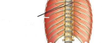
Diagnosis of median nerve neuropathy
When the C7-spinal nerve or the middle trunk of the brachial plexus is damaged, the function of the median nerve is partially affected, as a result there is a weakening of the flexion of the hand and its inward rotation in combination with damage to the radial nerve.
Almost the same loss of function of the median nerve occurs with damage to the external bundle of the brachial plexus, into which the fibers of the upper peduncle of the nerve pass from the middle trunk, but in combination with damage to the musculocutaneous nerve. When the spinal nerves C8–Th1 , the lower trunk and the internal bundle of the brachial plexus are damaged (Dejerine-Klumpke palsy), those fibers of the median nerve that make up its lower leg suffer in combination with damage to the ulnar nerve (weakening of the finger flexors and thenar muscles).
The motor function of the median nerve mainly consists of inward rotation of the hand, palmar flexion of the hand due to contraction of the corresponding muscles, flexion of fingers, mainly I, II and III, extension of the middle and terminal phalanges of II and III fingers.
The place of compression of the median nerve in carpal tunnel syndrome is at the level of the carpal tunnel.
Sensitive fibers of the median nerve innervate the skin of the palmar surface of the I, II, III and radial half of the IV fingers, the corresponding part of the palm, as well as the skin of the rear of the terminal phalanges of these fingers.
When the median nerve is damaged (median nerve neuritis), inward rotation of the hand suffers, palmar flexion of the hand is weakened, flexion of the 1st, 2nd and 3rd fingers and extension of the middle phalanges of the 2nd and 3rd fingers are impaired.
Superficial sensitivity in case of median nerve neuritis is impaired on the hand in an area free from the innervation of the ulnar and radial nerves. Articular-muscular sensation with neuritis of the median nerve is always impaired in the terminal phalanx of the index, and often in the second finger.
Diagnosis of the level of damage to the median nerve during neuritis is made using electroneurography (ENG).
Muscle atrophy with damage to the median nerve is most pronounced in the thenar region. The resulting flattening of the palm and bringing the thumb close and in one plane to the index finger create a peculiar position of the hand, which is called “monkey”. Pain when the median nerve is damaged, especially partial, is quite intense and often takes on a causal nature. In the latter case, the position of the hand may become bizarre.
Vasomotor-secretory-trophic disorders are also common and characteristic of lesions of the median nerve: the skin, especially the 1st, 2nd and 3rd fingers, becomes bluish or pale in color; nails become “dull”, brittle and streaked; skin atrophy, thinning of fingers (especially II and III), sweating disorders, hyperkeratosis, hypertrichosis, ulcerations, etc. are observed. These disorders, as well as pain, are more pronounced with partial rather than complete damage to the median nerve.
The median nerve, like the ulnar nerve, gives off its first branches only to the forearm, so the clinical picture with high damage along the entire length from the axillary fossa to the upper parts of the forearm is the same. When the median nerve is damaged in the middle third of the forearm, the functions of internal rotation of the hand, palmar flexion of the hand and flexion of the middle phalanges are not affected.
The use of acupuncture is very effective in the treatment of median nerve neuritis.
With lower lesions of the nerve, the function of flexion of the terminal phalanges of the I, II and III fingers may be preserved, and then all symptoms of the lesion are limited to damage to the thenar and lumbrical muscles and sensory disturbances in the typical zone.
The main tests to determine movement disorders that occur with damage to the median nerve (median nerve neuritis) are the following:
- When the hand is clenched into a fist, fingers I, II and partly III do not bend
- Bending the terminal phalanges of the thumb and index fingers is impossible, as is scratching the index finger on the table with the hand tightly adjacent to it.
- During the thumb test, the patient cannot hold a strip of paper with a bent thumb and will hold it by adducting the straightened thumb with the adductor muscles from the preserved ulnar nerve
Ultrasound examination of the peripheral nervous system
Ultrasound scanner RS80
A benchmark for new standards!
Unparalleled clarity, resolution, ultra-fast data processing, and a comprehensive suite of advanced ultrasound technologies to solve the most challenging diagnostic problems.
Ultrasound examination of the peripheral nervous system first began to be used to diagnose diseases of the nerve trunks in the late 90s of the last century [1]. With the beginning of the use of this method, its undeniable advantages over other diagnostic methods became clear. Electrophysiological methods, such as electromyography and neuromyography, are traditionally recognized as the “gold standard” for identifying pathology of the peripheral nervous system. However, it should be noted that the information obtained during the above examinations does not provide an idea of the state of the surrounding tissues, does not indicate the nature and cause of damage to the nerve trunk, and does not always accurately reflect the localization of changes [2, 3]. At the same time, it is this information that helps determine the tactics of conservative or surgical treatment.
The introduction of ultrasound sonography into clinical practice has successfully filled the gaps in the diagnosis of peripheral nerve diseases. This article presents the experience of ultrasound examination of peripheral nerves of the upper and lower extremities, accumulated in our clinic.
Ultrasound anatomy of normal peripheral nerves
For ultrasound studies, sensors with a frequency of 7-17 MHz are used, but in some cases it is necessary to use transducers with a lower frequency - 3-5 MHz. During the scanning process, the anatomical integrity of the nerve trunk, its structure, the clarity of the contours of the nerve and the condition of the surrounding tissues are assessed. All of the above points must be reflected in the study protocol. If pathological changes in the structure of the nerve are detected, the type of damage (complete or partial), the zone and degree of compression of the nerve trunk are indicated (the decrease in the diameter of the nerve and the cause of the compression are noted). If a space-occupying formation is detected, its size and structure, contours, relationship with surrounding soft tissues, and the presence or absence of blood flow are described.
It is advisable to begin ultrasound examination of peripheral nerves with a transverse projection at the point where the nerve trunk is most easily identified, then moving in the proximal and distal directions, assessing the structure of the nerve along its length [3-5].
The image of a nerve has a number of characteristic features. In the transverse projection, it looks like an oval or round formation with a clear hyperechoic contour and an internal heterogeneous ordered structure (“salt and pepper”, “honeycomb”) [4, 6, 7]. In the longitudinal projection, the nerve is located in the form of a linear structure with a clear echogenic contour, in which hypo- and hyperechoic stripes regularly alternate - “electric cable” [7]. The thickness of peripheral nerves is variable and ranges from 1 mm for the digital nerves to 8 mm for the sciatic nerve.
The key to a successful ultrasound examination is a good knowledge of the anatomy of the area being examined.
The main nerve trunks accessible to ultrasound examination in the upper limb are the radial, median and ulnar nerves.
The radial nerve is the largest branch of the posterior portion of the brachial plexus. The nerve is visualized on the posterior and lateral surfaces of the shoulder, where it accompanies the brachial artery. In the middle third of the shoulder, the radial nerve bends around the humerus and is directly adjacent to it in the spiral canal (Fig. 1).
Rice. 1.
Transverse sonogram of the radial nerve (short arrows) at the level of the spiral canal of the humerus (long arrows - outline of the humerus).
It is from the spiral canal that it is most advisable to begin the process of scanning the radial nerve. As a rule, sensors with a frequency of 9-17 MHz are used for this, and the study is carried out mainly in the transverse projection. Further, immediately anterior to the lateral epicondyle of the humerus, n. radialis is divided into sensory (or superficial) and motor (deep) branches and the posterior interosseous nerve (Fig. 2).
Rice. 2.
Transverse sonogram at the level of the distal shoulder. Division of the radial nerve into superficial and deep branches (arrows).
The superficial branch runs along the medial edge of the brachioradialis muscle and is accompanied by the radial artery and vein. In this place, the nerve is most accessible to ultrasound examination, but only if high-frequency sensors are used (over 15 MHz), since the diameter of this branch is very small.
The deep branch of the radial nerve passes directly into the supinator, here the nerve is also accessible to visualization due to the difference in sonographic structure between it and the surrounding muscle.
In the distal section on the extensor surface of the forearm n. radialis (its superficial branch) ends in division into 5 dorsal digital nerves. Ultrasound examination of digital nerves can only be performed using high-frequency transducers, and even then, clear sonographic images of these structures are rarely obtained.
The median nerve is formed from the lateral and medial bundles of the brachial plexus. On the shoulder n. medianus is located in the medial groove of the biceps muscle anterior to the brachial artery. The median nerve is the largest nerve of the upper extremity, so its visualization is not difficult, but the easiest way to obtain an ultrasound image of the nerve is in the carpal tunnel, where it is located superficially, and also at the level of the elbow joint. In the latter case, it is advisable to use a vascular bundle as a marker. In the area of the elbow joint, the median nerve is located medial to the more deeply located brachial artery and vein (Fig. 3).
Rice. 3.
The median nerve at the level of the elbow joint in transverse view (short arrows). The brachial artery is visualized nearby (long arrow).
In the proximal forearm, the nerve usually passes between the two heads of the pronator teres muscle. In the area of the wrist joint, the median nerve is located under the palmaris longus tendon and between the flexor tendons, passing under the flexor retinaculum onto the hand through the so-called carpal tunnel. The common palmar digital nerves (there are three of them) are formed by branching the main trunk of the median nerve at the level of the distal end of the flexor retinaculum.
The ulnar nerve is the main branch of the medial bundle of the brachial plexus. On the shoulder n. ulnaris does not produce branches. In the area of the elbow joint, the nerve passes through the cubital canal formed by the medial epicondyle of the humerus and the olecranon process. Here the ulnar nerve is adjacent directly to the bone and is covered on top only by fascia and skin. When performing an ultrasound examination of the elbow joint, care should be taken to ensure that the patient’s arm is positioned freely and is not bent. This is important because when the elbow is flexed to 90 degrees, the diameter of the nerve decreases due to its stretching.
On the forearm n. ulnaris is usually located between the two heads of the flexor carpi ulnaris, and in the distal forearm the nerve lies between the flexor carpi ulnaris tendon medial and lateral to the ulnar artery and vein. The ulnar nerve enters the hand through the ulnar nerve canal called Guyon's canal. As it passes through the canal, the ulnar nerve is accompanied by the artery and vein of the same name. In the distal part of Guyon's canal, the nerve divides into a deep motor branch and a superficial sensory branch, and it is the superficial branch that continues to accompany the ulnar artery, which makes it easier to navigate during ultrasound examination.
In the lower extremity, ultrasound scanning can easily identify the sciatic nerve and its branches. Foreign literature also describes sonographic examination of the femoral nerve. It should be noted that visualization of this peripheral nerve is difficult and the best acoustic window is the groin area, where the nerve accompanies the femoral artery and vein.
The sciatic nerve is the largest peripheral nerve in the human body. In fact, it consists of two large trunks: the common peroneal nerve is located outside, and the tibial nerve is medial. The sciatic nerve exits the pelvic cavity through the greater sciatic foramen under the piriformis muscle.
Already in the gluteal region, the nerve is accessible to visualization; you just need to correctly determine the frequency of the sensor used: if there is sufficient muscle mass, it is advisable to use sensors with a frequency of 2-5 MHz; if the muscle mass in the gluteal region is not expressed, you can use sensors with a higher frequency - 5-9 MHz. In the area of the gluteal fold, the sciatic nerve is located close to the lata fascia of the thigh, moves laterally and then lies under the long head of the biceps femoris muscle, located between it and the adductor magnus muscle (Fig. 4).
Rice. 4.
Sciatic nerve (longitudinal view, panoramic scan) in the middle third of the thigh (arrows).
In the distal parts of the thigh, usually in the upper corner of the popliteal fossa, the nerve is divided into two branches: a thicker medial one - the tibial nerve and a thinner lateral one - the common peroneal nerve. It is from this area that it is best to begin an ultrasound examination of the sciatic nerve and its branches.
The common peroneal nerve, having separated from the main trunk, descends laterally under the biceps femoris muscle to the head of the femur. In the area of the head of the fibula, the nerve is located superficially, covered only by fascia and skin, here it is also easily accessible for visualization (Fig. 5).
Rice. 5.
Longitudinal sonogram of the common peroneal nerve (arrows) at the level of the fibular head (F).
Next, the common peroneal nerve penetrates the thickness of the proximal part of the peroneus longus muscle and divides into its two terminal branches - the superficial peroneal nerve and the deep peroneal nerve. Visualization of the terminal branches of the common peroneal nerve is difficult due to their small diameter and the lack of anatomical markers as they pass through the thickness of the calf muscles. The superficial peroneal nerve divides into terminal branches (dorsal branches of the foot) on the lateral surface of the lower third of the leg. The deep peroneal nerve passes to the anterior surface of the leg and here, located laterally, accompanies the anterior peroneal vessels. On the dorsum of the foot, the nerve enters under the lower extensor retinaculum and under the tendon of the long extensor of the first finger. Here it divides into terminal branches. To visualize the common peroneal nerve and its branches, it is more convenient to use sensors with a frequency of 9-17 MHz.
The tibial nerve in its direction is a continuation of the sciatic nerve. In the popliteal fossa, the nerve is located above the popliteal vein and artery and somewhat outward from them (Fig. 6).
Rice. 6.
Sonograms of the tibial nerve in the popliteal fossa (arrows). The popliteal vascular bundle is visualized - vein (V) and artery (A).
A)
Longitudinal sonogram.
b)
Transverse sonogram.
On the lower leg, the tibial nerve enters between the heads of the gastrocnemius muscle and accompanies the posterior tibial vessels, passing under the soleus muscle. The tibial nerve enters the foot through the so-called “tarsal canal” or medial malleolar canal, formed medially by the medial malleolus and laterally by the fascia of the flexor retinaculum. This fibrous tunnel is similar in structure to the carpal tunnel on the hand. At the exit from the tarsal canal, the nerve divides into terminal branches - the medial and lateral plantar nerves. The tibial nerve is best examined in the popliteal fossa and proximal tibia, as well as at the level of the medial malleolus (Fig. 7). In the middle third of the leg, the nerve is located quite deep and its image is difficult to differentiate from the surrounding tissues.
Rice. 7.
Transverse sonogram of the tibial nerve at the level of the medial malleolus (arrows). The posterior tibial veins (V) and artery (A) are visualized.
In the lower leg, the tibial nerve gives off cutaneous and muscular branches. Of all the branches, the sural (sural) nerve is most often accessible to visualization (Fig. 8). It is located outward from the small saphenous vein and accompanies it to the lateral malleolus, where it divides into terminal cutaneous branches.
Rice. 8.
Sural nerve (arrows). Projections at the level of the middle third of the leg.
A)
Longitudinal projection.
b)
Transverse projection.
To study the tibial nerve and its branches, sensors with a frequency of up to 9-17 MHz are used.
Ultrasound diagnosis of peripheral nerve diseases
Nerve damage
Traumatic nerve injuries can be divided into two large groups: damage with complete or partial disruption of the anatomical integrity of the nerve and damage to the internal structure of the nerve trunk while maintaining the integrity of the outer sheath of the nerve. The ultrasound picture of nerve injuries has characteristic signs depending on the type of damage and is common to any peripheral nerve. The reasons for the violation of the integrity of the nerve trunk may be different. In our practice, we most often encounter the consequences of injuries: intersection of a nerve as a result of an incised wound, damage by bone fragments or pinching of the nerve between them during displaced fractures, compression of the nerve by scar tissue or callus. In addition, iatrogenic damage to the nerve can occur during closed or open reposition of fragments with subsequent fixation with a plate, during surgery on soft tissues adjacent directly to the nerve trunk.
In the upper extremity, the most common injuries to the radial nerve are associated with a fracture of the humerus, which is primarily explained by the close apposition of the nerve to the bone as it passes through the spiral canal of the humerus. On the lower limb, the most vulnerable area in this regard is the head of the fibula, where the common peroneal nerve is directly adjacent to it.
A conclusion about a violation of the anatomical integrity of the nerve can be made on the basis of visualization of the distal and proximal ends of the nerve with a clearly visible diastasis between them. Moreover, in the first days after the injury, the severed nerve segments, as a rule, are not changed, and only after some time (from 1 to 12 months) does a post-traumatic neuroma most often form at the proximal end of the damaged nerve trunk (Fig. 9). The distal end of a completely damaged nerve becomes thinner, and in some cases traumatic neuromas can form in it.
Rice. 9.
Terminal post-traumatic neuroma of the ulnar nerve. The arrow indicates the proximal end of the damaged nerve, ending in an oval hypoechoic formation with a clear contour—neuroma. Longitudinal sonogram.
Traumatic neuromas, depending on the location of the formation and the cause that caused them (complete or partial rupture), are divided into terminal and intra-trunk. The structure of the neuroma is hypoechoic and homogeneous, the size of the neuroma depends on the size of the damaged nerve and the amount of nerve tissue involved in the damage, the formation has clear contours and is avascular. If the integrity of the nerve is partially damaged in the damaged nervous tissue, as mentioned above, an intra-trunk neuroma can form (Fig. 10). In this case, the formation is visualized directly in the nerve trunk; it has the same ultrasound characteristics as with a complete break of the trunk; the size of the neuroma is variable and can reach several centimeters in length. The ultrasound report must indicate the diastasis between the ends of the damaged nerve and the structure of the proximal and distal ends, the size of the neuroma, and its location.
Rice. 10.
Intrastem neuroma of the median nerve. Short arrows indicate the proximal and distal ends of the damaged nerve; the hypoechoic formation is surrounded by perineurium and has clear contours—neuroma. Longitudinal sonogram.
In case of contusion of a nerve or its traction, if the outer sheath remains intact, the internal structure of the nerve trunk changes. There is a loss of differentiation into individual fibers, the nerve becomes hypoechoic, thickened, with an unclear contour. The ultrasound signs listed above are detected directly at the site of damage; in the proximal and distal directions, the nerve trunk, as a rule, is not changed. At the site of pinching of the nerve trunk between bone fragments or metal structures, thinning of the nerve directly at the site of the lesion and loss of an ordered echostructure are noted (Fig. 11). The same picture can be seen when compressed by scar tissue or callus (while maintaining the integrity of the nerve). Proximal to the place of compression, the diameter of the nerve increases due to the thickening of individual nerve bundles in its composition. In this case, the trunk has unclear contours and a structure of reduced echogenicity. The described ultrasound signs are caused by swelling of the nerve segment proximal to the compressed area. Distal to the site of injury, the structure of the nerve may not be changed.
Rice. eleven.
Entrapment of the deep branch of the radial nerve (short arrows) at the level of the proximal forearm by a bone fragment (long arrow). The nerve has a hypoechoic homogeneous structure, thickened, and there is no differentiation into individual fibers. Panoramic scanning.
Peripheral nerve compression syndromes (tunnel syndromes)
Peripheral nerves of the extremities can be subject to compression in natural fibrous canals, when located in the thickness of muscle tissue and when adjacent to bone. The following localizations of potential compression of nerve trunks have been described in the upper limb. For the radial nerve, this is the spiral canal and m. supinator of the forearm. The median nerve can become entrapped as it passes between the heads of the pronator teres and in the carpal tunnel. For the ulnar nerve, the most likely sites for carpal tunnel syndrome are the cubital canal and Guyon's canal. In the lower extremity, the common peroneal nerve is most often compressed at the level of the fibular head; the tibial nerve may be compressed distally as it passes through the tarsal canal.
Carpal tunnel syndrome is the most common peripheral nerve compression syndrome. This pathology has a characteristic clinical picture and is easily diagnosed. Ultrasound can help confirm compression of the median nerve in the carpal tunnel. The main ultrasound signs of this tunnel syndrome include: thickening of the median nerve proximal to the carpal tunnel, flattening or decreased height of the nerve in the distal carpal tunnel, and curvature of the flexor retinaculum. In the proximal parts, the median nerve loses its differentiation into fibers, and its structure becomes hypoechoic (Fig. 12). A number of foreign studies devoted to the problem of ultrasound diagnosis of “carpal tunnel” syndrome emphasize the need for quantitative assessment of changes in the median nerve. In our practice, we use two main criteria: an increase in the cross-sectional area of the median nerve over 0.11 cm², measured at the level of the pisiform bone, and a flattening coefficient, defined as the ratio of the maximum width of the nerve to its height (values above 3.3 are considered pathological) . The appearance of intraneural hypervascularization at the site of compression of the median nerve when examined in color-coding mode may also indicate the development of “carpal tunnel” syndrome.
Rice. 12.
Compression of the median nerve in the carpal tunnel. The location of nerve compression is indicated by a light arrow. Above the place of compression (dark arrow), the nerve is thickened, its contours are unclear, and there is a thickening of individual nerve bundles within the nerve. Longitudinal sonogram.
The second most common is ulnar nerve compression syndrome in the cubital tunnel. True entrapment in the ulnar groove occurs when the nerve is compressed by scar tissue, callus, exostoses or soft tissue formations, such as an organized hematoma, intraneural ganglia and intraarticular ganglia, and accessory ulnar muscle. External nerve compression may develop in the presence of predisposing factors: a shallow groove of the ulnar nerve, prolonged pressure on the cubital canal area, or subluxation of the nerve in patients who are in a coma or during prolonged anesthesia. Repeated dislocation of the ulnar nerve with its displacement towards the medial epicondyle of the humerus can cause damage to the nerve or provoke permanent trauma.
Ultrasound examination of the ulnar nerve if compression of the nerve in the cubital canal is suspected begins in the transverse projection from the distal shoulder. Usually in this area the nerve has an oval shape; when passing through the cubital canal it becomes round. It should be emphasized again that patients may experience a slight decrease in the echogenicity of the ulnar nerve in this area and a slight increase in its size without clinical symptoms of neuropathy. Unlike healthy individuals, in patients with cubital tunnel syndrome, an increase in the diameter of the ulnar nerve is found at the level of the medial humeral condyle.
After examining the nerve in the cubital canal in the transverse and longitudinal projections, the sensor is moved distally and the structure of the nerve in the proximal forearm is assessed. The main ultrasound signs of ulnar nerve compression syndrome in the cubital canal include flattening of the nerve directly at the site of compression, thickening above this zone, loss of internal differentiation of the nerve into separate bundles, swelling of the surrounding soft tissues and hypervascularization.
With chronic trauma to the ulnar nerve in the cubital canal, the clinical manifestations do not differ from those with compression of the nerve in this area, and ultrasound data will have other characteristic signs. The main one is diffuse thickening of the nerve at the level of the cubital canal (Fig. 13). In addition, one can detect an increase in the size of individual nerve bundles within the nerve, blurring of the contour of the nerve due to swelling of the surrounding soft tissues.
Rice. 13.
Neuropathy of the ulnar nerve at the level of the cubital canal. Longitudinal sonogram.
Compression of the ulnar nerve in Guyon's canal is much less common than cubital tunnel syndrome. The main causes of entrapment of the ulnar nerve in Guyon's canal are external compression by various formations: lipoma, intra-articular ganglion, aneurysm of the ulnar artery. This type of tunnel syndrome is rare, and the ultrasound findings of compression of the ulnar nerve in Guyon's canal are consistent with those described above for other compression syndromes.
When examining the medial ankle joint, it is necessary to remember about such a type of pathology as tarsal tunnel syndrome. This type of tunnel syndrome is associated with compression of the tibial nerve in the tarsal tunnel. The tarsal canal is similar in structure to the carpal canal on the hand. The flexor tendons and neurovascular bundle are contained in a fairly tight space between the medial malleolus and the flexor retinaculum. When the pressure in this space increases, compression of the tibial nerve occurs, which is clinically manifested by pain and paresthesia in the medial part of the foot. Ultrasound diagnosis of this tunnel syndrome is based on identifying additional formations in the tarsal canal: this is an accumulation of fluid, an intraarticular ganglion, which causes compression and flattening of the tibial nerve. Above the compression zone, there is thickening of the nerve trunk with ultrasound signs of its edema.
Space-occupying formations of peripheral nerves
Schwannomas and neurofibromas are the most common peripheral nerve tumors. It should be noted that their ultrasound signs are similar. Hypoechoic formations of oval or fusiform shape are identified, oriented along the long axis of the nerve and giving the effect of dorsal enhancement (Fig. 14). The contours of the formation are clear, even, and sometimes its capsule can be located. Ultrasound examination can reveal heterogeneity and fluid inclusions in the structure of the tumor. The size of the formations ranges from 2 to 5 cm. In the color Doppler mode, abundant vascularization is usually detected in tumors. Since, as noted above, neurofibromas and schwannomas have similar sonographic characteristics, the histological diagnosis is not indicated in the ultrasound report, limiting itself to a detailed description of the identified formation.
Rice. 14.
Neurofibroma of the tibial nerve at the level of the popliteal fossa is a formation with clear contours, fusiform shape, hypoechoic structure.
A)
Panoramic scanning.
b)
With color Doppler mapping, blood flow is determined in the formation.
Malignant peripheral nerve tumors typically affect large nerve trunks, such as the sciatic nerve or brachial plexus. In addition, patients with malignant tumors are more likely to have definite neurological symptoms compared to patients with benign tumors. Ultrasound signs such as a tumor size of more than 5 cm, blurred contours, heterogeneity of the structure with the presence of calcifications and the reaction of surrounding tissues in the form of edema and infiltration most likely indicate a malignant process. The characteristics listed above (with the exception of indications of invasive growth) are not specific and do not allow us to draw an unambiguous conclusion about the nature of the tumor.
Morton's neuroma (perineural fibrosis, focal traumatic neuritis of the plantar nerve) is a fibrous thickening of the interdigital nerve and belongs to tumor-like lesions of the nerve trunks. The predominant localization of this tumor between the 3rd and 4th metatarsal bones has an anatomical basis: a kind of nerve plexus is formed here from the branching of the common plantar nerve of the third interdigital space and the branches of the anastomosis from the lateral plantar nerve. Ultrasound examination of the interdigital spaces between the toes is best done from the plantar surface, with the sensor installed in the transverse plane at the level of the metatarsal heads. The experience of our research shows that a transverse examination of the foot alone is not enough, therefore longitudinal ultrasound scanning in this area is also necessary. For research, it is advisable to use sensors with a frequency of at least 12 MHz. The normal interdigital space is characterized by the presence of echogenic material, including fat and connective tissue. The neuroma has a round or fusiform shape and a hypoechoic structure, defined in the lower part of the intermetatarsal space between the heads of the metatarsal bones (Fig. 15). An attempt should always be made to establish a connection with the interdigital nerve, which immediately increases the specificity of the sonographic examination. Due to the small size of the plantar nerves, this is not always achieved. A neuroma can be confused with inflammation in the adjacent metatarsal bursa. Ultrasound differences are that the metatarsal bursa is located anterior to the interdigital nerve between the metatarsal heads and when the bursa is inflamed, there is usually a fluid component in it. In addition, the clinical symptoms of Morton's neuroma are characteristic enough to suspect this particular disease.
Rice. 15.
Morton's neuroma. A hypoechoic formation (dark arrow) is located between the metatarsal heads (white arrows). Longitudinal sonogram.
Conclusion
Ultrasound examination of the peripheral nervous system is becoming increasingly important in clinical practice every year. The undeniable advantages of sonography compared to other imaging methods are the relative cheapness of the study and the ability to repeat it the required number of times. During the examination, a specialist can assess the structure of the nerve trunk along its length and the condition of the surrounding tissues, and conduct a series of dynamic tests. The main disadvantage of the ultrasound method is the subjectivity in assessing the data obtained, associated with various practical skills and experience of specialists. We hope that this publication will make a modest contribution to the introduction of ultrasound neurology into widespread clinical practice and will help doctors better navigate the issues of diagnosing diseases of the peripheral nervous system.
Literature
- Fornage BD Peripheral nerves of the extremities: imaging with US // Radiology. 1988. V. 167. N1. R. 179-182.
- Gruber H., Peer S., Meirer R. et al. Peroneal Nerve Palsy Associated with Knee Luxation: Evaluation by Sonography-Initial Experiences // Am. J. Roentgenol. 2005. V. 185. P. 1119-1125.
- Peer S., Bodner G. High-Resolution Sonography of the Peripheral Nervous System // 2003. Springer. 140 p.
- Eskin N.A., Golubev V.G., Bogdashevsky D.R. and others. Echography of nerves, tendons and ligaments // SonoAce International. 2005. Vol. 13. pp. 82-94.
- Bodner G., Buchberger W., Schocke M. et al. Radial Nerve Palsy Associated with Humeral Shaft Fracture: Evaluation with US-Initial Experience // Radiology. 2001. V. 219. N3. P. 811-816.
- Mironov S.P., Eskin N.A., Golubev V.G. and others. Ultrasound diagnosis of pathology of tendons and nerves of the extremities // Bulletin of Traumatology and Orthopedics. 2004. N3. pp. 3-4.
- Stewart JD Peripheral nerve fascicles: anatomy and clinical relevance // Muscle Nerve. 2003. V. 28. N5. P. 525-541.
Ultrasound scanner RS80
A benchmark for new standards!
Unparalleled clarity, resolution, ultra-fast data processing, and a comprehensive suite of advanced ultrasound technologies to solve the most challenging diagnostic problems.
Treatment of median nerve neuropathy
Treatment for median nerve neuritis is selected individually in each specific case. It includes a set of conservative procedures:
- acupuncture
- nerve and muscle stimulation
- vitamins of group “B”, “C” and “E”
- antiviral drugs
- homeopathic remedies
- surgical treatment (neurolysis, suturing of the nerve trunk, etc.)
Elimination of pain, tingling and restoration of sensitivity in the fingers in the treatment of median nerve neuritis is accelerated by the use of physical therapy.
Elimination of pain, tingling and restoration of sensitivity in the fingers in the treatment of median nerve neuritis is accelerated by the use of physiotherapy.