Copy of the Encyclopedia = Copy of the encyclopedia Translate: Englisch ↔ Russian Library = Library
SPINAL NERVES: CHARACTERISTICS
[spinal nerves: characteristics]
Spinal nerves, n. spinales are paired nerve trunks extending from both sides of the segments (neuromeres) of the spinal cord. Thirty-one pairs of spinal nerves correspond to thirty-one pairs of spinal cord segments: 8 pairs of cervical, 12 pairs of thoracic, 5 pairs of lumbar, 5 pairs of sacral and a pair of coccygeal nerves. Each spinal nerve innervates a specific myomere of the body, a segment developing from a given somite. Such a segment represents a certain area of skin (dermatome derivative), certain muscles (myotome derivative) and bones (sclerotome derivative). Connecting branches (gray) approach all spinal nerves, rr. communicantes (grisei). The connecting branches consist of postganglionic nerve fibers coming from all nodes of the sympathetic trunk. Consisting of all
spinal nerves, postganglionic sympathetic nerve fibers are sent to blood vessels, glands, muscles that lift the hair, striated muscles, and all other tissues. These efferent fibers carry signals from the regulators of the nervous system that control the structures and functions of all these listed objects. The table below presents the characteristics of the spinal nerves: names, main purpose (predominant types of nerve fibers: afferent, efferent, somatic, motor, sympathetic), belonging to spinal cord segments, belonging to somatic nerve plexuses, innervated structures (regions, tissues, organs, systems). See: spinal cord: anatomy, spinal cord: histology.
Table. Characteristics of spinal nerves
.
| Table number Names of spinal nerves | Characteristics | |||
| Type of nerve fibers that make up a nerve | Spinal cord segments | Nerve plexus | Innervated structures | |
| 1 Neck loop , ansa cervicalis | Afferent (sensitive), efferent sympathetic | CI ÷ III | Cervical plexus, plexus cervicalis | Muscles below the hyoid bone |
| 2 Muscular branches of the cervical plexus , rr. musculares plexus cervicalii | Efferent: somatic, (motor), sympathetic | CI ÷ III | Cervical plexus, plexus cervicalis | Long muscles of the neck and head, mm. longi colli et capitis , scalene muscles, |
| 3 Lesser occipital nerve , n. occipitalis minor | Afferent (sensitive), efferent sympathetic | CII ÷ CIII | Cervical plexus, plexus cervicalis | Skin of the lateral part of the occipital region |
| 4 Greater auricular nerve , n. auricularis magnus | Afferent (sensitive), efferent sympathetic | III | Cervical plexus, plexus cervicalis | Skin of the auricle and external auditory canal |
| 5 Transverse nerve of the neck , n. transversus colli | Afferent (sensitive), efferent sympathetic | III | Cervical plexus, plexus cervicalis | Skin of the anterior and lateral areas of the neck |
| 6 Supraclavicular nerves , nn. supraclaviculares | Afferent (sensitive), efferent sympathetic | CIII ÷ CIV | Cervical plexus, plexus cervicalis | Skin of the lateral neck and clavicle area, as well as the supradeltoid and pectoralis major muscles |
| 7 Phrenic nerve , n. phrenicus | Afferent (sensitive), efferent: somatic (motor) and sympathetic | CIII ÷ CIV, sometimes CV | Cervical plexus, plexus cervicalis | Diaphragm, diaphragma , pleura, |
| 8 Dorsal nerve of the scapula , n. dorsalis scapulae | Efferent: somatic (motor) and sympathetic | CIV ÷ CV | Brachial plexus, plexus brachialis | The levator scapulae muscle m. levator scapulae , rhomboid muscles, |
| 9 Long thoracic nerve , n. thoracicus longus | Efferent: somatic (motor) and sympathetic | CV ÷ CVI | Brachial plexus, plexus brachialis | Serratus anterior muscle, m. serratus anterior |
| 10 Subclavian nerve , n. subclavius | Efferent: somatic (motor) and sympathetic | CV | Brachial plexus, plexus brachialis | Subclavius muscle, m. subclavius |
| 11 Suprascapular nerve , n. suprascapularis | Afferent (sensitive) and efferent: somatic (motor) and sympathetic | CV ÷ CVI | Brachial plexus, plexus brachialis | Motor fibers innervate the supraspinatus muscle, |
| 12 Subscapular nerve , n. subscapularis | Afferent (sensitive) and efferent: somatic (motor) and sympathetic | CVI ÷ CVII | Brachial plexus, plexus brachialis | Subscapularis muscle m. subscapularis , teres major muscle, |
| 13 Thoracospinal nerve , n. thoracodorsails | Efferent: somatic (motor) and sympathetic | CVII ÷ CVIII | Brachial plexus, plexus brachialis | Latissimus dorsi muscle, m. latissimus dorsi |
| 14 Lateral and medial thoracic nerves , nn. pectorales lateralis et medialis | Efferent: somatic (motor) and sympathetic | CV ÷ CVIII, ThI | Brachial plexus, plexus brachialis | pectoralis minor muscle, m. pectoralis minor , pectoralis major muscle, |
| 15 Axillary nerve , n. axillaris | Afferent (sensitive) and efferent: somatic (motor) and sympathetic | CV ÷ CVII | Brachial plexus, plexus brachialis | Motor fibers innervate the deltoid muscle, |
| 16 Musculocutaneous nerve , n. musculocutaneus | Afferent (sensitive) and efferent: somatic (motor) and sympathetic | CV ÷ CVIII | Brachial plexus, plexus brachialis | Motor fibers innervate the coracobrachialis muscle, |
| 17 Median nerve , n. medianus | Afferent (sensitive) and efferent: somatic (motor) and sympathetic | CVI ÷ CVII | Brachial plexus, plexus brachialis | Motor fibers innervate pronator teres, |
| 18 Ulnar nerve , n. ulnaris | Afferent (sensitive) and efferent: somatic (motor) and sympathetic | CVII ÷ ThI | Brachial plexus, plexus brachialis | Motor fibers innervate the medial part of the deep flexor digitorum, |
| 19 Medial cutaneous nerve of the shoulder , n. cutaneus brachii medialis | Afferent (sensitive), efferent sympathetic | CVIII ÷ TI | Brachial plexus, plexus brachialis | Skin of the medial surface of the shoulder |
| 20 Medial cutaneous nerve of the forearm I, n. cutaneus antebrachii medialis | Efferent: somatic (motor) and sympathetic | CVIII ÷ TI | Brachial plexus, plexus brachialis | Skin of the medial part of the anterior forearm |
| 21 Radial nerve , n. radialis | Afferent (sensitive) and efferent: somatic (motor) and sympathetic | CV ÷ CVIII, TI | Brachial plexus, plexus brachialis | Motor fibers innervate: triceps brachii muscle, |
| 22 Intercostal nerves , n. intercostales | Afferent (sensitive) and efferent: somatic (motor) and sympathetic | TI÷TXII | Anterior branches of the thoracic nerves, rr. ventrales nervi thoracici | Motor fibers innervate: external and internal intercostal muscles, |
| 23 Muscular branches of the lumbar plexus , rr. musculares plexus lumbalii | Efferent: somatic (motor) and sympathetic | ThXII, LI ÷ LIV | Lumbar plexus, plexus lumbalis | Quadratus lumborum muscle, m. quadratus lumborum , psoas major muscle, |
| 24 Infrahypogastric nerve , n . iliohypogastricus | Afferent (sensitive) and efferent: somatic (motor) and sympathetic | TXII ÷ LI | Lumbar plexus, plexus lumbalis | Motor fibers innervate: transverse abdominal muscle, |
| 25 Suspensory inguinal nerve , n. ilioinguinalis | Afferent (sensitive) and efferent: somatic (motor) and sympathetic | TXII ÷ LIV | Lumbar plexus, plexus lumbalis | Motor fibers innervate: transverse abdominal muscle, |
| 26 Femoral-genital nerve , n. genitofemoralis | Afferent (sensitive) and efferent: somatic (motor) and sympathetic | LI ÷ LII | Lumbar plexus, plexus lumbalis | Motor fibers innervate: muscle that lifts the testicle, m. |
| 27 Lateral cutaneous nerve of the thigh , n. cutaneus femoris lateralis | Afferent (sensitive), efferent sympathetic | LI ÷ LII | Lumbar plexus, plexus lumbalis | Skin of the lateral thigh to the level of the knee joint |
| 28 Obturator nerve , n. obturatorius | Afferent (sensitive) and efferent: somatic (motor) and sympathetic | LII ÷ LIV | Lumbar plexus, plexus lumbalis | Motor fibers innervate: short adductor muscle of the thigh, |
| 29 Femoral nerve , n. femoralis | Afferent (sensitive) and efferent: somatic (motor) and sympathetic | LI ÷ LIV | Lumbar plexus, plexus lumbalis | Motor fibers innervate: sartorius muscle, |
| 30 Muscular branches of the sacral plexus , rr. muscilares plexus sacralii | Efferent: somatic (motor) and sympathetic | LIV ÷ SII | Sacral plexus, plexus sacralis | Obturator internus muscle m. obturatorius internus , piriformis muscle, |
| 31 Superior gluteal nerve , n. gluteus superior | Efferent: somatic (motor) and sympathetic | LIV ÷ LV, SI | Sacral plexus, plexus sacralis | gluteus minimus, m. gluteus minimus , gluteus medius muscle, |
| 32 Inferior gluteal nerve , n. gluteus inferior | Efferent: somatic (motor) and sympathetic | LV ÷ SII | Sacral plexus, plexus sacralis | Gluteus maximus muscle, m. gluteus maximus |
| 33 Genital nerve , n. pudendus | Afferent (sensitive) and efferent: somatic (motor) and sympathetic | SI÷SIV | Sacral plexus, plexus sacralis | Motor fibers innervate the muscles of the perineum: external anal sphincter, |
| 34 Posterior cutaneous nerve of the thigh , n. cutaneus femoris posterior | Afferent (sensitive) and efferent sympathetic | SI ÷ SIII | Sacral plexus, plexus sacralis | Skin of the posteromedial thigh to the popliteal fossa, perineum and lower part of the gluteal region |
| 35 Sciatic nerve , n. ischiadicus | Efferent: somatic (motor) and sympathetic | LIV ÷ SIII | Sacral plexus, plexus sacralis | semitendinosus muscle, m. semitendinosus , semimembranosus muscle, |
| 36 Tibial nerve , n. tibialis | Afferent (sensitive) and efferent: somatic (motor) and sympathetic | SI ÷ SIII | Sacral plexus, plexus sacralis | Motor fibers innervate: |
| 37 Common peroneal nerve , n. peroneus communis | Afferent (sensitive) and efferent: somatic (motor) and sympathetic | LIV ÷ SIII | Sacral plexus, plexus sacralis | Motor fibers innervate: peroneus longus muscle, |
| 38 Anal-coccygeal nerves , nn. aposossugei | Afferent (sensitive) and efferent sympathetic | SV ÷ CoI | Coccygeal plexus, plexus coccygeus | Skin in the area of the coccyx and anus |
See: Neurology: Dictionary, Neurology: Internet Resources.
| LIBRARY = LIBRARY Human Physiology = Human Physiology, Human Anatomy = Human Anatomy, Human Biochemistry = Human Biochemistry, Human Psychology = Human Psychology, Medicine = Medicine, Mathematics = Mathematics, Chemistry = Chemistry, Physics = Physics, General Scientific Literature = General Science Lexis. Click here to access any source in the site's library! Click here and receive access to the any reference of the library! “I AM LEARNED. . . N E D O U C H A ?” T E S T V A S H E G O I N T E L L E C T A Premise : The effectiveness of the development of any branch of knowledge is determined by the degree of compliance with the methodology of knowledge - the knowable entity. |
Error? Click here and fix it! Search on the site E-mail of the author (author)
Anatomy of the lumbosacral plexus and nerves of the lower limb
Anatomy of the lumbosacral plexus and nerves of the lower limb
A good knowledge of the anatomy of the lumbosacral plexus and the anatomical relationships of the plexus with various structures of the human body is important for the regional anesthesiologist to perform successful blocks.
The nerves of the lower limb originate from the lumbar and sacral plexuses [1-7].
Lumbar plexus
The lumbar plexus is formed by the anterior branches of the first four lumbar nerves (Fig. 1).
| Figure 1. The lumbar plexus is formed by the anterior rami of the first four lumbar nerves. |
Structure
The first lumbar nerve receives a branch from the last thoracic nerve (DXII), and divides into superior and inferior branches. The superior branch divides into the iliohypogastric and ilioinguinal nerves. The second lumbar nerve receives an inferior branch from the first lumbar nerve and gives rise to two nerves: the femorogenital nerve and the femoral cutaneous nerve. The remaining second, third and fourth nerves are divided into ventral and dorsal branches. The ventral rami of the second, third, and fourth nerves unite to form the obturator nerve; and the dorsal branches of the same lumbar nerves unite to form the femoral nerve.
Formation and localization
The plexus has the shape of a triangle with the apex located along the vertebral bodies, the base along the line of connection of the anterior superior iliac spine, inguinal ligament and pubic tubercle. The plexus runs between two bundles of the psoas major muscle.
Anastomoses
In addition to the nerves described above, the lumbar nerves connect to the sympathetic trunk through gray and white connecting branches (rami communicantes).
Branches of the lumbar plexus (Figure 2)
The lumbar plexus gives rise to collateral and terminal branches. Collateral branches innervate the quadratus lumborum muscle, as well as the psoas major and minor muscles. The terminal branches are the iliohypogastric, ilioinguinal, genital femoral, femoral cutaneous, obturator and femoral nerves.
| Figure 2. Branches of the lumbar plexus. |
Iliohypogastric nerve (T12-L1)
The iliohypogastric nerve emerges from the superior portion of the lateral border of the psoas major muscle and runs obliquely to the iliac crest along the anterior surface of the quadratus lumborum muscle. It then pierces the posterior portion of the transverse abdominis muscle and divides into lateral and anterior cutaneous branches. The lateral branch (iliac) pierces the internal and external oblique muscles and is distributed in the skin of the gluteal region. The anterior cutaneous branch (hypogastric) runs anteriorly between the internal oblique and transverse muscles. It then passes through the internal oblique and becomes cutaneous after passing through the fascia of the external oblique muscle. The cutaneous branch innervates the skin of the hypogastric region.
Ilioinguinal nerve (T12-L1)
The origin, course and location of the ilioinguinal nerve are the same as that of the iliohypogastric nerve, only the ilioinguinal nerve is located below the iliohypogastric nerve.
Femoral cutaneous nerve (L2)
The femoral cutaneous nerve emerges from the lateral border of the psoas major muscle and crosses the inferior aspect of the quadratus lumborum muscle and the iliacus muscle, turning into its fascia towards the anterior superior iliac spine. It then exits under the inguinal ligament onto the thigh along the anterior surface of the sartorius muscle, where the nerve divides into anterior and posterior branches. The anterior branch is divided into branches supplying the skin of the anterior and lateral surface of the thigh. The posterior branch (gluteal) innervates the skin of the gluteal region and the back of the thigh.
Femorogenital nerve (L2)
The femorogenital nerve passes along the anterior surface of the psoas major muscle in its fascia; then it continues along the outer border of the common iliac artery and external iliac vessels (directing vasomotor threads to them). It then divides into two branches at different levels above the inguinal ligament: the genital branch, which passes through the inguinal ring and supplies the scrotum or labia majora, and the femoral branch, which sends branches to the epigastric and external iliac arteries, the internal oblique and transverse muscles . The nerve then passes below the inguinal ligament, lying lateral to the femoral artery and passes through the cribriform fascia to supply the skin of the Scarpa triangle.
Obturator nerve (L2-L3-L4 anterior sections)
The obturator nerve descends along the medial border of the psoas major muscle; crosses the anterior surface of the sacroiliac joint, runs along the lateral wall of the small pelvis in front of the obturator vessels. It then enters the obturator canal, where the nerve divides into terminal and collateral branches. The collateral branches are two articular nerves that innervate the hip joint, and the muscular branch supplies the obturator externus muscle.
Terminal branches
Anterior branch
The anterior branch descends in front of the obturator externus and adductor brevis muscles; it then divides into three muscle branches, innervating the gracilis muscle, the adductor brevis muscle and the adductor longus muscle; before the nerve branches to the last muscle, the anterior branch gives off a large cutaneous branch, which descends to the knee along the medial edge of the long adductor muscle, then passes through its fascia and connects with the saphenous nerve, distributed in the skin along the tibial surface of the limb and knee joint.
Posterior branch
The posterior branch passes through the obturator externus muscle and descends between this muscle and the adductor brevis muscle; it innervates the obturator externus muscle, the adductor brevis muscle and the knee joint.
Accessory obturator nerve (L3-L4)
The accessory obturator nerve is present in 10% of cases. It goes along with the obturator nerve to the obturator canal, and then separates from it and runs along the inner side of the iliopectineal eminence. The ending of the nerve varies: it can connect with the femoral nerves, supplying the skin of the medial thigh; and sometimes ends in the capsule of the knee joint.
Femoral nerve (L2-L3-L4 posterior sections)
The femoral nerve is formed from the dorsal sections of L 2, L 3, L 4 in the body of the psoas major muscle. It is the largest branch of the lumbar plexus. The nerve exits the muscle along its lateral surface, descends into a recess bounded by the psoas major and iliacus muscles, covered by the iliac fascia. This depression disappears near the inguinal ligament. The femoral nerve then runs down the surface of the psoas major muscle; it becomes thinner and passes under the inguinal ligament laterally to the femoral artery, exiting onto the thigh. Crossing the inguinal ligament, the nerve gives off four terminal branches; sometimes these branches are not clearly distinguished, but the branches may differ in the area innervated.
The terminal branches of the femoral nerve are located in two different planes:
- Superficial branch: external and internal musculocutaneous nerve.
- Deep branch: nerve for the quadriceps femoris and cutaneous nerve.
External musculocutaneous nerve
The external musculocutaneous nerve divides into muscular branches for the upper half of the sartorius muscle and into three cutaneous branches: the superior cutaneous perforating branch, the middle cutaneous perforating branch, and the internal cutaneous branch of the saphenous nerve.
Superior and middle cutaneous perforating branches: these branches originate under the sartorius muscle; then they pass through its fascia, the first in the upper third, the last in the middle third of the muscle. They innervate the skin of the anterior thigh.
Internal cutaneous branch of the saphenous nerve: This branch divides into two more branches: a) Superficial branch or accessory branch of the internal saphenous vein. This branch passes through the fascia lata in the mid-thigh and runs along the saphenous vein on the medial side of the knee; b) Deep branch or accessory branch of the femoral artery. The branch enters the sheath of the femoral vessels and travels with the femoral artery to the Hunter canal, where it becomes saphenous and distributes along the medial side of the knee.
These two branches (superficial and deep) connect with the cutaneous branch of the obturator and saphenous nerves, forming a plexus that innervates the skin of the leg and the lateral surface of the knee.
Internal musculocutaneous nerve
The internal musculocutaneous nerve divides into cutaneous and muscular branches. Muscular branches pass behind the femoral artery, innervating the pectineus muscle and adductor longus muscle. The cutaneous branches further divide into two more branches that run anterior to the femoral artery; passing through the cribriform fascia, innervating the medial surface of the proximal femur. They also give branches to the knee joint.
Quadriceps nerve
The quadriceps nerve supplies the four heads of the quadriceps femoris muscle. Namely:
- Nerve to the rectus femoris muscle.
- Nerve to the vastus lateralis muscle.
- Nerve to vastus intermedius (femoris).
- Nerve to the vastus medialis muscle, which descends with the saphenous nerve to the apex of the Scarpa triangle, where the nerve leaves the saphenous nerve and enters the muscle; giving branches to the vastus intermedius muscle and branches to the knee joint.
Saphenous nerve
The saphenous nerve runs anterior to the femoral artery from Scarpa's triangle to the Hunter canal. It then passes in the lower part of the aponeurotic lining of the canal and descends along the medial surface of the knee behind the sartorius muscle to the articular line of the knee. The nerve then passes through the fascia lata, dividing into collateral and terminal branches.
Collateral branches
- Femoral cutaneous branch (medial side of the thigh and knee).
- Tibial cutaneous branch (medial surface of the leg).
- Articular branch (medial surface of the knee joint).
Terminal branches
- Popliteal branch (anterior surface of the knee).
- Tibial branch - goes along with the saphenous vein, innervates the medial side of the forefoot, medial malleolus and tibiotarsal joint.
Collateral branches of the femoral nerve
- Branches to the iliacus and psoas muscles.
- Branches to the femoral artery, which originate just above the inguinal ligament and go with the artery to the middle of the thigh.
- The superior external cutaneous nerve of the thigh is unstable; usually connects to the lateral cutaneous nerve of the thigh.
Anastomoses of the femoral nerve: The cutaneous branches of the femoral nerve unite:
- Cutaneous nerve of the thigh.
- Obturator nerve (cutaneous branch).
- Superficial peroneal nerve (branch of the common peroneal nerve).
Sacral plexus
The sacral plexus is formed by the lumbosacral trunk and the first, second and third sacral nerves (Figure 3). The lumbosacral trunk runs down the wing of the sacrum and the sacroiliac joint to the greater sciatic foramen, where it connects with the first sacral nerve. The sacral nerves decrease in size from top to bottom; they enter the pelvis through the anterior sacral foramen (the first sacral nerve passes through the superior border of the piriformis muscle, the second sacral nerve passes in front of the muscle, and the third passes along its inferior border) heading towards the flat ligament. The plexus is shaped like a triangle with its base at the sacral foramina and its apex at the sciatic foramen.
| Figure 3. The sacral plexus is formed by the lumbosacral trunk and the first, second, and third sacral nerves. |
Localization and relationships
The sacral plexus is clearly connected to the reproductive plexus. It is located on the anterior surface of the body of the piriformis muscle and is covered by the pelvic fascia. The pelvic fascia separates the plexus from the hypogastric vessels and pelvic organs.
Anastomosis
- With lumbar plexus.
- With the genital plexus.
- With a sympathetic trunk.
- With hypogastric plexus.
Branches of the sacral plexus
There are 6 collateral branches and one terminal branch of the sacral plexus.
Collateral branches
- Nerve to obturator internus: originates from the abdominal divisions of the fourth and fifth lumbar nerves, the first sacral nerve. It leaves the pelvis through the greater sciatic foramen and returns through the lesser sciatic foramen; it then extends onto the pelvic surface of the obturator internus muscle, where it ends.
- Superior gluteal nerve: Originates from the dorsal division of the fourth and fifth lumbar nerves and the first sacral nerve. It leaves the pelvis through the greater sciatic foramen above the piriformis muscle along with the superior gluteal vessels, and is divided into the superior (innervates the gluteus medius and minimus muscles) and inferior branches (innervates the same muscles, ends in the tensor fascia lata muscle).
- Nerve to the piriformis muscle: originates from the dorsal portions of the second sacral nerve and runs along the anterior surface of the muscle.
- Nerve to superior gemellus: originates from the ventral plexus, leaves the pelvis through the greater sciatic foramen, descends and enters the muscle.
- Nerve to quadratus femoris and gemellus inferior: originates from the ventral divisions of the fourth and fifth lumbar nerves and the first sacral nerve; it leaves the pelvis through the greater sciatic foramen, passes along the anterior surfaces of the muscles and gives a branch to the hip joint.
- Posterior cutaneous nerve of the thigh, also known as the “lesser sciatic nerve”: originates from the dorsal divisions of L4-S2, exits the pelvis through the greater sciatic foramen below the piriformis muscle. In the gluteal region it is divided into two branches:
A. Inferior gluteal nerve (LS-S1-S2): runs along the deep surface of the gluteus maximus muscle.
B. Branch to the posterior thigh and leg (S2): The branch descends under the gluteus maximus with the inferior gluteal artery, runs down the posterior thigh under the fascia lata and over the long head of the biceps femoris to the popliteal fossa. Here the nerve branch penetrates the deep fascia and goes down the middle of the back of the leg. Along the way, the nerve gives off the cutaneous gluteal branch, which bends around the lower edge of the gluteus maximus muscle and goes upward, innervating the skin of the lower gluteal region, and gives off the perineal branch, innervating the skin of the perineum, the skin of the scrotum in men and the skin of the labia majora in women; ends with a terminal branch that connects to the sural nerve.
Terminal branch of the sacral plexus
The greater sciatic nerve is a continuation of the apex of the sacral plexus. It is the largest nerve in the human body, measuring 1 to 2 cm in width.
Course of the nerve: the nerve leaves the pelvis through the greater sciatic foramen below the piriformis muscle; descends along the posterior surface of the thigh to the popliteal fossa, where it divides into two terminal branches: the common peroneal nerve and the tibial nerve.
Relationship: in the greater sciatic foramen, the nerve is located lateral to the inferior gluteal artery, internal pudendal vessels and nerves, obturator internal and lesser sciatic nerve. In the gluteal region, the nerve descends between the tuberosity of the ischium and the greater trochanter, together with the inferior gluteal artery and the cutaneous branch of the lesser sciatic nerve. The nerve is located on the twin muscles, obturator internus and quadratus femoris, and is covered by the gluteus maximus muscle. On the thigh it is accompanied by the inferior gluteal artery, lies on the adductor magnus muscle, and crosses obliquely the long head of the biceps femoris muscle. It then lies anteriorly on the fascia that unites this muscle with the semimembranosus muscle. At the upper edge of the popliteal fossa it divides into two terminal branches.
Collateral branches
The nerve gives off seven collateral branches to innervate the muscles of the hamstrings.
- Superior nerve to the semimembranosus muscle.
- Nerve to the long head of the biceps femoris muscle.
- Inferior nerve to semitendinosus muscle.
- Nerve to the semimembranosus muscle.
- Nerve to the adductor magnus muscle.
- Nerve to the short head of the biceps femoris (it leaves the greater sciatic nerve mid-thigh).
- Nerve to the knee joint (it also originates on the anterior side from the greater sciatic nerve or from the nerve to the short head of the biceps femoris muscle, ending on the lateral side of the knee joint).
Terminal branches
The sciatic nerve divides into two terminal branches: the tibial nerve and the common peroneal nerve. This separation occurs in most cases at the apex of the popliteal fossa, but can occur at any point between the sacral plexus and the lower third of the thigh.
Common peroneal nerve (external popliteal nerve)
The common peroneal nerve is a laterally located terminal branch of the greater sciatic nerve; it innervates the muscles and skin of the anterolateral surface of the leg and dorsum of the foot.
Nerve course and relationships: the nerve descends obliquely along the lateral side of the popliteal fossa to the fibula closer to the medial border of the biceps femoris muscle; bends around the neck of the fibula between the long peroneal muscle and the bone, and divides into the superficial and deep peroneal nerves.
Collateral branches
- Articular branch to the knee joint: begins at the level of the popliteal fossa and spreads along the lateral surface of the knee joint.
- Lateral sural cutaneous nerve: innervates the skin of the posterior and lateral surfaces of the leg.
- Peroneal anastomotic nerve (accessory sural nerve): runs from the lateral head of the gastrocnemius muscle to the mid-limb and connects to the sural nerve.
- Superior nerve to tibialis anterior muscle: supplies the superior ligament of the tibialis anterior muscle.
Terminal branches
A. _ Superficial peroneal nerve (musculocutaneous nerve)
Nerve Course and Relationships: The nerve descends the lateral surface of the fibula between the insertions of the peroneus longus muscle. In the lower third of the leg it passes through the deep fascia and then divides into two terminal branches: the internal and external branches. Along the course of the nerve, the nerve also gives off collateral branches.
Collateral branches
- Branch to the peroneus longus muscle.
- Branch to the peroneus brevis muscle.
- Cutaneous branch: to the lateral malleolus, connects with the accessory sural nerve.
Terminal branches
- Internal branch: Divides at the lower ankle into three branches (medial, intermediate and lateral), forming the dorsal collateral nerves of the great toe, second toe and medial collateral branch of the third toe; connects to the deep peroneal nerve.
- External branch: Divides into dorsal digital branches supplying the lateral collateral nerve of the third toe, the collateral nerves of the fourth toe, and the medial collateral branch of the fifth toe. The nerve connects with the sural nerve and sends cutaneous branches to the skin of the dorsum of the foot.
B. Deep peroneal nerve (anterior tibial nerve)
Nerve course and relationships: The nerve begins at the bifurcation of the common peroneal nerve between the fibula and the upper part of the peroneus longus muscle; runs obliquely anteriorly below the extensor digitorum longus to the interosseous membrane, where the nerve passes along with the anterior tibial artery. The nerve then descends with the artery in the space formed by the tibialis anterior and extensor digitorum longus muscles, after passing below the annular ligament, the nerve divides into two terminal branches: the external and internal branches.
Collateral branches
- Muscular branches to the anterior group of muscles of the limb (tibialis anterior, extensor digitorum longus, peroneus tertis and extensor pollicis intrinsic).
- Articular branch: to the anterior surface of the ankle joint.
Terminal branches
The external branch passes externally below the dorsal artery of the foot. It then crosses the dorsal tarsal artery below the extensor digitorum brevis muscle, where it ends. The nerve gives off articular branches and thin branches that enter each interosseous space. The ending is variable: it can connect with the superficial peroneal nerve, it can connect with the first interosseous branch, or it can end in the skin proximal to the first interdigital space.
Tibial nerve (internal popliteal nerve)
The tibial nerve is the largest branch of the two terminal branches of the sciatic nerve; it supplies the muscles of the back of the leg and the plantar surface of the foot. The nerve runs down the back of the thigh, overlapping the hamstring muscles; passes through the center of the popliteal fossa. Within the popliteal fossa, it is located below the deep fascia and lateral to the popliteal vessels. The nerve is crossed by the vault of the external saphenous vein. It then travels with the popliteal vessels below the soleus arch and with the posterior tibial vessels to the space between the medial malleolus and the calcaneus, where the nerve divides below the dissected ligament into the medial and lateral plantar nerves. In the upper section along the course of the nerve, it is covered by the gastrocnemius muscles; in the lower part - covered with skin, superficial and deep fascia.
Collateral branches
The nerve gives off several muscle branches:
- Medial and lateral branches to the gastrocnemius muscle.
- Nerve to the soleus muscle e.
- Nerve to the plantaris muscle (appears at the apex of the popliteal fossa).
- Nerve to the popliteus and to the interosseous membrane (leaves the tibial nerve at the level of the interarticular line; passes below the popliteus and then gives off a branch to the proximal fibotibial joint, as well as several small vascular branches. The nerve ends as the nerve to the interosseous membrane near the distal end limbs.)
- Nerve to tibialis posterior muscle.
- Nerve to flexor digitorum longus.
- Nerve to flexor pollicis longus.
Articular branches
- To the knee and ankle joints.
Cutaneous branches
- The gastrocnemius nerve (tibial saphenous nerve) descends between the two heads of the gastrocnemius muscle and pierces the deep fascia approximately halfway down the back of the leg along with the external saphenous vein. It sends the peroneal anastomotic branch from the common peroneal nerve.
The nerve then courses down near the lateral edge of the calcaneal tendon into the space between the lateral malleolus and the calcaneus. Runs anteriorly below the lateral malleolus ending on the lateral side of the foot.
The nerve innervates the skin in the area of the lateral malleolus, the lateral calcaneus, the skin of the foot and the fifth toe.
- Medial calcaneal branch
This is a large branch that leaves the tibial nerve proximal to the medial malleolus; it passes through the dissected ligament to innervate the skin of the medial and posterior aspect of the heel.
Terminal branches: Divisions of the tibial nerve at the level of the medial malleolus or slightly further below the calcaneal canal.
Internal plantar nerve
From the beginning, under the dissected ligament, the nerve passes under the sheath of the abductor hallucis muscle, along with the plantar artery. At the level of the scaphoid it divides into two terminal branches.
Internal branch: follows the internal plantar artery (the artery is located lateral to the nerve) along the medial edge of the flexor pollicis longus tendon. The branch ends as the internal collateral plantar nerve.
External branch: passes below the tendon of the flexor pollicis longus and ends to give off the digital plantar nerves to the first, second and third interosseous spaces (the first and second digital nerves give branches to the first and second lumbrical muscles, respectively). The first digital nerve innervates the adjacent surfaces of the big and second toes, the second digital nerve innervates the adjacent surfaces of the second and third toes, and the third digital nerve innervates the third and fourth toes. The third digital nerve receives a branch from the lateral plantar nerve.
Collateral branches of the internal plantar nerve
- Cutaneous branches: to the skin of the heel and to the skin of the medial plantar region.
- Articular branches: These innervate the joints of the tarsus and metatarsus.
- Muscle branches: innervate the abductor hallucis, flexor digitorum brevis, and flexor hallucis brevis muscles.
External plantar nerve
The external plantar nerve runs obliquely anteriorly, accompanied by the lateral plantar artery, to the lateral side of the foot, located between the flexor digitorum brevis muscle and the quadratus plantaris muscle. At the level of the fifth metatarsal, the nerve divides into superficial and deep branches. Before its division, the nerve innervates the quadratus plantae muscle and the abductor little toe muscle.
Terminal branches of the external plantar nerve
Superficial branch: It splits into inner and outer branches.
a) Internal branch: innervates the lateral surface of the fourth toe and the medial surface of the fifth toe.
b) External branch: innervates the lateral surface of the fifth toe, the flexor little toe brevis, the abductor little toe and the oppons little toe muscle.
Deep branch: It runs medial to the deep surface of the flexor tendons. Innervates the third and fourth lumbrical muscles, all interosseous muscles and the adductor pollicis muscle (oblique and transverse heads).
Conclusion
The internal plantar nerve follows a similar course to the median nerve of the arm, and the lateral plantar nerve follows a course similar to that of the ulnar nerve.
Links
- Testut L., Jacob O. Tratado de Anatomia Topografica con Aplicaciones Medicoquirurjicas, Tomo II. Barcelona, Espana, Salvat, 1982, pp. 307-356.
- Sunderland S. Nervios Perifuricos y sus Lesiones. Barcelona, Espana, Salvat, 1985, pp. 932-1016.
- Brown DL Regional Anesthesia and Analgesia. Philadelphia, PA, WB Saunders Company, 1996, pp. 279-291.
- Cousins MJ, Bridenbaugh PO Neural Blockade in Clinical Anesthesia and Management of Pain (ed 2). Philadelphia, PA, JB Lippincott Company, 1998, pp 419-441.
- Bernstein RL, Rosemberg AD Manual of Orthopedic Anesthesia and Related Pain Syndromes. Philadelphia, PA, Churchill Livingstone, 1993, pp. 201-242.
- Rouviere H., Delmas A. Anatomia Humana, Tomo 3. Masson, 1996, pp 469-492.
- Rohen JW, Yokochi C. Atlas Fotogr a fico de Anatomia Humanaqq. Madrid, Espana, Harcout Brace SA Capitulo IX, 1994, pp 444-472.
- Carlos Sforsini, Jaime A. Wikinski. Anatomical review of the lumbosacral plexus and nerves of the lower extremity // Techniques in Regional Anesthesia and Pain Management (2006) 10, 138-144
Nervous plexuses
Nerve plexuses - This is the largest initial section of the peripheral nervous system. They contain a large number of thick nerve fibers, as well as connections between them. Nerve plexuses are formed as follows. The anterior (motor) and posterior (sensory) nerve roots emerge directly from the spinal cord. The anterior and posterior roots on each side then fuse to form the spinal nerve trunk, which exits through the bony intervertebral foramen. Then the individual trunks split into a large number of branches, already outside the spinal canal, and they, in turn, are also closely intertwined, forming many connections. The largest nerves then depart from the resulting nerve plexus, which are directly sent to various organs and tissues. Thus, the nerve plexuses include both sensory and motor nerve fibers. Accordingly, in the human body there are several large nerve plexuses that are located on the sides of the spinal cord.The cervical plexus is formed from the branches of the spinal nerves 1–4 segments of the spinal cord. Nerve fibers depart from it, which are solely responsible for motor, sensory function, or are mixed in nature. They innervate the muscles and skin of the neck, and partly the head and face. One of the thickest and longest branches of the cervical nerve plexus is the phrenic nerve, which includes both sensory and motor fibers. The motor ones are responsible for the work of the diaphragm - the muscle that separates the chest and abdominal cavities, and the sensory ones end with receptors on the pleura (see the section “lungs”) and the pericardium - the connective tissue membrane of the heart.
The brachial plexus is formed from the spinal nerves of both the cervical (segments 4–8) and thoracic (first thoracic segment) parts of the spinal cord. It is located in the space between the scalene muscles, which connect the neck and chest. Here the plexus is already clearly demarcated into three large bundles - external, internal and posterior. They are located next to the axillary artery, as if surrounding it from different sides. These bundles include motor and sensory nerves, which are then directed to the upper limb, innervating its muscles, skin, bones, etc.
The lumbar plexus is formed by spinal nerves that arise from the first four lumbar segments of the spinal cord, as well as from the twelfth thoracic segment. On the right and left, the plexus is located on the transverse processes of the lumbar vertebrae and is covered by the massive muscles of the lumbar group. The nerves that arise from this plexus have a very important function of innervation of the muscles, organs and skin of the pelvis. In addition, a very large number of small muscle fibers depart from the plexus, which directly innervate the lumbar muscles. It is very important that it is from the lumbar plexus that the bladder and, accordingly, the act of urination are innervated. It occurs consciously, since the conductive sensory and motor bundles within the spinal cord connect the sacral plexus to the brain, passing through the entire length of the spinal cord. If the latter is damaged at one level or another, the act of urination is disrupted to one degree or another. This is a very important diagnostic sign for various injuries of the spine and spinal cord.
The sacral plexus is formed by the first four pairs of spinal nerves arising from the sacral segments of the spinal cord, as well as by the spinal nerves of the fifth and partly the fourth lumbar segment of the spinal cord. The plexus contains nerve fibers that are motor, sensory, and autonomic in nature. They innervate the skin, bones and muscles of the lower extremities.
The coccygeal plexus is the smallest in the body. It is formed by the trunks of the spinal nerves, which arise from the last sacral segment of the spine and the first coccygeal segment. These nerves innervate the coccygeus muscle and also send nerve receptors to the skin around the anus.
17.07.2011
Human Spinal Nerve Anatomy – Information:
Spinal nerves, nn. spinales, are located in the correct order (neuromeres), corresponding to the myotomes (myomeres) of the body and alternating with the segments of the spinal column; Each nerve has a corresponding skin area (dermatome).
A person has 31 pairs of spinal nerves, namely: 8 pairs of cervical, 12 pairs of thoracic, 5 pairs of lumbar, 5 pairs of sacral and 1 pair of coccygeal.
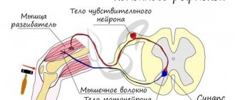
Each spinal nerve leaves the spinal cord with two roots: posterior (sensitive) and anterior (motor); both roots are connected into one trunk, truncus n. spinalis, emerging from the spinal canal through the intervertebral foramen. Near and somewhat outward from the junction, the dorsal root forms a node, ganglion spinale, in which the anterior motor root does not participate. Due to the connection of both roots, spinal nerves are mixed nerves: they contain sensory (afferent) fibers from the cells of the spinal ganglia, motor (efferent) fibers from the cells of the anterior horn, as well as vegetative fibers from the cells of the lateral horns, emerging from the spinal cord as part of the anterior root. Vegetative fibers are also present in the dorsal root.
Autonomic fibers entering the animal nerves through the roots provide processes in the soma such as trophism, vasomotor reactions, etc. In cyclostomes (lampreys), both roots continue into separate nerves - motor and sensory. In the further course of evolution, starting with transverse fish, the roots come closer together and merge, so that the separate course is maintained only for the roots, and the nerves become mixed.
Each spinal nerve, upon exiting the intervertebral foramen, is divided into two branches, respectively, into two parts of the myotome (dorsal and ventral):
- posterior, ramus dorsalis, for the autochthonous muscles of the back and the skin covering it, developing from the dorsal part of the myotome;
- anterior, ramus ventralis, for the ventral wall of the trunk and limbs, developing from the ventral parts of the myotomes.
In addition, two more types of branches depart from the spinal nerve:
- for the innervation of the viscera and blood vessels - connecting branches to the sympathetic trunk, rr. communicantes;
- for innervation of the spinal cord membranes - r. meningeus, going back through the intervertebral foramen.
Posterior branches of the spinal nerves. The posterior branches, rami dorsales, of all spinal nerves go back between the transverse processes of the vertebrae, bending around their articular processes. All of them (with the exception of the I cervical, IV and V sacral and coccygeal) are divided into ramus medialis and ramus lateralis, which supply the skin of the back of the head, the back of the neck and back, as well as the deep spinal muscles.
Posterior branch of the 1st cervical nerve, n. suboccipitalis emerges between the occipital bone and the atlas and then divides into branches supplying mm. recti capitis major et minor, m. semispinalis capitis, mm. obliqui capitis. To skin n. suboccipitalis does not produce branches. Posterior branch of the II cervical nerve, n. occipitalis major, emerging between the posterior arch of the atlas and the second vertebra, then pierces the muscles and, becoming subcutaneous, innervates the occipital region of the head. The rami dorsales of the thoracic nerves are divided into medial and lateral branches, giving branches to the autochthonous muscles; the cutaneous branches of the upper thoracic nerves arise only from the rami mediales, and those of the lower ones - from the rami laterales.
The cutaneous branches of the three upper lumbar nerves go to the upper part of the gluteal region called the nn. clunium superiores, and the cutaneous branches of the sacral ones are called nn. Clunium mediai. Anterior branches of the spinal nerves. The anterior branches, rami ventrales, of the spinal nerves innervate the skin and muscles of the ventral wall of the body and both pairs of limbs. Since the skin of the abdomen in its lower part takes part in the development of the external genitalia, the skin covering them is also innervated by the anterior branches. The latter, except for the first two, are much larger than the hind ones.
The anterior branches of the spinal nerves retain their original metameric structure only in the thoracic region (nn. intercostales). In the remaining sections associated with the limbs, during the development of which segmentation is lost, the fibers extending from the anterior spinal rami are intertwined. This is how nerve plexuses, plexus, are formed, in which fibers of different neuromeres are exchanged. A complex redistribution of fibers occurs in the plexuses: the anterior branch of each spinal nerve supplies its fibers to several peripheral nerves, and, therefore, each of them contains fibers from several segments of the spinal cord.
It is therefore clear that damage to one or another nerve is not accompanied by dysfunction of all muscles receiving innervation from the segments that gave rise to this nerve. Most of the nerves arising from the plexuses are mixed; therefore, the clinical picture of the lesion consists of motor disturbances, sensory disturbances and autonomic disorders. There are three large plexuses: cervical, brachial and lumbosacral. The latter is divided into lumbar, sacral and coccygeal.