The limbic system within the brain performs the task of organizing the coordinated activity of all brain regions. Encircling the brain stem, this structure closely interacts with other parts of the brain, directing complex behavioral reactions, participating in the formation of emotional manifestations and motivation. Developed functional connections support various forms of body activity - preservation of life and species, somatovegetative integration, processes of concentration, memorization, and emotional response. The system is equipped with numerous afferent and efferent inputs and outputs.
Definition
The limbic system consists of a number of brain structures responsible for processing feelings and emotions to lead to new memories and bodily changes. It processes these stimuli as electrical signals throughout the central nervous system, allowing memory formation as well as autonomic and behavioral changes. Ultimately, the limbic system communicates with the endocrine system to allow the release of hormones so that the body can properly respond to presented stimuli.
How it works?
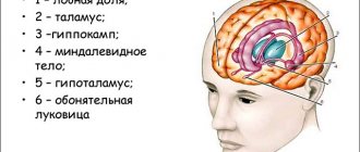
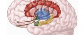
The limbic system spans multiple regions of the brain, and the full functionality of the system remains to be fully understood. Despite this, scientists have identified many structures that are important to the limbic system, such as the limbic lobe, hippocampal formation, amygdala, thalamus, and hypothalamus. These structures are able to communicate with each other, stimulating the individual cells of the structures, known as neurons. Eventually, connections are established between neurons that enable rapid processing from the initial stimulus to the final physical change.
Almond nuclei
In a series of brain autopsies of patients with schizophrenia, no significant increase in the volume of the amygdala nuclei was detected (Steven A. et al., 2002). However, data obtained using MRI currently refers to rather crude imaging methods, not to mention CT, and may be less reliable compared to autopsy.
In a comparative review by Wright et al (2000), who studied the size of 44 brain regions in schizophrenia, it was noted that the amygdala of the left and right hemispheres was reduced in volume by 10%, and this was noticeable to a greater extent than the reduction in volume of other brain regions. Loss of gray matter in the amygdala and changes in the shape of the amygdala have also been demonstrated through studies that looked at ventricular-brain index scores. At the same time, many authors note methodological difficulties in studying the amygdala of the limbic system of patients with schizophrenia.
Return to Contents
Overview of the Nervous System
The nervous system is made up of individual nerve cells known as neurons, which are specialized in their ability to interact with electrical signals at speeds of up to 120 meters per second. These high speeds are critical to the functioning of the nervous system because it allows a single neuron to send information to and from the brain almost instantly.
- Dendrites, which convert and receive electrical signals
- cell body (or soma) that contains the nucleus and processes signal
- An axon that transmits a signal to the next neuron
At rest, neurons have a membrane potential, which is determined by the concentration of ions on either side of the neuron's membrane. When a stimulus is received, neurons create a rapid and temporary change in this potential, now called an action potential. These action potentials quickly change the polarization of the neuron, allowing signals to be transmitted between neurons.
Neurons form the entire nervous system, which is divided into two subsystems: the central nervous system and the peripheral nervous system. The central nervous system (CNS) includes the brain and spinal cord, and the peripheral nervous system (PNS) includes all other neurons distributed throughout the body. When discussing the limbic system we will primarily focus on the CNS brain, and when discussing the hypothalamus we will primarily focus on the PNS.
Clinical manifestations. Disturbances in the limbic system
1- Dementia
The limbic system is associated with the causes of neurodegenerative diseases, in particular Alzheimer's disease and Pick's disease. These pathologies are accompanied by atrophy in the limbic system, especially in the hippocampus. In Alzheimer's disease, senile plaques and neurofibrillary tangles (tangles) appear.
2- Anxiety
Anxiety disorders result from disturbances in the regulation of amygdala activity. The scientific literature has detailed a fear circuit involving the amygdala, prefrontal cortex, and anterior cingulate cortex of the brain. (Cannistraro, 2003).
3- Epilepsy
Epilepsy may manifest itself as a consequence of changes in the limbic system. Temporal lobe epilepsy is most common in adults, and occurs as a result of sclerosis in the hippocampus. It is believed that this type of epilepsy is associated with dysfunction at the level of the limbic system.
Structures, connections and functions of the limbic system
Limbic lobe
The dentate cortex is the inner part of the brain that is located superior (above) and dorsal (behind) the corpus callosum. It consists of an anterior (frontal) and posterior (posterior) division, which both receive information from the thalamus and hippocampus formation.
The main function of this area is to regulate both autonomic function and conscious function. Autonomic functions involve involuntary changes, such as increasing heart rate to a threat in the environment, while conscious responses involve voluntary choices, such as deciding to respond to a threat (such as taking a step back). Conscious response is also important in controlling emotions. In other words, rather than perceiving the emotion itself (such as feeling pain), the cingulate cortex controls what a person thinks about the emotion (such as how you feel about pain).
The parahippocampal gyrus consists of several distinct areas in the medial temporal lobe. The most significant of these areas includes the entorhinal cortex, which the parahippocampal gyrus uses to send information from the cortex to the hippocampal formation.
Hippocampal formation
The hippocampus is a horn-like structure that surrounds the lateral ventricles. The main function is to process new information and form long-term memories, which are then stored in other areas of the brain. If the damaged person is unable to create new long-term memories. However, a person can remember long-term memories that have already been stored.
The hippocampus most often receives information from the entorhinal cortex of the parahippocampal gyrus. When received, information is usually mediated through execution. An efficient pathway sends signals using fibers throughout the subiculum of the specific complex and into the dentate gyrus, where fibers then extend from the dentate gyrus to CA3 axons. Schaffer collaterals then connect to dendrites on CA1 axons. CA1 axons contain fibers that extend from the hippocampus to subsequent brain regions.
amygdala
The amygdala supports two main information processing pathways: dorsal and ventral. Both routes send information to the hypothalamus, while the ventral route also sends information to the thalamus.
thalamus
*Note: Smell is not processed by the thalamus. Smell is received by the olfactory bulbs and is directly sent for emotional processing and memory formation.
Hyopthalamus
The hypothalamus is also located in the diencephalon, which is inferior to the thalamus and both inferior and lateral to the third ventricle. The main function is homeostatic regulation, as the hypothalamus directly controls the autonomic nervous system (ANS). The ANS is part of the PNS, which controls the involuntary actions and rhythms of internal organs, including heart rate, blood pressure, and digestive movements. The ANS covers two subsystems – the Sympathetic Nervous System (“Fight or Flight”) and the Parasympathetic Nervous System (“Rest and Digest”).
The hypothalamus is the central structure that must be stimulated to create physical changes in the body. This is because the hypothalamus is directly connected to the pituitary gland and thus immediately influences the hormones released by the pituitary gland. This connection is a crosstalk between the nervous system and the endocrine system, translating neural stimuli received by the brain into hormonal changes throughout the body.
Alternatively, the hypothalamus may release neurosecretory cells into blood vessels that penetrate and activate the anterior pituitary gland. The anterior pituitary gland then increases or decreases the hormones distributed in the blood depending on whether neurosecretory cells from the hypothalamus are directed to stimulate or inhibit a particular hormone.
The part of the pituitary gland with which the hypothalamus interacts - anterior or posterior - depends on the signal received by the brain and the needs of the body.
Hippocampus
In a study by C. McDonald et al. (2006) revealed a decrease in the volume of the right and left hippocampus, 2.47 ml and 2.50 ml, versus 2.54 ml and 2.61 ml in healthy individuals, which corresponds to approximately a decrease in the volume of this brain structure by approximately 6%.
Some authors note that a decrease in the volume of the hippocampus, as part of the limbic system, is noticeable after the first psychotic episode, however, according to other researchers, these changes are recorded before the manifestation of schizophrenia and progress after its onset. Note that in relatives of patients with schizophrenia, a decrease in the volume of the hippocampus and amygdala can also be detected.
In schizophrenia, the functioning of the amygdala is impaired; this fact is discovered when studying patients with schizophrenia using the method of evoked potentials (P300).
In the hippocampus of patients with schizophrenia, with a decrease in the volume of neurons and “sparseness” of their mutual arrangement, an increase in the number of pathologically altered myelinated axons with thinned myelin sheaths, swollen perioxal glial processes and wrinkled axons was found. The proportion of these fibers in the total number of myelinated axons and their numerical density in schizophrenia is 2 times greater than in the control group. At the same time, it is known that pathological and reparative changes in axons depend on the reaction of the microglial cells surrounding them (Kolomeets N.S., 2007).
Peculiar memory impairments, primarily working memory, in schizophrenia, affecting learning difficulties in persons suffering from this disease, may indicate the involvement of the hippocampus in the pathological process.
According to Shenton et al. (2001), the volumes of the medial temporal lobes, which usually include the hippocampus and amygdala, are significantly reduced in size. These changes were noted in 70% of patients with schizophrenia.
Bibliography
Show hide
Functions
The limbic system is responsible for the following functions:
- Olfactory.
- Communicative.
- Short-term and long-term memory.
- Regulates sleep.
- Regulates the functioning of the internal organs of the body.
- Forms motivation and emotions.
- Participates in intellectual processes.
- Forms the vegetative and endocrine activity of the body.
- Partly forms sexual and food instincts.
The functions of the limbic system are not limited to those listed.
Due to its anatomical structure, it is the main structure in coordinating the vital functions of the body. Summarizing signals from the external and internal environment, it analyzes them and sends commands, thereby activating a number of somatic and autonomic reactions. This structure helps regulate the body’s adaptive reactions to external stimuli and maintain internal balance at an optimal level. That is why its dysfunction is so significant for humans.
When some of its structures are irritated, the functions of internal organs are significantly disrupted. For example, when the tonsils are affected, cardiac activity is disrupted, intestinal paresis occurs or peristalsis is accelerated, the secretion process of the stomach changes, disruptions are also observed in the hormonal sphere, the pituitary gland is especially susceptible to them.
In addition, the limbic system is responsible for the adequate functioning of the wakefulness-sleep chain. It also regulates metabolic processes in the body, affects water-salt metabolism and temperature balance.
The main social significance of the visceral brain system is the formation of emotions . In experiments on animals, it was proven that when part of its structures, namely the tonsils, is removed, it leads to uncertainty, anxiety, and a decrease in aggression. When performing electrical stimulation of the tonsils, people, on the contrary, experienced irritability, aggression, fear, and panic attacks.
When the frontal cortex is damaged, a person experiences emotional lability, this is especially evident when assessing emotions aimed at satisfying one’s needs. All these studies prove the significant role of the visceral brain in the emotional, and therefore social, spheres.
Another important feature of the visceral brain is participation in the learning process . The main role in this is played by the posterior regions of the frontal cortex and the hippocampus. Their importance in the transformation of short-term memory into long-term memory can hardly be overestimated. Dysfunction of these structures leads to the inability to learn new knowledge and the absence of long-term memory formation.
It was previously believed that, due to its anatomical structure, the visceral brain was responsible only for processing data received from the olfactory organs. Nowadays, scientists have proven that this is not so and it is capable of analyzing signals received from various sources.
The limbic system is responsible for a person’s social adaptation in the outside world and his adaptation to changes in society.
Limbic system disorders
When there are disorders of the visceral brain, memory is primarily affected. And although the limbic system is not an archive, the processes of reproduction and restoration of knowledge and skills are disrupted, memories remain, but become scattered.
There are many reasons for its violation, the main ones include the following:
- head injuries;
- infections affecting the nervous system;
- neurotoxins;
- diseases of the vascular system of the brain;
- psychiatric pathology;
- alcohol poisoning.
As a result of these diseases, disorders of the visceral brain manifest themselves in the form of frequent mood swings, loss of orientation, psychiatric pathologies (visual, olfactory and auditory hallucinations), confusion (sporosis), disorders of the gastrointestinal tract, cardiovascular, endocrine and immune systems, in special cases – epileptoid conditions (depending on the location of the pathological process).
The visceral brain, like the nervous system as a whole, has not yet been fully studied. Scientists are still conducting research to reliably establish all the functions and methods of correcting conditions caused by dysfunction.