Types of neurons
The anatomical and histological unit of the nervous system is the neuron - a nerve cell and its processes.
Types of neurons.
By localization:
- central (located in the central nervous system);
- peripheral (located outside the central nervous system - in the spinal, cranial ganglia, in the autonomic ganglia, in the plexuses and intraorgan).
Functionally:
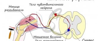
- receptor (afferent, sensitive) are those nerve cells through which impulses travel from receptors to the central nervous system. They are divided into: primary afferent neurons - their bodies are located in the spinal ganglia, they have a direct connection with receptors and secondary afferent neurons - their bodies lie in the visual thalamus, they transmit impulses to the overlying sections, they are not connected to receptors, they receive impulses from others neurons;
- Efferent neurons transmit impulses from the central nervous system to other organs. Motor neurons are located in the anterior horns of the spinal cord (alpha, beta, gamma motor neurons) and provide a motor response. Neurons of the autonomic nervous system: preganglionic (their bodies lie in the lateral horns of the spinal cord), postganglionic (their bodies lie in the autonomic ganglia);
- intercalary (interneurons) - ensure the transmission of impulses from afferent to efferent neurons. They make up the bulk of the gray matter of the brain and are widely represented in the brain and its cortex. Types of interneurons: excitatory and inhibitory neurons.
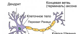
A neuron consists of a body, an axon, and dendrites.
The neuron body contains all the components of cellular structures and is capable of generating nerve impulses and performing a trophic function. At the origin of the axon there is a section of unmyelinated fiber (about 50-100 nm) - this is the initial segment. It is here that the highest activity (level of excitability) is the trigger zone, here the difference between the membrane potential and Ek is 7-10 mV.
An axon is a long extension that carries impulses from the nerve cell body. It can be myelinated or unmyelinated and ends at various synapses.
Dendrites are short, highly branched processes that conduct impulses to the neuron body and ensure interaction between neurons of the central nervous system.
Neuron sizes: diameter from 4-6 microns to 130 microns. Membrane potential - 50-90 mV; action potential amplitude 80-120 mV. The neuron membrane at rest is highly permeable to K+, and when excited it is highly permeable to Na+ and Ca2+.
Functions of neuroglia:
- supporting (prevents neuron deformation);
- trophic (regulates metabolic processes in nervous tissue);
- regulation of ionic composition (concentration of ions on both sides of the membrane);
- regulation of blood supply to the central nervous system.
Central neurons have their own characteristics.
The ability for spontaneous depolarization is the spontaneous generation of nerve impulses. The reason is that neurons form complex closed circuits within the central nervous system, where spontaneous release of the transmitter occurs.
Long period of trace hyperpolarization. After excitation occurs, neurons remain in a state of reduced excitability for a long time and, as a consequence, low lability.
Interneurons have a short period of trace hyperpolarization and, as a consequence, lability increases to 1000 impulses/s. Motor neurons have a longer period of trace hyperpolarization, so their lability is 500 impulses/s for alpha motor neurons, and 50-100 impulses/s for gamma motor neurons.
Identification of various mediators. Depending on the type of mediators, there are 2 types of nerve cells: cholinergic and adrenergic.
Central and peripheral motor neuron
Beginning in the motor cortex, the pyramidal tract goes deep into the hemisphere, gradually gathers into a compact bundle, passes through the anterior two-thirds of the posterior limb of the internal capsule, descending into the brainstem (corticonuclear tract) and the spinal cord (corticospinal tract). In the lower part of the medulla oblongata, most of the pyramidal fibers of the corticospinal tract cross over - they pass into the lateral cord of the spinal cord of the opposite side, a smaller part (about 10%) remains in the anterior cord (Turk's bundle) on its side.
In the dorsal parts of the brain stem and the anterior horns of the spinal cord, the pyramidal tract ends on the dendrites of the corresponding motor neurons. The axons of the latter, in the form of motor roots of the brain stem or anterior roots of the spinal cord, come out, forming the motor (efferent) part of the cranial and spinal nerves, and innervate all the muscles of the head, neck, torso and limbs of a person. In this case, the motor neurons of the anterior horns of the spinal cord have predominantly cross connections, and the motor neurons of the motor nuclei of the brain stem have bilateral connections with the motor area of the cortex.
Rice. Nuclei of the oculomotor and trochlear nerves (III, IV pairs of cranial nerves). 1 - trochlear nerve (nervus trochlearis); 2 - oculomotor nerve (nervus oculomotorius); 3-7 - nucleus of the oculomotor nerve (nucl. nervi oculomotorii); from the nuclear cells the fibers go to the following muscles: 3 - musculus rectus inferior; 4 - musculus rectus internus; 5 - musculus rectus superior; 6 - musculus obligus inferior; 7 - musculus levator palpebrae superior; 8 - accessory nucleus of the oculomotor nerve (nucl. oculomotorius accessorius, small cell nucleus of Edinger-Westphal-Yakubovich).
In the precentral gyrus there is a well-defined projection of the opposite half of the human body. In the uppermost sections the leg is represented, in the middle sections there are the torso and arm, in the lower sections there are the facial muscles, the muscles of the tongue, and the pharynx, i.e. the person is positioned upside down, as it were.
In each hemisphere there are about 25-40 thousand Betz cells; the pyramidal tract at the level of the cerebral pons contains more than 1 million axons, i.e., a significant part of its fibers originates from the neurons of the cerebral cortex, which are part of the extrapyramidal-cerebellar and afferent systems.
In the brain stem and spinal cord there are several hundred thousand motor cells, i.e. one pyramidal fiber starting from Betz cells controls several motor neurons through a system of interneurons.
In the anterior horns of the spinal cord, motor neurons are located in groups (in the upper cervical and thoracic regions there are 3, in the cervical and lumbar enlargement - 5), and each of them innervates certain muscles of the trunk, shoulder and pelvic girdles, limbs, etc. In each of the groups cells of the anterior horns of the spinal cord and in the motor nuclei of the cranial nerves, three types of neurons differ in function - alpha large cells, alpha small cells and gamma neurons. Alpha large cells receive impulses from the pyramidal system and ensure the execution of voluntary movements, alpha small cells - from the extrapyramidal system and are involved in maintaining muscle tone, gamma neurons - from the reticular formation and influence the excitability of the neuromuscular spindle and the maintenance of muscle tone.
There are especially many motor neurons in the cervical and lumbar enlargements of the spinal cord, since innervation of the limbs—the muscles of the arms and legs—is provided from these levels. The axillary nerve (p. axillaris) innervates the deltoid muscle (m. deltoideus), which raises the arm to a horizontal level; musculocutaneous nerve (n. musculocutanens) - biceps brachii muscle (m. biceps brachii), which flexes the arm at the elbow joint; radial nerve (n. radialis) - triceps brachii muscle (m. triceps brachii), which extends the arm at the elbow joint. The median (n. te-dianus), ulnar (n. ulnaris) and radial (n. radialis) nerves innervate the muscles that flex and extend the hand, etc.
With the help of the femoral nerve (n. femoralis), the iliopsoas muscle (m. iliopsoas), which flexes the leg at the hip joint, and the quadriceps femoris muscle (m. guadriceps femoris), which extends the leg at the knee joint, are innervated; inferior gluteal nerve (n. glutaens inferior) - the gluteus maximus muscle (m. gluteus maximas), which extends the leg at the hip joint; peroneal nerve (n. peronaeus) - tibialis anterior muscle (m. tibialis anterior), which extends the foot; tibial nerve (n. tibialis) - triceps surae muscle (m. triceps surae), flexor of the foot, etc.
The facial muscles are innervated by cranial nerves. In structure and function, the neurons of the nuclei of the motor cranial nerves are analogues of the cells of the anterior horns of the spinal cord. Motor functions are performed by nine pairs of cranial nerves (six pairs - III,
IV, VI, VII, XI, XII are motor and three pairs - V, IX, X - mixed). The nuclei of the cranial nerves are located in the brain stem, mainly in the middle or dorsal parts of it (at the bottom of the rhomboid fossa). The axons of the cells of these nuclei form motor roots, and then nerves that emerge from the brain stem and innervate the muscles of the face, larynx, pharynx, tongue and partly the neck.
The nuclei of the cranial nerves that control the movements of the eyeball are located: oculomotor and trochlear (III, IV) in the tegmentum of the midbrain at the level of the quadrigeminal at the bottom of the aqueduct; abducens nerve (VI) - in the dorsal part of the pons and the bottom of the fourth ventricle.
Article on the topic Central and peripheral motor neuron
Related pages:
- Nerves of the pyramidal system Article content1 Nerves of the pyramidal system 1.1 Trochlear nerve 1.2 Abducens nerve 1.3 Nucleus of the facial nerve 1.3.1 Nucleus of the accessory nerve 1.4 Nucleus…
- PYRAMIDAL SYSTEM
PYRAMIDAL SYSTEM The term “pyramidal system” is similar to the concept of “motor analyzer”, which was introduced into neurology by I. P. Pavlov, trying to show that the motor... - Sympathetic part
Sympathetic part Sympathetic cells are located in the lateral horns of the spinal cord at the level from CVII to LII-LIV segments. The peripheral processes of these... - The structure of the spinal cord
The structure of the spinal cord The spinal cord is a long cord (its length in an adult is about 45 cm), somewhat flattened in front... - Human autonomic nervous system
Contents of the article1 Human autonomic nervous system1.1 General plan of the structure of the autonomic nervous system1.2 Sympathetic part1.3 Function of the autonomic nervous system Autonomic nervous system… - Central peripheral paralysis
Contents of the article1 Peripheral paralysis is 1.1 Symptoms of central peripheral paralysis 1.2 Symptoms of peripheral paralysis 1.3 Diagnosis of peripheral paralysis Peripheral paralysis is In severe ...
Peripheral neuron of the motor pathway. Symptoms of defeat. The main path of voluntary movements.
This is a two-neuron pathway
connecting the cerebral cortex with the skeletal (striated) muscles (corticomuscular pathway).
There are central and peripheral neurons
of this pathway.
The central
neuron is located in the Y layer (layer of Betz's large pyramidal cells) of the anterior central gyrus, in the posterior parts of the superior and middle frontal gyri and in the paracentral lobule.
There is a clear somatic distribution of these cells. The cells located in the upper part of the precentral gyrus and in the paracentral lobule innervate the lower limb and trunk, located in its middle part - the upper limb. In the lower part of this gyrus there are neurons that send impulses to the face, tongue, pharynx, larynx, and masticatory muscles. The axons of these cells in the form of two conductors (the corticospinal tract
(otherwise called the pyramidal tract) - from the upper two-thirds of the anterior central gyrus and the
cortico-bulbar tract
- from the lower part of the anterior central gyrus) go from the cortex deep into the hemispheres, passing through the internal capsule (the corticobulbar tract is in the knee area, and the corticospinal tract is through the anterior two-thirds of the posterior thigh of the internal capsule).
Then the cerebral peduncles, pons, and medulla oblongata pass through, and at the border of the medulla oblongata and spinal cord, the corticospinal tract undergoes an incomplete decussation. The large, crossed part of the tract passes into the lateral column of the spinal cord and is called the main, or lateral, pyramidal fasciculus. The smaller uncrossed part passes into the anterior column of the spinal cord and is called the direct uncrossed fasciculus. The fibers of the corticobulbar tract end in the motor nuclei of the cranial nerves (Y, YII, IX, X, XI, XII
), and the fibers of the corticospinal tract end in
the anterior horns of the spinal cord
. Moreover, the fibers of the corticobulbar tract undergo decussation sequentially as they approach the corresponding nuclei of the cranial nerves (“supranuclear” decussation). For the oculomotor, masticatory muscles, muscles of the pharynx, larynx, neck, trunk and perineum, there is bilateral cortical innervation, i.e. fibers of central motor neurons approach part of the motor nuclei of the cranial nerves and some levels of the anterior horns of the spinal cord not only from the opposite side, but also to some levels of the anterior horns of the spinal cord. but also with one’s own, thus ensuring the approach of impulses from the cortex not only of the opposite, but also of one’s hemisphere. The limbs, tongue, and lower part of the facial muscles have unilateral (only from the opposite hemisphere) innervation. The axons of the spinal cord motor neurons are directed to the corresponding muscles as part of the anterior roots, then the spinal nerves, plexuses and, finally, the peripheral nerve trunks.